Dear Editor,
Transoesophageal echocardiography (TEE) allows imaging of unparalleled quality because of the proximity of esophagus to cardiac structures without interposition of pulmonary or parietal structures.[1] The main indications for TEE include acute aortic endocarditis, thromboembolic accidents, cryptogenic stroke, and valvular heart disease.[1] TEE also plays an invaluable role in diagnosing and monitoring the patient's hemodynamics during cardiac and noncardiac surgery.[2] Guidelines for performing a comprehensive TEE have been made by the American Society of Echocardiography and the Society of Cardiovascular Anesthesiologists.[3] TEE is usually done by cardiologists and anesthesiologists who have described incidental extracardiac findings such as liver abnormalities, inferior vena cava filling defects, and mediastinal masses.[4] The route of esophagus is significant for endoscopic ultrasonography (EUS) of mediastinum for gastroenterologists and pulmonologists. Normal cardiovascular anatomy of mediastinum has been described by endosonographers as well as by cardiologists.[5] However, the assumption that entire heart can be visualized by TEE/EUS is wrong, as some interference in the pathway of ultrasound beam is always present due to tracheobronchial and pulmonary structures between the esophagus and the heart. The images given in this letter define the blind areas of cardiac imaging during TEE/EUS [Figures 1–8 and Table 1]. This knowledge can be of importance for endosonographers who perform TEE/EUS.
Figure 1.
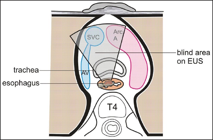
The trachea lies in front of the esophagus and the presence of air in trachea interfere with imaging during TEE/EUS. As shown in this figure the joining of the azygos vein with the superior vena cava is a blind area of imaging. The part of the ascending aorta lying in front of the left bronchus and left lower part of the trachea is also a blind area of imaging
Figure 8.
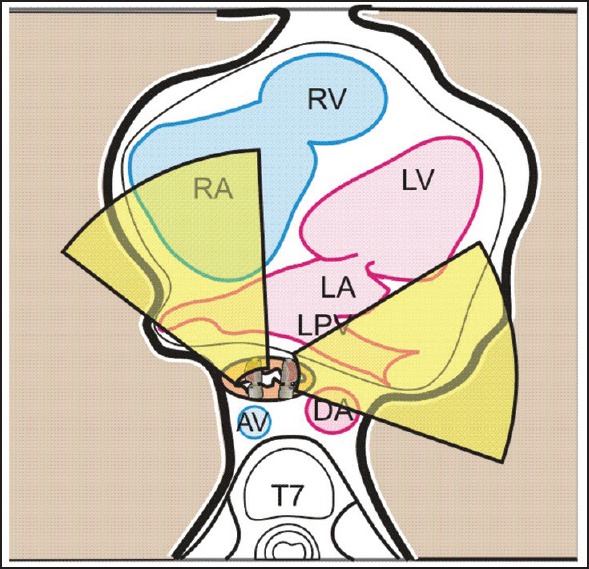
Left atrium is identified in the subcarinal area. From the left atrium, the left ventricular outflow tract and from the pulmonary artery, the right ventricular outflow tract can be followed. In this area no blind area is seen
Table 1.
Blind areas of cardiac imaging during TEE/EUS
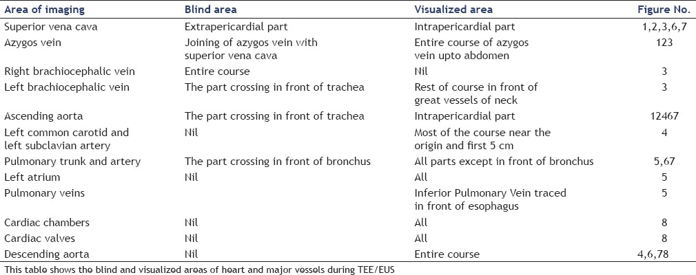
Figure 2.
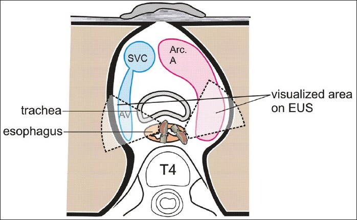
The visualized area of the azygos vein and aorta at the level of the arch
Figure 3.
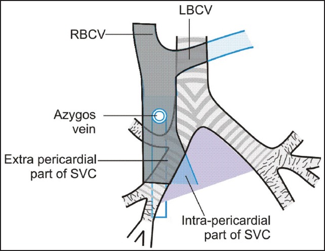
The shaded portion is a blind area of the TEE/EUS imaging (the extrapericardial part of the superior vena cava, right brachiocephalic, and the part of the left brachiocephalic vein)
Figure 4.
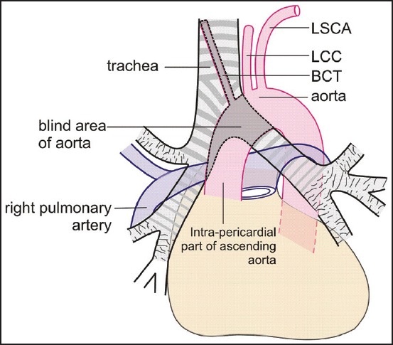
The shaded portion is the blind area of imaging of the ascending aorta and its branch (brachiocephalic trunk)
Figure 5.
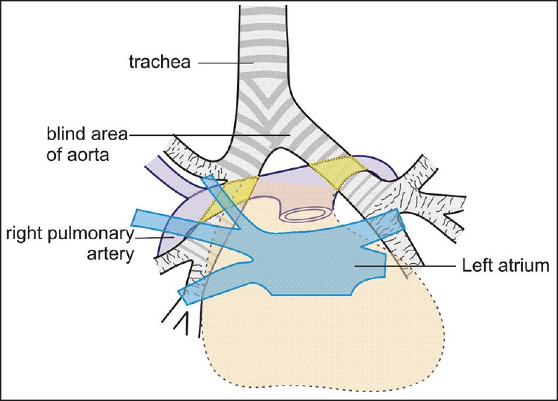
The shaded portion (yellow) is a blind area of imaging of the right and left pulmonary artery
Figure 6.
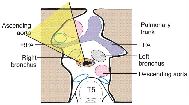
The area within the zone of yellow beam is a blind area of imaging (of the ascending aorta and superior vena cava) in front of the right pulmonary artery due to obstruction from the right bronchus
Figure 7.
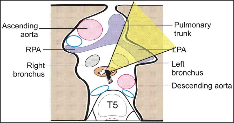
The area within the zone of yellow beam is a blind area of imaging in front of the left bronchus (left pulmonary artery)
Financial support and sponsorship
Nil.
Conflicts of interest
There are no conflicts of interest.
REFERENCES
- 1.Lesbre JP. The main indications for transesophageal echocardiography. Ann Cardiol Angeiol (Paris) 1995;44:547–51. [PubMed] [Google Scholar]
- 2.Marymont J, Murphy GS. Intraoperative monitoring with transesophageal echocardiography: Indications, risks, and training. Anesthesiol Clin. 2006;24:737–53. doi: 10.1016/j.atc.2006.08.007. [DOI] [PubMed] [Google Scholar]
- 3.Hahn RT, Abraham T, Adams MS, et al. American Society of Echocardiography; Society of Cardiovascular Anesthesiologists. Guidelines for performing a comprehensive transesophageal echocardiographic examination: Recommendations from the American Society of Echocardiography and the Society of Cardiovascular Anesthesiologists. Anesth Analg. 2014;118:21–68. doi: 10.1213/ANE.0000000000000016. [DOI] [PubMed] [Google Scholar]
- 4.Alkhouli M, Sandhu P, Wiegers SE, et al. Extracardiac findings on routine echocardiographic examinations. J Am Soc Echocardiogr. 2014;27:540–6. doi: 10.1016/j.echo.2014.01.026. [DOI] [PubMed] [Google Scholar]
- 5.Hahn RT, Abraham T, Adams MS, et al. Guidelines for performing a comprehensive transesophageal echocardiographic examination: Recommendations from the American Society of Echocardiography and the Society of Cardiovascular Anesthesiologists. J Am Soc Echocardiogr. 2013;26:921–64. doi: 10.1016/j.echo.2013.07.009. [DOI] [PubMed] [Google Scholar]


