Abstract
Background:
Noncontact anterior cruciate ligament (ACL) injury in adolescent female athletes is an increasing problem. The knee-ankle separation ratio (KASR), calculated at initial contact (IC) and peak flexion (PF) during the drop vertical jump (DVJ), is a measure of dynamic knee valgus. The Microsoft Kinect V2 has shown promise as a reliable and valid marker-less motion capture device.
Hypothesis:
The Kinect V2 will demonstrate good to excellent correlation between KASR results at IC and PF during the DVJ, as compared with a “gold standard” Vicon motion analysis system.
Study Design:
Descriptive laboratory study.
Level of Evidence:
Level 2.
Methods:
Thirty-eight healthy volunteer subjects (20 male, 18 female) performed 5 DVJ trials, simultaneously measured by a Vicon MX-T40S system, 2 AMTI force platforms, and a Kinect V2 with customized software. A total of 190 jumps were completed. The KASR was calculated at IC and PF during the DVJ. The intraclass correlation coefficient (ICC) assessed the degree of KASR agreement between the Kinect and Vicon systems.
Results:
The ICCs of the Kinect V2 and Vicon KASR at IC and PF were 0.84 and 0.95, respectively, showing excellent agreement between the 2 measures. The Kinect V2 successfully identified the KASR at PF and IC frames in 182 of 190 trials, demonstrating 95.8% reliability.
Conclusion:
The Kinect V2 demonstrated excellent ICC of the KASR at IC and PF during the DVJ when compared with the Vicon system. A customized Kinect V2 software program demonstrated good reliability in identifying the KASR at IC and PF during the DVJ.
Clinical Relevance:
Reliable, valid, inexpensive, and efficient screening tools may improve the accessibility of motion analysis assessment of adolescent female athletes.
Keywords: anterior cruciate ligament, injury prevention, Microsoft Kinect, knee-ankle separation ratio, drop vertical jump
An anterior cruciate ligament (ACL) tear is a major life event, requiring surgical reconstruction and months of rehabilitation.17 Biomechanical and neuromuscular imbalances have been studied as factors influencing an increased risk of an ACL injury, especially in young female athletes.4,29 The primary mechanism of injury in female athletes occurs during noncontact activities such as landing, pivoting, and cutting during sport play, while ACL injuries in male athletes occur more often through contact mechanisms.2 This difference may reflect the severity of the biomechanical and neuromuscular imbalances relating to ACL injury in the female athletic population. Dynamic knee valgus has been specifically described as altered hip and knee kinematics in the frontal and transverse planes and has been linked in some studies to a biomechanical and neuromuscular imbalance related to ACL injuries in female athletes.10,18,34 It has been established as a movement pattern characterized by excessive femoral adduction, femoral internal rotation, knee abduction, and external tibial rotation.1,26,28 The early identification and investigation of such poor movement patterns through specific movement analysis has gained significant attention in an attempt to address the risk of ACL injury in female athletes.7,16,20
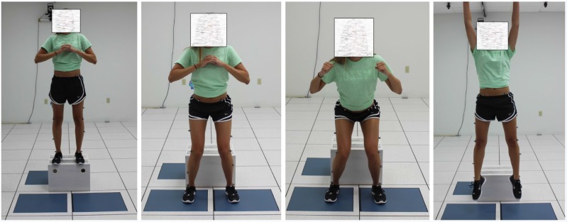
Previous investigators have examined the role of altered kinematics during movements such as the drop vertical jump (DVJ).11-13,16,20,25 The DVJ is performed with a participant standing on a 31-cm-high platform and dropping from the platform to the ground, immediately followed by a maximal vertical jump (similar to the movement needed to get a rebound in basketball) (Figure 1).12,13,16 Some studies have linked the DVJ as a functional movement for identifying high school female athletes with an elevated risk for ACL injury.11,12,16 Multiple kinematic and kinetic risk factors, including but not limited to knee valgus at initial contact (IC), peak knee abduction moment, peak knee flexion angle, peak vertical ground-reaction force, and medial knee displacement, have been analyzed during the DVJ.7,12,13,16,20 It has shown high within-session reliability values for kinematics and kinetics, with intraclass correlation coefficients (ICCs) of 0.93 to 0.99 and 0.66 to 0.93, respectively.11 In 205 female athletes participating in high-risk sports, the kinematic analysis of the DVJ showed that those who suffered an ACL injury had significantly different knee abduction angles between ACL-injured and uninjured groups.16 The female athletes who went on to suffer an ACL injury had an increased abduction angle of 8.4° at IC and 7.6° at peak flexion (PF). The injured group also lacked 10° of knee flexion at PF as compared with the uninjured group.16 Although the DVJ has shown early promise, controversies remain in its reliability and predictability of future ACL injury.3,19,24,31 The predictive value of the DVJ appears to be population specific, demonstrating the highest potential predictive value to identify noncontact ACL injury risk in untrained middle and high school female athletes.7,12,13,16,20
Figure 1.
Stages of the drop vertical jump (left to right): (1) initial stance, (2) initial contact, (3) peak flexion, and (4) vertical jump.
If high-risk biomechanics and altered neuromuscular control are identified early, these poor movement patterns can be corrected with specific ACL injury prevention programs, leading possibly to a relative risk reduction of 40% to 73% for overall ACL injuries and noncontact injuries, respectively.33 Unfortunately, the current methods of accurate, objective movement analysis can only be performed in select elaborate motion analysis laboratories with the use of expensive ($100,000 to $150,000) high-speed motion analysis cameras and force plates, which are labor intensive and time consuming.
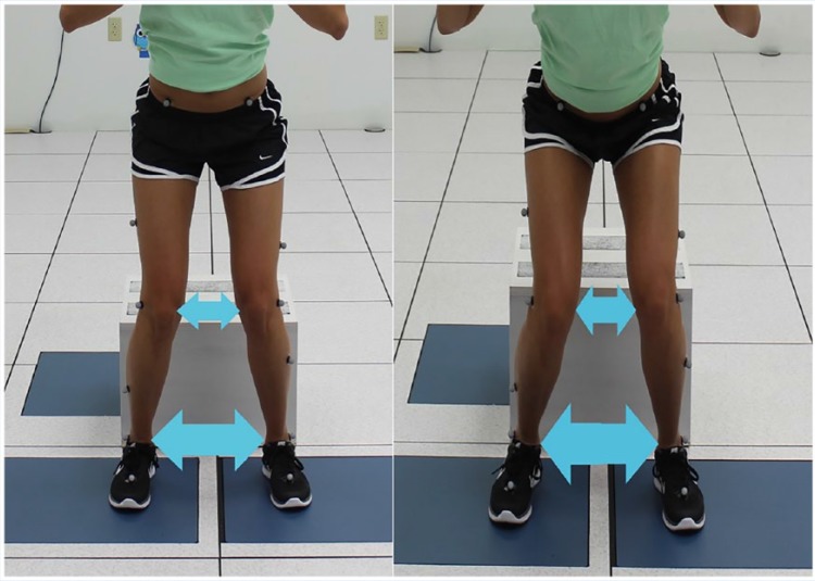
Subsequently, alternative technologies and outcome measures have been proposed to assess dynamic knee valgus during the DVJ.25,27,28,33 The Kinect V2 (Kinect for Windows Sensor; Microsoft) motion sensor is an efficient, cost-effective, reliable, and validated portable tool for calculating 3-dimensional motion analysis.21,23,32 In 2013, investigators examined the Kinect (version 1.0) sensor and the device’s applicability to accurately measure the knee-ankle separation ratio (KASR) at IC and PF during the DVJ.15,32 Using customized Microsoft Kinect–based software, these investigators found excellent correlation between the Kinect (version 1) and the frontal plane projection of the KASR as produced by an 8-camera Vicon motion analysis system (Vicon Motion System Ltd).15,32 The KASR has been proposed as an alternative outcome measure to assess dynamic knee valgus during the DVJ.25,28 The KASR is a ratio of the distances between the knees and ankles, measured individually at the time points of IC and PF during the DVJ (Figure 2). A KASR ratio of 1.0 represents the knee directly above the ankles, a ratio of <1.0 represents the knees medial to the ankles, and a ratio of >1.0 represents the knees lateral to the ankles.28 A moderate association between knee abduction angle and KASR has been shown during the DVJ, with further evaluation of this association recommended.25 Previous investigators used a 2-dimensional camera-based system, and although this is a cost-effective and portable option, efficiency and accuracy limitations persist.29
Figure 2.
Visual representation of the knee-ankle separation ratio at initial contact (left) and peak flexion (right).
The purpose of this study was to compare KASR measures from the Kinect V2 to an 8-camera Vicon motion capture system. We hypothesized that the Kinect V2 will demonstrate a good to excellent correlation between KASR measurements at IC and PF during the DVJ as compared with a “gold standard” marker-based Vicon motion capture system.
Methods
With institutional review board approval, 38 healthy volunteer subjects (20 men: mean age, 24.79 ± 2.68 years; mean weight, 83.79 ± 14.25 kg; mean height, 1.80 ± 0.07 m and 18 women: mean age, 24.10 ± 3.68 years; mean weight, 65.01 ± 9.58 kg; mean height, 1.64 ± 0.05 m) were recruited. Each participant signed an approved consent form prior to participation. Participants were excluded if a history of neurological illness or lower extremity injury within the past 12 months was present. All subjects wore tight-fit clothing or shorts and self-selected athletic shoes during data collection. Anthropometric measurements included body mass index, height, interanterior superior iliac spine distance, leg length, knee width, and ankle width for both limbs.
The DVJ was performed with the participant standing on a 31-cm-high platform, dropping from the platform to the ground, and immediately performing a maximal vertical jump.12,13,16 The 2 specific time points of interest during the DVJ were IC and PF.
An 8-camera Vicon MX-T40S retroreflective motion capture system was used, with data acquired at 100 Hz. Sixteen skin-surface retroreflective markers were placed on the trunk and legs according to the protocol of the lower body Plug-in-Gait model.8 Four additional markers were placed on the medial epicondyle of the knee and medial malleoli of the ankle to estimate the thigh rotation offset, shank rotation offset, and tibial torsion. These markers were removed after a static pose was acquired. Vicon kinematic data were filtered using a fourth-order zero-lag low-pass Butterworth filter with 6-Hz cutoff and processed using the Vicon Nexus software, and the Plug-in-Gait model was applied to estimate 3-dimensional joint centers. Additionally, 2 AMTI force platforms (Optima; Advanced Mechanical Technology Inc) were used to find IC during the DVJ by setting the force threshold to 10 N, with resultant KASR measurement by the Vicon. The KASR at PF was recorded using the Vicon system. With medial-lateral motion acting along the x-axis, the equation used for the Vicon KASR measure was:
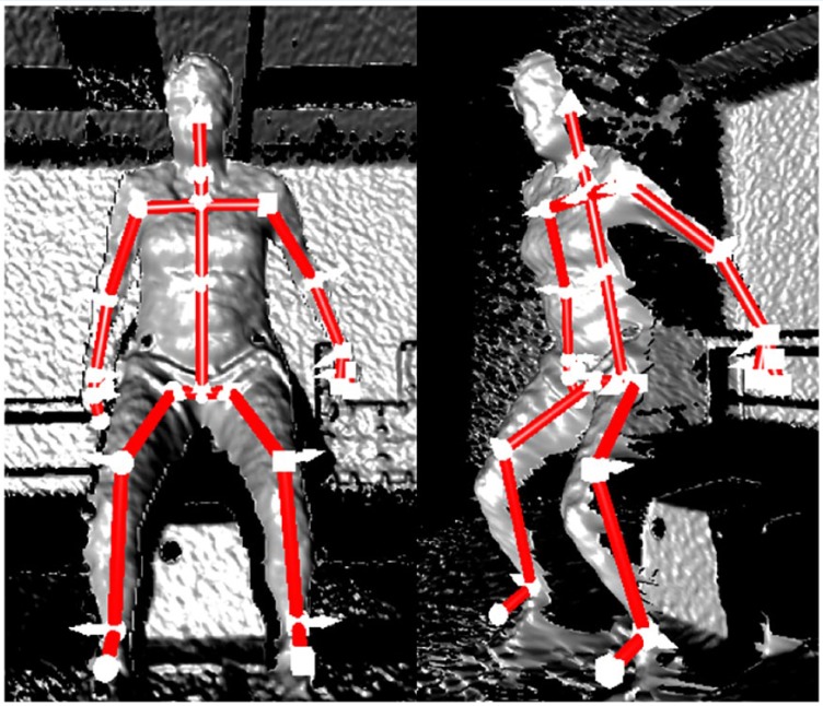
A Kinect V2 sensor was simultaneously used with the 8-camera Vicon system and 2 force platforms (Figure 3). The Kinect V2 uses an infrared time-of-flight depth sensor for 3-dimensional tracking at 30 frames per second.5 In a human detection task by the Kinect sensor, a 512 × 424 pixel image is first captured by the depth sensor. The Kinect V2 software then identifies and segments an individual from the depth image, matching each body to a 25-joint skeletal model (Figure 4). An output of the Kinect skeletal model is the 3-dimensional location of these 25 joints, including the knee and ankle joint centers. The Microsoft Kinect Software Development Kit, which is a customizable development package for the device, allowed the investigators to develop a software application to obtain a 1-dimensional KASR measure during the DVJ at IC and PF, obtained from the knee and ankle joint centers of the Kinect skeletal model. Two key frames were extracted, determining the IC and PF by the Kinect V2. IC was identified as the moment when the foot first contacted the floor after the initial drop from the platform. The software calculated this by evaluating the ankle and foot joint centers and finding the first frame where the velocity of the joints decreases, indicating that the foot has reached the ground. PF was determined as the moment when the knees were at a maximum angle of flexion after IC and prior to leaving the floor for the vertical jump. The software calculated this by taking the mean position of the hip joint centers and the spine base and identified the frame with the lowest position of these joint centers relative to the floor. The Kinect V2 was positioned 2.5 m from the front of the 31-cm-high jumping platform. With the x-axis aligned in the medial-lateral direction, the Kinect system measured the KASR by using the joint coordinates according to the following equation:
Figure 3.
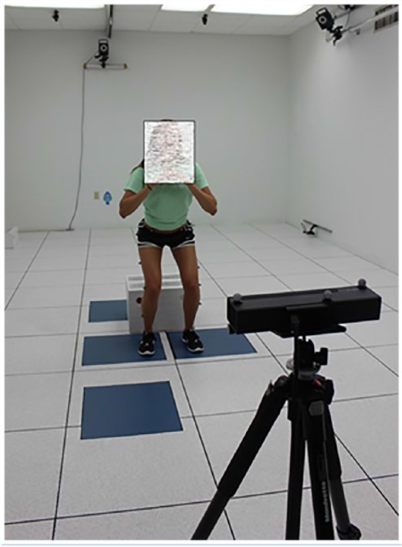
The Kinect V2 system as it is used in the laboratory space.
Figure 4.
The Kinect V2 system establishes a skeletal model overlay during the drop vertical jump.
Verbal instructions regarding proper technique of the DVJ were given to each subject according to previous study procedures.13,16 Subjects were able to perform 1 practice DVJ to become comfortable with the movement task. The 2 force platforms were aligned on the floor in front of the 31-cm box. Subjects performed the DVJ for 5 recorded trials, resulting in a total of 190 recorded jumps. The Kinect software recorded still images of the frames identified as IC and PF. The jump was considered unsuccessful when manual review of the images clearly did not identify either time point of interest. The KASR at IC and PF was then independently calculated for each individual jump by the Kinect and Vicon systems. The ICC (2-way, single measure, absolute agreement) was used to assess the degree of KASR agreement between the Kinect and Vicon systems at IC and PF. Standard interpretations of the ICC have suggested that values greater than 0.75 indicate excellent agreement between measures.35
Results
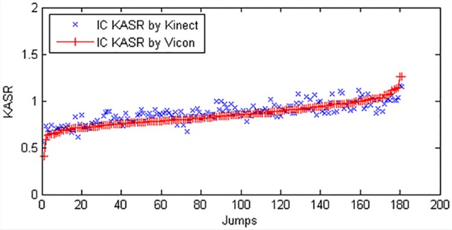
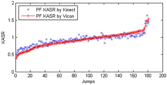
The Kinect customized software did not accurately detect the IC or PF in 8 of 190 (4.2%) jumps. The success rate was therefore 95.8%, demonstrating a high overall reliability of the Kinect V2 system. There were no injuries suffered during the DVJ jump trials. There was excellent agreement between the Kinect and Vicon KASR measure at IC and PF during 182 DVJ jump trials (Table 1). These findings demonstrated that the ICCs (P ≤ 0.05) of the Kinect V2 and Vicon KASR at IC and PF were 0.84 and 0.95, respectively (Figures 5 and 6). The mean KASR at IC contact was 0.84 ± 0.12 for the Vicon and 0.87 ± 0.11 for the Kinect system. The mean KASR at PF was 0.92 ± 0.20 for the Vicon and 0.93 ± 0.16 for the Kinect system.
Table 1.
Knee-ankle separation ratio during the drop vertical jump measured by the Kinect V2 and Vicon at initial contact and peak flexion
| Initial Contact |
Peak Flexion |
|||
|---|---|---|---|---|
| Mean ± SD | ICC (P ≤ 0.05) | Mean ± SD | ICC (P ≤ 0.05) | |
| Vicon | 0.84 ± 0.12 | 0.84 | 0.92 ± 0.20 | 0.95 |
| Kinect | 0.87 ± 0.11 | 0.93 ± 0.16 | ||
ICC, intraclass correlation coefficient.
Figure 5.
Comparison of Kinect V2 and Vicon: Knee-ankle separation ratio at initial contact (IC KASR).
Figure 6.
Comparison of Kinect V2 and Vicon: Knee-ankle separation ratio at peak flexion (PF KASR).
Discussion
The investigators’ primary hypothesis was that the Microsoft Kinect V2 sensor would demonstrate a good to excellent correlation between KASR measurements at IC and PF during the DVJ as compared with a “gold standard” marker-based Vicon system. Kinect V2 demonstrated excellent correlation (ICC > 0.75)35 with the Vicon motion system; additionally, the Kinect V2 was able to successfully calculate the KASR in 182 of 190 jumps, demonstrating a reliability of 95.8%.
Initial studies examining the Kinect’s capabilities used the first-generation Kinect motion sensor, whereas the Kinect V2 sensor provides improved depth and image sensor capabilities.32 Previous work compared the Kinect V1 with a simple frontal plane projection measure produced by the Vicon system without formal joint center estimation.32 The current study compared the KASR from the Kinect V2 correlated data with data from an 8-camera Vicon motion capture system capable of performing more advanced 3-dimensional joint center estimations with the Plug-in-Gait model. This enhances the validity of the Kinect V2 by examining its ability to estimate the knee and ankle joint centers and resultant KASR during the DVJ.
Multiple studies have shown inconsistencies on the predictive value of the DVJ as a screening tool for ACL injury risk, with its greatest utility in 13- to 19-year-old female athletes.7,14,16,19,20,24,31 Krosshaug et al20 studied the correlation of the DVJ to noncontact ACL injury in elite female soccer and handball players aged 21 ± 4 years. Previously proposed kinematic and kinetic measurements showed no association with noncontact ACL injury risk in previously uninjured athletes.20 Additionally, Smith et al31 found that a modified DVJ, as measured by the Landing Error Scoring System, was not predictive of noncontact ACL injuries. As a result, the authors acknowledge the importance of 3 essential methodological steps as proposed by Bahr3 to improve development of screening tests for injury risk assessment. These steps include initial prospective cohort studies to identify risk factors and cutoff values, followed by validation testing of the cutoff value in multiple cohorts, and finally the implementation of randomized controlled trials to test the effect of combined screening and intervention programs.3 Although the DVJ has shown early promise, implementation of such suggestions with reliable and validated tools to assess the DVJ need to be considered.3,30
A limitation of the Kinect V2 is the 30-Hz sample rate, which may be insufficient to capture peak motion during explosive movements. This study used the Kinect V2 to calculate the KASR at IC and PF. Measurement errors due to an insufficient sample rate would manifest from the medial-lateral motion of the knee while the feet are stationary and from inexact identification of the IC and PF events. Although data were acquired simultaneously between the Vicon and Kinect V2 systems, identification of IC and PF events were independent. The high correlation in KASR measurements between the 2 systems indicates that although the low sample rate is a limitation, the Kinect system may still accurately measure KASR during the 2 event time frames. Additionally, a well-known limitation of marker-based motion analysis is the interaction of soft tissue mobility between the markers and bones, producing an unpredictable effect and potentially altering the precision and accuracy of measurements.6,9,22
Several other limitations of this study exist. These include the small sample size, limited target population, and need for further examination of the KASR as a predictive measure for noncontact ACL injury. Finally, although the KASR has shown a moderate correlation to the knee abduction angle, a previously identified measure linked to an increased risk for noncontact ACL injuries, further investigation into the predictive value of the KASR is warranted.25
The Kinect V2 showed excellent correlation to a “gold standard” Vicon motion capture system in the identification of the KASR at IC and PF during the DVJ.35 Such results provide promise that portable, low-cost, and easy-to-use motion sensors may play an important role in the future of motion analysis.
Footnotes
The following author declared potential conflicts of interest: Seth L. Sherman, MD, is a paid consultant for Arthrex, Neotis, Regeneration Technologies Inc, and Vericel.
References
- 1. Alentorn-Geli E, Myer GD, Silvers HJ, et al. Prevention of non-contact anterior cruciate ligament injuries in soccer players. Part 1: mechanisms of injury and underlying risk factors. Knee Surg Sports Traumatol Arthrosc. 2009;17:705-729. [DOI] [PubMed] [Google Scholar]
- 2. Arendt EA, Agel J, Dick R. Anterior cruciate ligament injury patterns among collegiate men and women. J Athl Train. 1999;34:86-92. [PMC free article] [PubMed] [Google Scholar]
- 3. Bahr R. Why screening tests to predict injury do not work—and probably never will . . .: a critical review. Br J Sports Med. 2016;50:776-780. [DOI] [PubMed] [Google Scholar]
- 4. Carson DW, Ford KR. Sex differences in knee abduction during landings: a systematic review. Sports Health. 2011;3:373-382. [DOI] [PMC free article] [PubMed] [Google Scholar]
- 5. Castaneda V, Navab N. “Time-of-flight and kinect imaging.” Computer aided medical procedures. Kinect Programming for Computer Vision. Summer Program 2011. http://campar.in.tum.de/twiki/pub/Chair/TeachingSs11Kinect/2011-DSensors_LabCourse_Kinect.pdf. Accessed September 30, 2016.
- 6. Chiari L, Della Croce U, Leardini A, Cappozzo A. Human movement analysis using stereophotogrammetry. Part 2: instrumental errors. Gait Posture. 2005;21:197-211. [DOI] [PubMed] [Google Scholar]
- 7. Cruz A, Bell D, McGrath M, Blackburn T, Padua D, Herman D. The effects of three jump landing tasks on kinetic and kinematic measures: implications for ACL injury. Res Sports Med. 2013;21:330-342. [DOI] [PubMed] [Google Scholar]
- 8. Davis RB, 3rd, Öunpuu S, Tyburski D, Gage JR. A gait analysis data collection and reduction technique. Hum Movement Sci. 1991;10:575-587. [Google Scholar]
- 9. Della Croce U, Leardini A, Chiari L, Cappozzo A. Human movement analysis using stereophotogrammetry. Part 4: assessment of anatomical landmark misplacement and its effects on joint kinematics. Gait Posture. 2005;21:226-237. [DOI] [PubMed] [Google Scholar]
- 10. Earl JE, Monteiro SK, Snyder KR. Differences in lower extremity kinematics between a bilateral drop-vertical jump and a single-leg step-down. J Orthop Sports Phys Ther. 2007;37:245-252. [DOI] [PubMed] [Google Scholar]
- 11. Ford KR, Myer GD, Hewett TE. Reliability of landing 3D motion analysis: implications for longitudinal analyses. Med Sci Sports Exerc. 2007;39:2021-2028. [DOI] [PubMed] [Google Scholar]
- 12. Ford KR, Myer GD, Hewett TE. Valgus knee motion during landing in high school female and male basketball players. Med Sci Sports Exerc. 2003;35:1745-1750. [DOI] [PubMed] [Google Scholar]
- 13. Ford KR, Myer GD, Toms HE, Hewett TE. Gender differences in the kinematics of unanticipated cutting in young athletes. Med Sci Sports Exerc. 2005;37:124-129. [PubMed] [Google Scholar]
- 14. Goetschius J, Smith HC, Vacek PM, et al. Application of a clinic-based algorithm as a tool to identify female athletes at risk for anterior cruciate ligament injury: a prospective cohort study with a nested, matched case-control analysis. Am J Sports Med. 2012;40:1978-1984. [DOI] [PMC free article] [PubMed] [Google Scholar]
- 15. Gray AD, Marks JM, Stone EE, Butler MC, Skubic M, Sherman SL. Validation of the Microsoft Kinect as a portable and inexpensive screening tool for identifying ACL injury risk. Orthop J Sports Med. 2014;2(2 suppl):2325967114S00106. [Google Scholar]
- 16. Hewett TE, Myer GD, Ford KR, et al. Biomechanical measures of neuromuscular control and valgus loading of the knee predict anterior cruciate ligament injury risk in female athletes: a prospective study. Am J Sports Med. 2005;33:492-501. [DOI] [PubMed] [Google Scholar]
- 17. Ireland ML. The female ACL: why is it more prone to injury? Orthop Clin North Am. 2002;33:637-651. [DOI] [PubMed] [Google Scholar]
- 18. Kadaba MP, Ramakrishnan HK, Wootten ME, Gainey J, Gorton G, Cochran GV. Repeatability of kinematic, kinetic, and electromyographic data in normal adult gait. J Orthop Res. 1989;7:849-860. [DOI] [PubMed] [Google Scholar]
- 19. Krosshaug T, Steffen K, Kristianslund E, et al. Screening tests for ACL injury: response. Am J Sports Med. 2016;44:NP26-NP27. [DOI] [PubMed] [Google Scholar]
- 20. Krosshaug T, Steffen K, Kristianslund E, et al. The vertical drop jump is a poor screening test for ACL injuries in female elite soccer and handball players: a prospective cohort study of 710 athletes. Am J Sports Med. 2016;44:874-883. [DOI] [PubMed] [Google Scholar]
- 21. LaBelle K. Evaluation of Kinect Joint Tracking for Clinical and In-Home Stroke Rehabilitation Tools [bachelor’s thesis]. Notre Dame, IN: University of Notre Dame; 2011. http://netscale.cse.nd.edu/cms/pub/Edu/KinectRehabilitation/Eval_of_Kinect_for_Rehab.pdf. Accessed September 1, 2016. [Google Scholar]
- 22. Leardini A, Chiari L, Della Croce U, Cappozzo A. Human movement analysis using stereophotogrammetry. Part 3. Soft tissue artifact assessment and compensation. Gait Posture. 2005;21:212-225. [DOI] [PubMed] [Google Scholar]
- 23. Livingston MA, Sebastian J, Ai Z, Decker JW. Performance measurements of the Microsoft Kinect skeleton. IEEE Virtual Reality Short Papers and Posters. 2012. https://pdfs.semanticscholar.org/5348/160d2ffc90d937102fcc5757f860799da028.pdf. Accessed September 1, 2016.
- 24. McCunn R, Aus der Fünten K, Fullagar HH, McKeown I, Meyer T. Reliability and association with injury of movement screens: a critical review. Sports Med. 2016;46:763-781. [DOI] [PubMed] [Google Scholar]
- 25. Mizner RL, Chmielewski TL, Toepke JJ, Tofte KB. Comparison of two-dimensional measurement techniques for predicting knee angle and moment during a drop vertical jump. Clin J Sports Med. 2012;22:221-227. [DOI] [PMC free article] [PubMed] [Google Scholar]
- 26. Myer GD, Ford KR, Brent JL, Hewett TE. An integrated approach to change the outcome part I: neuromuscular screening methods to identify high ACL injury risk athletes. J Strength Cond Res. 2012;26:2265-2271. [DOI] [PMC free article] [PubMed] [Google Scholar]
- 27. Myer GD, Ford KR, Hewett TE. New method to identify athletes at high risk of ACL injury using clinic-based measurements and freeware computer analysis. Br J Sports Med. 2011;45:238-244. [DOI] [PMC free article] [PubMed] [Google Scholar]
- 28. Ortiz A, Rosario-Canales M, Rodriguez A, Seda A, Figueroa C, Venegas-Rios H. Reliability and concurrent validity between two-dimensional and three-dimensional evaluations of knee valgus during drop jumps. Open Access J Sports Med. 2016;7:65-73. [DOI] [PMC free article] [PubMed] [Google Scholar]
- 29. Renstrom P, Ljungqvist A, Arendt E, et al. Non-contact ACL injuries in female athletes: an International Olympic Committee current concepts statement. Br J Sports Med. 2008;42:394-412. [DOI] [PMC free article] [PubMed] [Google Scholar]
- 30. Shrier I, Verhagen E, Stovitz SD. Screening tests for ACL injury [letter to the editor]. Am J Sports Med. 2016;44:NP26. [DOI] [PubMed] [Google Scholar]
- 31. Smith HC, Johnson RJ, Shultz SJ, et al. A prospective evaluation of the Landing Error Scoring System (LESS) as a screening tool for anterior cruciate ligament injury risk. Am J Sports Med. 2012;40:521-526. [DOI] [PMC free article] [PubMed] [Google Scholar]
- 32. Stone EE, Butler M, McRuer A, Gray A, Marks J, Skubic M. Evaluation of the Microsoft Kinect for screening ACL injury. Conf Proc IEEE Eng Med Biol Soc. 2013;2013:4152-4155. [DOI] [PubMed] [Google Scholar]
- 33. Sugimoto D, Myer GD, McKeon JM, Hewett TE. Evaluation of the effectiveness of neuromuscular training to reduce anterior cruciate ligament injury in female athletes: a critical review of relative risk reduction and numbers-needed-to-treat analyses. Br J Sports Med. 2012;46:979-988. [DOI] [PMC free article] [PubMed] [Google Scholar]
- 34. Willson JD, Davis IS. Utility of the frontal plane projection angle in females with patellofemoral pain. J Orthop Sports Phys Ther. 2008;38:606-615. [DOI] [PubMed] [Google Scholar]
- 35. Zaki R, Bulgiba A, Nordin N, Ismail N. A systematic review of statistical methods used to test for reliability of medical instruments measuring continuous variables. Iran J Basic Sci. 2013;16:803-807. [PMC free article] [PubMed] [Google Scholar]


