Abstract
Introduction
Live peripheral blood mononuclear cells (PBMCs) can be frozen and thawed for later analyses by adding and removing a cryoprotectant, such as dimethyl sulfoxide (DMSO). Laboratories across the world use various procedures, but published evidence of optimal thawing procedures is scarce.
Materials and methods
PBMCs were separated from blood collected from healthy Danish blood donors, and stored at -80°C after adding of DMSO. The essential steps in the thawing procedure were modified and performance was evaluated by flow cytometry with respect to the percentage and total yield of viable PMBCs.
Results
The best-performing washing medium was Roswell Park Memorial Institute (RPMI) 1640 at 37°C with 20% fetal bovine serum. When using 10 mL washing medium in a 15-mL Falcon tube, samples should be centrifuged for at least 10 minutes at 500 g. We failed to detect any differences between the tested methods of mixing PBMCs with washing medium. Likewise, neither the thawing duration nor centrifugation temperature (20°C and 37°C) had any effect. PBMCs could be incubated (rested) for up to eight hours in a 37°C 5% CO2 incubator without affecting cell counts, but incubating PBMCs for 16 hours significantly decreased viability and recovery. In general, high viability was not necessarily associated with high recovery.
Conclusion
Changing the thawing procedure significantly impacted PBMC viability and live cell recovery. Evaluating both viability and live PBMC recovery are necessary to evaluate method performance. Investigation of differential loss of PBMC subtypes and phenotypic changes during thawing and incubation requires further evaluation.
1. Introduction
Analyses of human peripheral blood mononuclear cells (PBMCs) can be done on freshly collected blood samples or on cryopreserved samples. Analysis of freshly collected samples is usually preferred because the cryopreservation could affect functional and phenotypic properties of cells [1–7]. On the other hand, using cryopreserved PBMCs could minimize operator-dependent inter-assay [8] and inter-laboratory variation [9], which is evident in multi-center clinical trials with local sample collection [10,11].
The freezing / thawing procedure consists of several steps. Although dimethyl sulfoxide (DMSO) is often used as cryoprotectant, a high concentration of DMSO is toxic to human leukocytes [12,13]. Therefore, cells are usually washed as part of the thawing process to minimize the duration of DMSO exposure [14]. Each step in the thawing process can be performed in several ways. Thawing time, type and temperature of washing medium, method of mixing cells and washing medium, centrifugation and incubation conditions vary between laboratories, and are all parameters that could be adjusted to optimize yield [15].
Published studies and protocols vary with respect to the method used to thaw PBMCs, and several publications fail to describe the method used [16]. There is a general lack of evidence-based cell thawing procedures that ensure maximum cell recovery. In this study, we systematically evaluated different steps in the procedure of thawing human PBMCs using paired samples from healthy blood donors. Our outcome measurements consisted of PBMC viability and absolute count of live PBMCs (PBMC recovery).
2. Materials and methods
2.1 Blood donor enrollment
Peripheral venous blood samples were collected from voluntary blood donors at the blood bank in Aarhus, Denmark [17]. Each donor contributed two filled 4-mL heparin tubes, and further sample processing was initiated within two hours.
2.2 Isolation of PBMCs
First, blood samples were diluted with an equal volume of tris-buffered (pH 7.5) saline solution (BSS) (Ampliqon A/S, Odense, Denmark). The diluted blood was then carefully layered on top of a volume of Lymphoprep (Stemcell Technologies Inc., Vancouver, Canada) equal to the original volume of the blood sample. The solutions were centrifuged at 1,000 g for 20 min at 20°C for formation of the PBMC layer. Meanwhile, a 15-mL polypropylene conical centrifuge tube (Corning Science, Tamaulipas, Mexico) with 2 mL BSS was prepared for each donor. After centrifugation, sterile glass pipettes were used to aspirate the PBMCs, and cells were transferred to the 15-mL tubes with BSS. PBMCs were then centrifuged again at 500 g for 10 min at 20°C followed by aspiration of the supernatant. Subsequently, PBMCs were resuspended, mixed with 4 mL locally produced heat-inactivated human serum pool (HSP) at 4°C from donors with blood type AB, and stored for 10 min at 4°C.
2.3 Freezing procedure
For each donor, 4 mL cryopreservation medium was prepared, consisting of 3.2 mL RPMI 1640 without L-Glutamine (ThermoFisher Scientific Inc., Waltham, USA) and 0.8 mL DMSO (OriGen Biomedical, Austin, USA). Mixing was performed by dropwise addition of the 4°C cryopreservation medium to the PBMCs suspended in HSP. Thus, the final concentration of DMSO was 10%. For each donor, 1 mL cell suspension was added to each of 8 cryotubes, with each tube thus containing a number of PMBCs corresponding to that obtained from 1 mL whole blood. The cryotubes were then immediately placed directly in a rack in a -80°C freezer, where they were stored until thawing. Samples were stored for up to 2 weeks before thawing and cell counting.
2.4 Evaluation of thawing methods
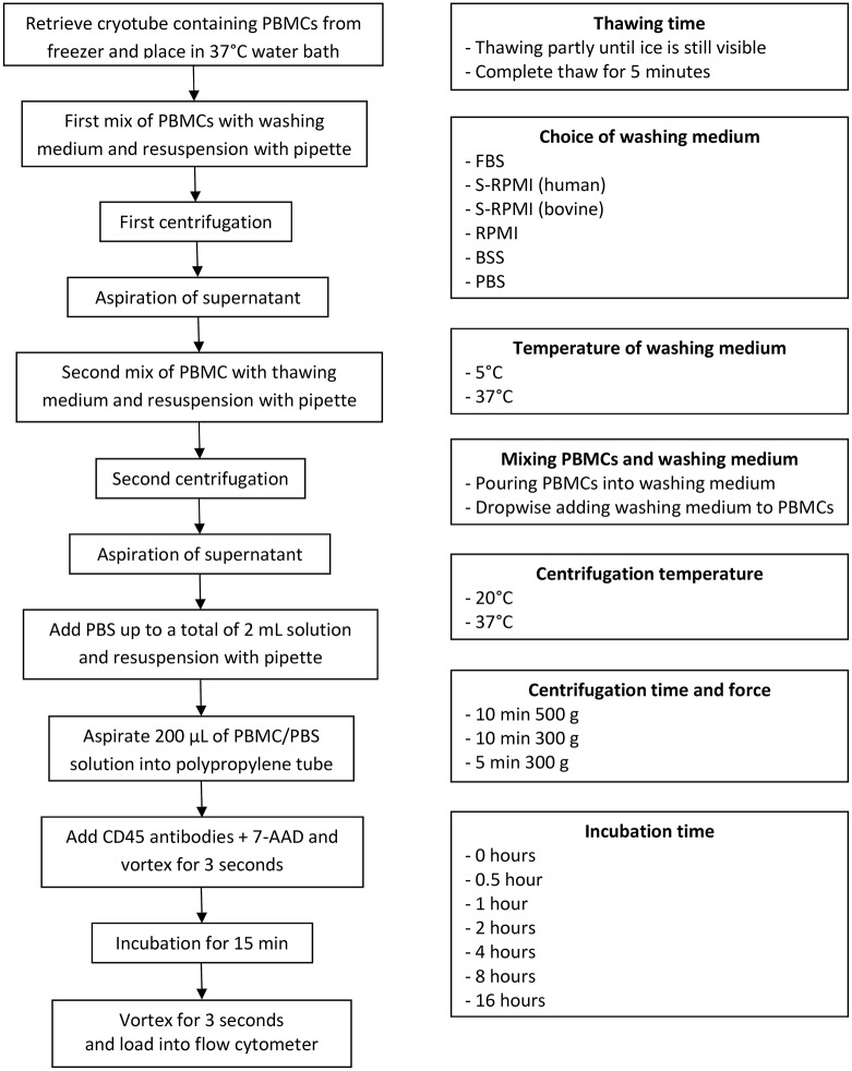
For each evaluation, PBMC viability and live PBMC count were analyzed on paired donor samples; i.e., when an evaluation contained six variations of a step in the thawing process, then six cryotubes with frozen PBMCs from each donor were included. For each evaluation, one parameter in the thawing process was adjusted while all other parameters were kept constant. If nothing else is stated, the constant parameters consisted of partly thawed PBMCs with ice is still visible, using 20% S-RPMI (FBS) as thawing medium at 37°C, mixing PBMCs and thawing medium by pouring PBMCs into the washing medium, leaving the centrifuge at room temperature (20°C), centrifuging for 10 minutes at 500 g, and not incubating PBMCs before staining.
In all evaluations, the thawing process consisted of retrieving the PBMC-containing cryotubes from the -80°C freezer and placing them in a 37°C water bath for rapid thawing (Fig 1). The procedure involved two washing steps. First, using a Pasteur pipette the thawed PBMCs were mixed with 10 mL washing medium in a 15-mL polypropylene tube and centrifuged. The supernatant was then aspirated. Second, an additional 10 mL of washing medium was added, and the PBMCs were resuspended using a Pasteur pipette. The tube was then centrifuged once more, and the supernatant aspirated. The resuspended cells were then transferred to polypropylene tubes (Beckham-Coulter, 12x75 mm round bottom).
Fig 1.
Flow chart of the thawing and staining process (on the left). Variations in the thawing procedure (on the right).
2.5 Flow cytometry
After the two washing steps, PBMCs were resuspended in phosphate buffered saline (PBS). Samples were stained with 1 uL FITC-conjugated mouse-anti-human-CD45 antibody (BD, clone 2D1, Catalogue number 345808) for leukocyte count and 1 uL 7-AAD (1 mM) solution (Nordic BioSite, catalogue number ABD-17501) for viability staining. Absolute cell counting was performed using a volumetric NovoCyte flow cytometer (ACEA Biosciences, San Diego, USA). Gating was performed using NovoExpress 1.2.1 software (ACEA Biosciences, Inc., San Diego, USA), as visualized in Fig 2.
Fig 2. Gating strategy to identify live/dead CD45+ leukocytes.
A) Events were triggered on FSC-H at a deliberately low threshold to avoid accidental exclusion of dead cells. B) Doublet exclusion C) Identification of CD45 positive cells D) Dead cells identified as 7-AAD positive events. Compensation for spectral overlap was not required.
2.6 Statistical analyses
The best-performing method for each evaluation was identified by comparing paired means of the percentage of viable cells and by comparing paired absolute counts of live leukocytes. The thawing procedures were compared using paired t-test after ensuring normal distribution in Stata IC 13.0 (StataCorp, Texas, USA). Figs 3–9 were made using PRISM (Graphpad Software, CA, USA).
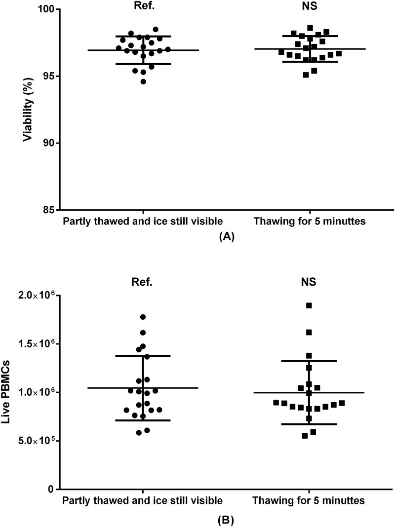
Fig 3. PBMC viability (A) and absolute recovery (B) as a result of varying thawing time in 37°C heated water bath.
Paired samples from 20 donors included. Ref.: reference group. NS: non-significant.
Fig 9. PBMC viability (A) and absolute live PBMC recovery (B) by incubation.
Samples were stored in a 37°C incubator with 5% CO2. Paired samples from 20 donors were included. Ref.: reference group. NS: non-significant. *: p<0.05. **: p<0.01.
2.7 Ethics
Blood samples were collected anonymized from volunteer donors during routine blood donation as part of a quality assurance program (project number 119). According to Danish law, this does not require separate ethical approval.
3. Results
3.1 Thawing time
As previously suggested [16], cryotubes containing frozen PBMCs can be kept in a 37°C heated water bath until either the content is partly thawed and ice is still visible (approximately 1 minute and 15 seconds), or until the content of the cryotubes has reached 37°C for several minutes. We evaluated the thawing time in heated water bath by comparing partly thawed samples with samples kept in the water bath for 5 minutes. The choice of method affected neither viability nor absolute PBMC count (Fig 3).
3.2 Choice of washing medium
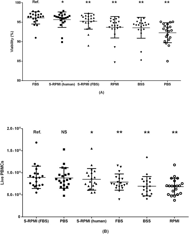
A total of six different washing media were evaluated; BSS, FBS, PBS, RPMI, S-RPMI (bovine) and S-RPMI (human). The washing medium producing highest mean PBMC viability was FBS (96.1%), followed closely by S-RPMI (human) (95.7%) and S-RPMI (bovine) (95.2%), Fig 4. However, the best performing washing medium in terms of live PBMC recovery was S-RPMI (bovine), which resulted in a significant higher live PBMC count than S-RPMI (human) (1.06 fold difference, p = 0.04), and the other media except PBS. This demonstrates that cell viability and absolute cell recovery are not necessarily associated. Indeed, a harsh thawing method could result in total disintegration of compromised cells to a degree where all “fragile” dead cells are no longer discernible as individual cells in flow cytometric analysis, and only the most robust, live cells “make it”. Such a method would have a high viability (inasmuch as no dead cells are left)–but a low yield.
Fig 4. Performance of six different washing media presented in order of declining performance.
Values presented with mean +/- SD. (A) PBMC viability (%) and (B) live PBMCs by thawing medium. Paired samples from 20 donors included. Ref.: reference group. NS: non-significant. *: p<0.05. **: p<0.01.
3.3 Temperature of washing medium
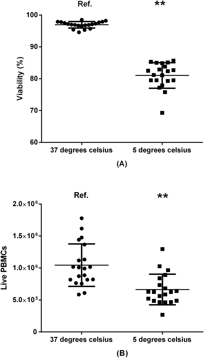
In the first washing step, washing media (20% S-RPMI (bovine)) were either pre-heated to 37°C in a water bath, or pre-cooled in a 4°C refrigerator (Fig 5). Mean viability was higher for cells washed with 37°C medium (96.9%) compared to 4°C Medium (81.0%, p<0.01). Similarly live PBMC recovery was greater in the samples washed with 37°C medium (1.62 fold difference, p<0.01).
Fig 5. Impact of washing-medium temperature on viability (A) and PBMC recovery (B).
20 donors included. Ref.: reference group. **: p<0.01.
3.4 Mixing PBMCs and thawing medium
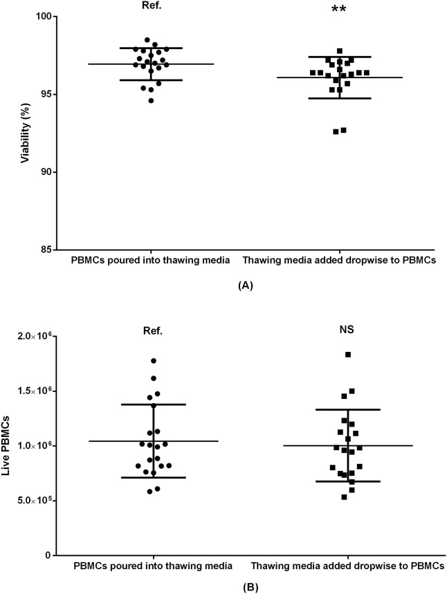
PBMCs and washing medium may be mixed in different ways; we compared swift pouring of partly thawed PMBCs directly into the washing medium, with dropwise addition of washing medium to the partly thawed PBMCs (Fig 6). Although the viability was marginally higher in the samples where the partly thawed PBMCs were poured into the washing medium (96.9%) than the samples where washing medium was added dropwise to PBMCs (96.1%, p = 0.01), there was no difference in the absolute count of live PBMCs (p = 0.17).
Fig 6. Comparing two ways of mixing PBMCs and washing medium by viability (A) and absolute count of live PBMCs (B).
Paired samples 20 donors included. Ref.: reference group. NS: non-significant. **: p<0.01.
3.5 Centrifugation temperature
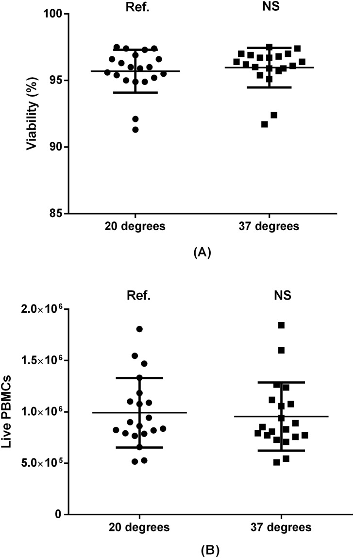
Because we had just demonstrated a significant positive effect of using warm washing medium, we hypothesized that heating the centrifuge could also improve PBMC viability and recovery. We therefore compared the effect of pre-heating the centrifuge to 37°C with leaving the centrifuge at room temperature (20°C). As presented in Fig 7, pre-heating the centrifuge did not affect outcomes significantly.
Fig 7. Temperature of centrifuge by PBMC viability (A) and absolute live PBMC recovery (B).
Paired samples from 20 donors included. Ref.: reference group. NS: non-significant.
3.6 Centrifugation time and force
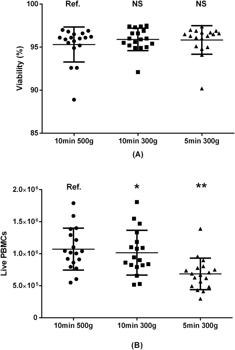
For this evaluation, PBMCs were resuspended in 10 mL washing medium consisting of S-RPMI (bovine) before the two rounds of centrifugation. There was no difference in average cell viability when comparing centrifugation time and force (Fig 8); 95.8% for 5 minutes centrifugation at 500 g, 95.9% for 10 minutes at 300 g, and 95.3% for 10 minutes at 500 g. Conversely, the absolute live PBMC count was significantly higher for a 10-minute centrifugation at 500 g than 10 minutes at 300 g (1.07 fold difference, p = 0.04) and 5 minutes at 300 g (1.60 fold difference, p<0.01).
Fig 8. Centrifugation time and force and PBMC viability (A) and absolute live PBMC count (B).
Paired samples from 18 donors included. Ref.: reference group. NS: non-significant. *: p<0.05. **: p<0.01.
3.7 Incubation time
After the second resuspension in washing media, we placed the PBMCs in an incubator at 37°C with 5% CO2 for different durations (Fig 9). For viability, we found a slight positive effect of incubating the samples for 30 minutes or 1 hour before the second centrifugation and staining. However, in terms of live PBMC count, there was no effect of incubation except a lower number of cells after 16 hours (1.09 fold difference, p = 0.02). This difference became non-significant (p = 0.10) after adjusting for multiple comparisons.
4. Discussion
We systematically evaluated seven steps in the process of thawing PBMCs by measuring cell viability and absolute live PBMC count. The live PBMC count depended significantly on the choice and temperature of washing medium, the centrifugation force, centrifugation duration, and on the incubation duration. Within the tested range, we found no effect of changing thawing time, method of mixing PBMCs and washing medium, or centrifugation temperature.
There is limited evidence-based recommendations for conducting the thawing of PBMCs for analyses by flow cytometry [15]. To fill this gap, we compared variations in several steps of the process. We evaluated the thawing step, during which PBMCs are placed in a heated water bath, but found no differences between thawing until ice was still visible, and thawing for a full 5 minutes. Our findings corroborate the findings reported by Ramachandran et al. In their study, cells were similarly thawed until only a small portion of the ice remained, or kept in the water bath for 10 minutes [16]. The investigators found no difference in cell viability when using pre-heated (37°C) washing medium, but when the wash medium was cooled, and mixed rapidly with PBMCs, the viability was lower. A lower cell viability was also described by Disis et al. [18] when using washing medium at 4°C. Thus, previous reports suggest the use of pre-heated, 37°C medium. Furthermore, we found that thawing PBMCs until only a small amount of ice remains produced results similar to those obtained when leaving the sample in a heated water bath for 5 minutes.
The choice of washing medium significantly affected our outcomes. In the study by Disis et al. [18] cell viability was higher when using FBS compared with human AB serum. We found highest viability using pure FBS, but the number of live PBMCs was higher in samples treated with S-RPMI (bovine). This illustrates that cell viability is not ideal as the primary endpoint when optimizing thawing procedures even though it was used as the primary endpoint in prior studies [13,16,18]. Owing to possible activation of T cells by xenoproteins, human serum from AB positive donors, or autologous serum, in washing media is recommended over FBS [15] but human serum is not as readily available. A general concern when using serum in washing media–human or bovine–is the risk of biologically significant batch-to-batch variations.
Studies using FBS as an ingredient in the washing medium have used a range of different concentrations. We tested 100% FBS, 20% FBS mixed with 80% RPMI, and 100% RPMI, and found the highest cell recovery using 20% FBS 80% RPMI. Others have used 100% RPMI [19,20], 10% FBS [3,7,9,13,21], 20% FBS [5], 30% FBS [22] or 50% FBS [23] mixed with RPMI. And some studies simply fail to mention the FBS concentration in the washing medium [24]. As FBS is relatively expensive compared to RPMI, a low FBS concentration would be preferable if performance is not compromised. This needs to be elucidated.
Because the temperature of the washing medium affected cell viability and live cell recovery, we hypothesized that increasing the centrifugation temperature to 37°C could improve results further; however, we failed to detect any such effect. Instead, lowering centrifugation force and time produced higher viability and lower live cell recovery. In a previous study [18] there was no difference in cell viability when adjusting centrifugation time (5 vs. 10 minutes) or force (280 vs. 450 rpm). Although cell viability is unaffected, adjusting these parameters could change the absolute live cell count. The purpose of centrifugation is to collect cells at the bottom of the tube allowing for aspiration of the supernatant without cell loss. The centrifugation time and force necessary to retain cells at the bottom of the tube may depend on the cell type and concentration, the type and volume of the washing medium, and the geometry of the centrifugation tube. The data we have generated in this study should therefore be considered specific for centrifugation of PBMCs in 10 mL S-RPMI (bovine) in a 15-mL Falcon tube. Using other settings may require separate evaluation.
Incubating, or “resting”, cells after thawing is a common practice when performing functional assays [2,25,26]. The rationale is that dead and dying cells are eliminated during the “rest period”, and only viable cells remain. For practical reasons, resting may also be performed before staining and analysis by flow cytometry [3,27]. However, there is limited evidence for the duration of this resting time in terms of optimizing cell recovery. We found no impact on live PBMC recovery when incubating cells for up to eight hours, but extending the period to 16 hours resulted in significantly lower cell counts. Our study was not designed to evaluate potential changes in cell phenotype during freezing/thawing or during incubation. Indeed, the functional capacity of cells changes during an “overnight resting” period [28], and both biased loss of certain cell types and changes in cell phenotypes could theoretically occur. This constitutes a limitation to our study.
This study did not evaluate freezing procedures, which also differ between laboratories. We used a standard DMSO concentration of 10% in the freezing medium, but other concentrations are described elsewhere [13,29]. After mixing the PBMC suspension with the freezing medium and dispensing the solution in cryotubes, we immediately placed the cryotubes directly in a rack in a -80°C freezer. Other laboratories have used devices to ensure a controlled freezing rate [30,31], and our freezing procedure may be a potential source of bias. Our samples were stored for up to 2 weeks in freezer -80°C freezer instead of moving samples to a liquid nitrogen container after 24 hours which is generally recommended. Whereas freezing temperature may affect PBMC functionality [32,33], there is little evidence to suggest that the PBMC phenotype is affected by freezing temperature. Variations in DMSO concentration, freezing procedure and storage time may have influence on which thawing procedure gives the highest PBMC viability and recovery.
Conclusion
Laboratories world-wide use different approaches when freezing and thawing leukocytes. We here show that the thawing procedure has crucial impact on the recovery of live cells. The best-performing washing medium was S-RPMI (FBS), which should be pre-heated to 37°C before use. Centrifugation force and time may depend on the washing volume, but when using 10 mL 20% S-RPMI (bovine) in a 15 mL Falcon tube, samples should be centrifuged for at least 10 minutes at 500 g. Other conditions such as thawing time, method of mixing PBMCs and washing medium, or centrifugation temperature, seemed to have little or no influence on cell viability and recovery. Incubating PBMCs for up to eight hours without consequences on cell counts allows for practical variations in the laboratory set-up. Potential impact of the thawing procedure, and use of FBS containing xeno-proteins, on cell phenotype and functional characteristics needs clarification.
Abbreviations
- BSS
balanced saline solution
- DMSO
dimethyl sulfoxide
- FBS
fetal bovine serum
- PBMC
peripheral blood mononuclear cell
- PBS
phosphate buffered saline
- RPMI
Roswell Park Memorial Institute
- SD
standard deviation
- S-RPMI (bovine)
RPMI 1640 with 20% bovine serum
- S-RPMI (human)
RPMI 1640 with 20% human serum
Data Availability
Raw data is available from“Open Science Network” (URL: https://osf.io/mdgx3/).
Funding Statement
The authors received no specific funding for this work.
References
- 1.Seale AC, de Jong BC, Zaidi I, Duvall M, Whittle H, Rowland-Jones S, et al. Effects of cryopreservation on CD4+ CD25+ T cells of HIV-1 infected individuals. J Clin Lab Anal. 2008;22: 153–158. doi: 10.1002/jcla.20234 [DOI] [PMC free article] [PubMed] [Google Scholar]
- 2.Ferreira V da S, Mesquita FV, Mesquita D, Andrade LEC. The effects of freeze/thawing on the function and phenotype of CD4+ lymphocyte subsets in normal individuals and patients with systemic lupus erythematosus. Cryobiology. 2015;71: 507–510. doi: 10.1016/j.cryobiol.2015.10.149 [DOI] [PubMed] [Google Scholar]
- 3.Shete A, Jayawant P, Thakar M, Kurle S, Singh DP, Paranjape RS. Differential modulation of phenotypic composition of HIV-infected and -uninfected PBMCs during cryopreservation. J Immunoassay Immunochem. 2013;34: 333–345. doi: 10.1080/15321819.2012.741087 [DOI] [PubMed] [Google Scholar]
- 4.Meijerink M, Ulluwishewa D, Anderson RC, Wells JM. Cryopreservation of monocytes or differentiated immature DCs leads to an altered cytokine response to TLR agonists and microbial stimulation. J Immunol Methods. 2011;373: 136–142. doi: 10.1016/j.jim.2011.08.010 [DOI] [PubMed] [Google Scholar]
- 5.Sattui S, de la Flor C, Sanchez C, Lewis D, Lopez G, Rizo-Patrón E, et al. Cryopreservation modulates the detection of regulatory T cell markers. Cytometry B Clin Cytom. 2012;82B: 54–58. doi: 10.1002/cyto.b.20621 [DOI] [PubMed] [Google Scholar]
- 6.Brooks-Worrell B, Tree T, Mannering SI, Durinovic-Bello I, James E, Gottlieb P, et al. Comparison of cryopreservation methods on T-cell responses to islet and control antigens from type 1 diabetic patients and controls. Diabetes Metab Res Rev. 2011;27: 737–745. doi: 10.1002/dmrr.1245 [DOI] [PubMed] [Google Scholar]
- 7.Elkord E. Frequency of human T regulatory cells in peripheral blood is significantly reduced by cryopreservation. J Immunol Methods. 2009;347: 87–90. doi: 10.1016/j.jim.2009.06.001 [DOI] [PubMed] [Google Scholar]
- 8.Costantini A, Mancini S, Giuliodoro S, Butini L, Regnery CM, Silvestri G, et al. Effects of cryopreservation on lymphocyte immunophenotype and function. J Immunol Methods. 2003;278: 145–155. doi: 10.1016/S0022-1759(03)00202-3 [DOI] [PubMed] [Google Scholar]
- 9.Trück J, Mitchell R, Thompson AJ, Morales-Aza B, Clutterbuck EA, Kelly DF, et al. Effect of cryopreservation of peripheral blood mononuclear cells (PBMCs) on the variability of an antigen-specific memory B cell ELISpot. Hum Vaccines Immunother. 2014;10: 2490–2496. doi: 10.4161/hv.29318 [DOI] [PMC free article] [PubMed] [Google Scholar]
- 10.Stevens VL, Patel AV, Feigelson HS, Rodriguez C, Thun MJ, Calle EE. Cryopreservation of Whole Blood Samples Collected in the Field for a Large Epidemiologic Study. Cancer Epidemiol Biomark Amp Prev. 2007;16: 2160–2163. doi: 10.1158/1055-9965.EPI-07-0604 [DOI] [PubMed] [Google Scholar]
- 11.Hensley-McBain T, Heit A, De Rosa SC, McElrath MJ, Andersen-Nissen E. Optimization of a whole blood phenotyping assay for enumeration of peripheral blood leukocyte populations in multicenter clinical trials. J Immunol Methods. 2014;411: 23–36. doi: 10.1016/j.jim.2014.06.002 [DOI] [PMC free article] [PubMed] [Google Scholar]
- 12.Kloverpris H, Fomsgaard A, Handley A, Ackland J, Sullivan M, Goulder P. Dimethyl sulfoxide (DMSO) exposure to human peripheral blood mononuclear cells (PBMCs) abolish T cell responses only in high concentrations and following coincubation for more than two hours. J Immunol Methods. 2010;356: 70–78. doi: 10.1016/j.jim.2010.01.014 [DOI] [PubMed] [Google Scholar]
- 13.Nazarpour R, Zabihi E, Alijanpour E, Abedian Z, Mehdizadeh H, Rahimi F. Optimization of human peripheral blood mononuclear cells (PBMCs) cryopreservation. Int J Mol Cell Med. 2012;1: 88 [PMC free article] [PubMed] [Google Scholar]
- 14.Riedhammer C, Halbritter D, Weissert R. Peripheral Blood Mononuclear Cells: Isolation, Freezing, Thawing, and Culture In: Weissert R, editor. Multiple Sclerosis. New York, NY: Springer New York; 2014. pp. 53–61. [DOI] [PubMed] [Google Scholar]
- 15.Mallone R, Mannering SI, Brooks-Worrell BM, Durinovic-Belló I, Cilio CM, Wong FS, et al. Isolation and preservation of peripheral blood mononuclear cells for analysis of islet antigen-reactive T cell responses: position statement of the T-Cell Workshop Committee of the Immunology of Diabetes Society: PBMC preparation for T cell assays. Clin Exp Immunol. 2011;163: 33–49. doi: 10.1111/j.1365-2249.2010.04272.x [DOI] [PMC free article] [PubMed] [Google Scholar]
- 16.Ramachandran H, Laux J, Moldovan I, Caspell R, Lehmann PV, Subbramanian RA. Optimal Thawing of Cryopreserved Peripheral Blood Mononuclear Cells for Use in High-Throughput Human Immune Monitoring Studies. Cells. 2012;1: 313–324. doi: 10.3390/cells1030313 [DOI] [PMC free article] [PubMed] [Google Scholar]
- 17.Pedersen OB, Erikstrup C, Kotzé SR, Sørensen E, Petersen MS, Grau K, et al. The Danish Blood Donor Study: a large, prospective cohort and biobank for medical research. Vox Sang. 2012;102: 271 doi: 10.1111/j.1423-0410.2011.01553.x [DOI] [PubMed] [Google Scholar]
- 18.Disis ML, dela Rosa C, Goodell V, Kuan L-Y, Chang JCC, Kuus-Reichel K, et al. Maximizing the retention of antigen specific lymphocyte function after cryopreservation. J Immunol Methods. 2006;308: 13–18. doi: 10.1016/j.jim.2005.09.011 [DOI] [PubMed] [Google Scholar]
- 19.Keane KN, Calton EK, Cruzat VF, Soares MJ, Newsholme P. The impact of cryopreservation on human peripheral blood leucocyte bioenergetics. Clin Sci. 2015;128: 723–733. doi: 10.1042/CS20140725 [DOI] [PubMed] [Google Scholar]
- 20.Lockmann A, Schön MP. Phenotypic and functional traits of peripheral blood mononuclear cells retained by controlled cryopreservation: implications for reliable sequential studies of dynamic interactions with endothelial cells. Exp Dermatol. 2013;22: 358–359. doi: 10.1111/exd.12123 [DOI] [PubMed] [Google Scholar]
- 21.Sarzotti-Kelsoe M, Needham LK, Rountree W, Bainbridge J, Gray CM, Fiscus SA, et al. The Center for HIV/AIDS Vaccine Immunology (CHAVI) multi-site quality assurance program for cryopreserved Human Peripheral Blood Mononuclear Cells. J Immunol Methods. 2014;409: 21–30. doi: 10.1016/j.jim.2014.05.013 [DOI] [PMC free article] [PubMed] [Google Scholar]
- 22.Rasmussen SM, Bilgrau AE, Schmitz A, Falgreen S, Bergkvist KS, Tramm AM, et al. Stable phenotype of B-cell subsets following cryopreservation and thawing of normal human lymphocytes stored in a tissue biobank: Cryopreservation of Human B-Cell Subsets. Cytometry B Clin Cytom. 2015;88: 40–49. doi: 10.1002/cyto.b.21192 [DOI] [PubMed] [Google Scholar]
- 23.Zhang W, Nilles TL, Johnson JR, Margolick JB. The effect of cellular isolation and cryopreservation on the expression of markers identifying subsets of regulatory T cells. J Immunol Methods. 2016;431: 31–37. doi: 10.1016/j.jim.2016.02.004 [DOI] [PMC free article] [PubMed] [Google Scholar]
- 24.Mata MM, Mahmood F, Sowell RT, Baum LL. Effects of cryopreservation on effector cells for antibody dependent cell-mediated cytotoxicity (ADCC) and natural killer (NK) cell activity in 51Cr-release and CD107a assays. J Immunol Methods. 2014;406: 1–9. doi: 10.1016/j.jim.2014.01.017 [DOI] [PMC free article] [PubMed] [Google Scholar]
- 25.Kierstead LS, Dubey S, Meyer B, Tobery TW, Mogg R, Fernandez VR, et al. Enhanced rates and magnitude of immune responses detected against an HIV vaccine: effect of using an optimized process for isolating PBMC. AIDS Res Hum Retroviruses. 2007;23: 86–92. doi: 10.1089/aid.2006.0129 [DOI] [PubMed] [Google Scholar]
- 26.Boaz MJ, Hayes P, Tarragona T, Seamons L, Cooper A, Birungi J, et al. Concordant proficiency in measurement of T-cell immunity in human immunodeficiency virus vaccine clinical trials by peripheral blood mononuclear cell and enzyme-linked immunospot assays in laboratories from three continents. Clin Vaccine Immunol CVI. 2009;16: 147–155. doi: 10.1128/CVI.00326-08 [DOI] [PMC free article] [PubMed] [Google Scholar]
- 27.Nettenstrom L, Alderson K, Raschke EE, Evans MD, Sondel PM, Olek S, et al. An optimized multi-parameter flow cytometry protocol for human T regulatory cell analysis on fresh and viably frozen cells, correlation with epigenetic analysis, and comparison of cord and adult blood. J Immunol Methods. 2013;387: 81–88. doi: 10.1016/j.jim.2012.09.014 [DOI] [PMC free article] [PubMed] [Google Scholar]
- 28.Kutscher S, Dembek CJ, Deckert S, Russo C, Körber N, Bogner JR, et al. Overnight resting of PBMC changes functional signatures of antigen specific T- cell responses: impact for immune monitoring within clinical trials. PloS One. 2013;8: e76215 doi: 10.1371/journal.pone.0076215 [DOI] [PMC free article] [PubMed] [Google Scholar]
- 29.Liseth K, Abrahamsen JF, Bjørsvik S, Grøttebø K, Bruserud Ø. The viability of cryopreserved PBPC depends on the DMSO concentration and the concentration of nucleated cells in the graft. Cytotherapy. 2005;7: 328–333. doi: 10.1080/14653240500238251 [DOI] [PubMed] [Google Scholar]
- 30.Buhl T, Legler TJ, Rosenberger A, Schardt A, Schön MP, Haenssle HA. Controlled-rate freezer cryopreservation of highly concentrated peripheral blood mononuclear cells results in higher cell yields and superior autologous T-cell stimulation for dendritic cell-based immunotherapy. Cancer Immunol Immunother CII. 2012;61: 2021–2031. doi: 10.1007/s00262-012-1262-0 [DOI] [PMC free article] [PubMed] [Google Scholar]
- 31.Sputtek A, Lioznov M, Kröger N, Rowe AW. Bioequivalence comparison of a new freezing bag (CryoMACS(®)) with the Cryocyte(®) freezing bag for cryogenic storage of human hematopoietic progenitor cells. Cytotherapy. 2011;13: 481–489. doi: 10.3109/14653249.2010.529891 [DOI] [PubMed] [Google Scholar]
- 32.Yang J, Diaz N, Adelsberger J, Zhou X, Stevens R, Rupert A, et al. The effects of storage temperature on PBMC gene expression. BMC Immunol. 2016;17 doi: 10.1186/s12865-016-0144-1 [DOI] [PMC free article] [PubMed] [Google Scholar]
- 33.Germann A, Oh Y-J, Schmidt T, Schön U, Zimmermann H, von Briesen H. Temperature fluctuations during deep temperature cryopreservation reduce PBMC recovery, viability and T-cell function. Cryobiology. 2013;67: 193–200. doi: 10.1016/j.cryobiol.2013.06.012 [DOI] [PubMed] [Google Scholar]
Associated Data
This section collects any data citations, data availability statements, or supplementary materials included in this article.
Data Availability Statement
Raw data is available from“Open Science Network” (URL: https://osf.io/mdgx3/).


