Abstract
Background
Approximately one-third of women diagnosed with breast cancer undergo mastectomy with subsequent implant-based or autogenous tissue-based reconstruction. Potential complications include infection, capsular contracture, and leak or rupture of implants with necessity for explantation. Skin rashes are infrequently described complications of patients who undergo mastectomy with or without reconstruction.
Methods
A retrospective analysis of breast cancer patients referred to the Dermatology Service for diagnosis and management of a rash post-mastectomy and expander or implant placement or transverse rectus abdominis myocutaneous (TRAM) flap reconstruction was performed. Parameters studied included reconstruction types, time to onset, clinical presentation, associated symptoms, results of microbiologic studies, management, and outcome.
Results
We describe 21 patients who developed a rash on the skin overlying a breast reconstruction. Average time to onset was 25.7 months after expander placement or TRAM flap reconstruction. Clinical presentations included macules and papules or scaly, erythematous patches and plaques. Five patients had cultures of the rash, which were all negative. Skin biopsy was relatively contraindicated in areas of skin tension, and was reserved for non-responding eruptions. Treatments included topical corticosteroids and topical antibiotics, which resulted in complete or partial responses in all patients with documented follow-ups.
Conclusion
Our findings suggest that tension and post-surgical factors play a causal role in this hitherto undescribed entity: “post-reconstruction dermatitis of the breast.” This is a manageable condition that develops weeks to years following breast reconstruction. Topical corticosteroids and antibiotics result in restoration of skin barrier integrity and decreased secondary infection.
Keywords: Dermatitis, rash, breast reconstruction, breast cancer, topical corticosteroids
Introduction
It is estimated that 231,840 American women were diagnosed with breast cancer in 2015 and nearly 1 in 3 of these patients underwent mastectomy as part of their treatment 1. During this time period, over 106,338 breast reconstruction surgeries using either implants or autogenous tissues were performed as reported by the American Society of Plastic Surgeons2. Although both implant and autogenous tissue reconstructions have high success rates with low overall rates of complications, the most commonly reported complications of tissue expansion and permanent implants or autogenous tissue reconstruction include infection, hematoma, extrusion, capsular contracture, leak, flap necrosis, and donor site complications 3–5. The rates of complications with either reconstruction are substantially increased by radiation therapy 6,7.
In general, skin rashes are an infrequently described complication of patients who undergo mastectomy with or without reconstruction. When skin rashes do occur in these patients, they are often attributed to radiation therapy, allergic contact dermatitis to bandages, tape, or topical medication, or dry skin 8. Previous studies had also hypothesized that leaking silicone implants were associated with a variety of autoimmune disorders as well as cutaneous manifestations; however, these reports have more recently been largely disproven 9–15.
In our consult service at a tertiary care cancer center, we have observed 21 cases, over a 13-year period (1999–2012), of a rash overlying the affected breast or breasts weeks to years following breast reconstruction, unrelated to a contactant, radiation therapy, or the type of breast reconstruction. Currently, there is no published literature describing a similar rash on the skin overlying a breast in patients after breast reconstruction or augmentation. This current study describes the clinical features of rash and response to treatment in patients after implant-based or autogenous tissue reconstruction post-mastectomy for breast cancer. On the basis of clinical presentation and resolution of symptoms with topical steroids and antibiotics, we hypothesize that this represents an eczematous dermatitis of the breast. A better understanding of this previously undescribed rash will facilitate diagnostic accuracy and appropriate therapeutic interventions, including skin biopsy. Herein we describe our clinical findings and discuss this as a new entity: “post-reconstruction dermatitis of the breast.”
Methods
We conducted a retrospective analysis of 21 female patients with breast cancer referred to the Dermatology Service for diagnosis and management of rash post-mastectomy and tissue expander and implant placement or TRAM flap reconstruction. We included patients treated with total mastectomy, with or without axillary lymph node dissection, adjuvant chemotherapy, or radiation. Parameters studied included reconstruction types (tissue expanders with either silicone or saline implants, or transverse rectus abdominis myocutaneous flap (TRAM flap)), time to rash onset from mastectomy and either tissue expander placement or TRAM flap reconstruction, clinical presentation at the time of dermatologic evaluation, associated symptoms, results of microbiologic studies, management, and patient outcomes. Photographic images were obtained when available. It should be noted that, at MSKCC from 1999 to 2012, 10,793 implant based individual breast reconstructions and 1,490 autologous-based breast reconstructions were performed. An Institutional Review Board waiver was approved for this study.
Results
Demographics and baseline characteristics of the 21 patients are presented in Table 1. Our patients ranged in age from 34 to 65 years at the time of breast cancer diagnosis, mean of 48 years. All but one patient underwent immediate reconstruction after mastectomy with tissue expansion and permanent implants; the remaining patient underwent immediate reconstruction using a TRAM flap. Nine patients had axillary lymph node dissection, 11 received adjuvant chemotherapy, and 3 patients underwent radiation therapy. Five patients underwent bilateral mastectomies: 4 patients with unilateral breast cancer and prophylactic mastectomy of the contralateral breast, and one with pathology in bilateral breasts. Of the patients who received implant-based breast reconstruction, 11(58%) of the patients were reconstructed with saline implants while the remaining 8 (42%) had silicone gel implants. One patient had not yet undergone permanent implant replacement. All patients had uneventful post-operative recoveries without delayed healing or skin-limited infections and none had evidence of capsular contracture. One patient had a documented possible allergy to surgical tape, 3 patients had documented contact allergies to latex, one patient was allergic to bacitracin, and one patient had a contact allergy to the Jackson Pratt drain.
Table 1.
Patient Demographics and Baseline Characteristics
| Total Patients n=21 | |
|---|---|
|
| |
| Mean Age at Diagnosis of Breast Cancer, yrs (range) | 48 (34–65) |
|
| |
| Breast Cancer Diagnosis | n (%) |
|
| |
| Invasive Ductal Carcinoma with or without foci of DCIS | 12 (57) |
| Invasive Lobular Carcinoma with or without foci of DCIS | 2 (10) |
| Ductal Carcinoma In Situ | 7 (33) |
| Vascular Invasion Present | 3 (14) |
|
| |
| Surgical Management | |
|
| |
| Unilateral mastectomy | 16 (76) |
| Bilateral Mastectomy | 5a (24) |
|
| |
| Lymph Nodes | |
|
| |
| Patients with Sentinel Lymph Node Biopsies | 8 (38) |
| Patients with Axillary Lymph Node Dissection | 9 (43) |
|
| |
| Adjuvant Therapy for Breast Cancer | |
|
| |
| Chemotherapy | 11 (52) |
| Radiotherapy | 3 (14)* |
|
| |
| Type of Breast Reconstruction Post Mastectomy | |
|
| |
| Silicone Implants | 8 (38) |
| Saline Implants | 11(52) |
| Transverse Rectus Abdominis Myocutaneous Flap | 1 (5) |
|
| |
| Time to Onset of Initial Rash Post Expander Placement or TRAM Flap | |
|
| |
| ≤ 1 month | 2 (10) |
| 2 months – 6 months | 7 (33) |
| 7 months – 1 year | 5 (24) |
| >1 year | 7 (33) |
| Patients who Presented Post-Expander Placement but Prior to Permanent Implants | 10 (48) |
| Patients who Presented Post-Permanent Implants | 10 (48) |
Four patients with bilateral mastectomies had disease in one breast and a prophylactic mastectomy or no evidence or disease in the contralateral breast. One patient had disease in bilateral breasts
Two patients had silicone implants and one patient had a TRAM flap reconstruction
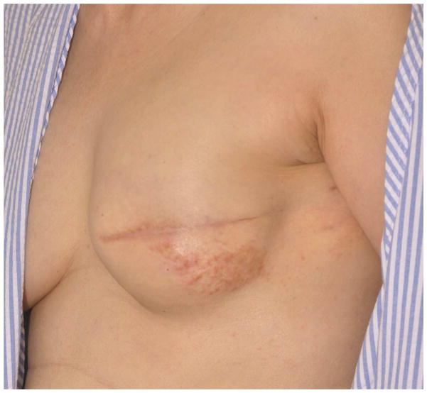
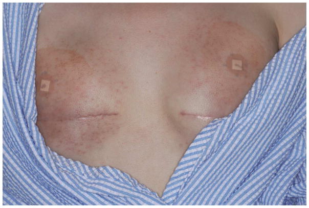
Average time to rash onset was 25.7 months after tissue expander placement or TRAM flap reconstruction (range one month to nine years). Nine patients presented in the first 6 months, 5 patients presented between 6 months and one year, and 7 patients presented greater than one year after tissue expander placement. Ten patients (48%) initially developed the rash with tissue expanders in place, prior to permanent implant replacement. In most patients, the rash presented clinically as pruritic or non-pruritic, scaly, erythematous patches or plaques or as asymptomatic macules and papules on the contours of the breast(s), adjacent or inferior to the mastectomy scars (Figure 1). Only one patient presented with ill-defined red papules distributed diffusely on the bilateral breasts (patient 15) (Figure 2). Of note, patients with unilateral mastectomies and reconstruction had more extensive dermatitis on the ipsilateral breast while patients with bilateral mastectomies and reconstruction showed dermatitis on both breasts. In 4 cases, the dermatitis was annular in configuration. A minority of patients had a “crackled” skin appearance without xerotic surrounding skin. Potassium hydroxide (KOH) examinations performed on 2 (10%) of the patients who had particularly annular appearances of the rash were negative. Additionally, bacterial and/or fungal cultures obtained in 6 (29%) of the patients were all negative. None of the patients had vesicles or bullae or a well-demarcated pattern. None of the cases were biopsied since all cases of dermatitis improved greatly with conservative therapy.
Figure 1.
Erythematous papules and plaque on the left breast inferior to the surgical site of a patient ten months post-tissue expander placement prior to permanent implant replacement.
Figure 2.
Bilateral ill defined red papules on the contours of the breasts of a patient two months post bilateral total mastectomy and expander placement, prior to permanent implant replacement.
Treatment for all rashes included a moderate to high-potency topical steroid. Topical antibiotics were also prescribed for all but one patient. Additionally, 2 patients were prescribed a topical antifungal and one patient was prescribed a 10-day course of oral antibiotics at the initial Dermatology visit. Treatment resulted in a complete response in 10 of the 13 patients (77%) with documented follow-ups typically between 4 and 8 weeks (Figure 3A, B). Two patients who initially had complete responses to treatment had recurrences of the rash several years later. One patient had a recurrence after surgical excision of a 3 centimeter liposarcoma on the ipsilateral shoulder to the breast reconstruction, which caused increased tension to the overlying skin (Figure 3C); the second patient experienced a 30-pound weight gain followed by a recurrence of her rash. The patient with liposarcoma did not have adjuvant radiation therapy while the patient with weight gain did. In neither case was it felt that radiation therapy played any significant role in the development of stasis dermatitis of the affected breast. Three patients had partial responses, with intermittent episodes of eczematous dermatitis on the affected breast(s) after treatment discontinuation. At time of data collection, one patient was still being followed for response to treatment but had no documented follow-up. Mild post-inflammatory hyperpigmentation was the only notable sequela. All 21 cases are summarized in Table 2.
Figure 3.
Figure 3a. Initial presentation of a patient with a several centimeter scaly patch on the right breast.
Figure 3b. Patient at follow-up three years later with resolution of post-reconstruction dermatitis of the breast and only mild hyperpigmentation.
Figure 3c. Patient with a mild flare of post-reconstruction dermatitis 7 years after initial presentation, following a surgical excision of a 3cm liposarcoma on her right shoulder. The recurred rash greatly improved with topical corticosteroids and topical antibiotics.
Table 2.
Summary of patient cases: Clinical descriptions, management, and outcomes
| Pt # | Description of Rash | Treatment Prescribed for Rash | Outcome of Treatment and Follow-up |
|---|---|---|---|
| 1 | Right breast with several centimeter erythematous scaly patch on and below the surgical site | Fluocinonide and mupirocin cream bid | At 1 month follow-up, post-reconstruction dermatitis was resolved with only mild hyperpigmentation. |
| 2 | Bilateral pink scaly patches over surgical sites | Fluocinonide and mupirocin cream bid | At 1 month follow-up, post-reconstruction dermatitis was improved bilaterally. Rash recurred 3 months following permanent implant exchange and was successfully retreated. |
| 3 | Right breast with a annular, scaly plaques near the surgical scar | Fluocinonide and mupirocin cream bid | At 1 month follow-up, post-reconstruction dermatitis was improved with only mild hyperpigmentation. 7 years later, the patient had surgical excision of a 3 cm liposarcoma on her right shoulder. A few months later the patient presented with erythema and scaling inferior to scar on right reconstructed breast and she was successfully retreated. |
| 4a | Right breast with 3×5 cm scaly, scalloped plaque with raised border and honey colored crust on the surgical scar | Fluocinonide and mupirocin cream bid and Clindamycin 150 mg po qid × 10 days | At 1 month follow-up, post-reconstruction dermatitis was much improved with only mild hyperpigmentation. A 30 pound weight gain seven years after her initial presentation resulted in rash recurrence that was successfully treated. |
| 5 | Right breast with a scaly and erythematous plaque near the surgical scar | Fluocinonide and mupirocin cream bid | At 2 months follow-up with breast oncologist, post-reconstruction dermatitis was resolved. |
| 6 | Left breast with a 4 cm annular eczematous patch with subtle crusting lateral to surgical site | Fluocinonide cream | At 8 months follow-up, post-reconstruction dermatitis was resolved. |
| 7 | Bilateral erythema and slight scaling inferior to mastectomy scars | Fluocinonide and mupirocin cream bid | At 1 month follow-up, there was improvement in erythema on the right and left breast. At her most recent visit, the lesions were completely resolved with only mild hyperpigmentation. |
| 8 | Right breast with hyperpigmented macules and scaly plaques, some annular in appearance, in the center of a slightly hypertrophic mastectomy scar | Fluocinonide and mupirocin cream bid and topical ketoconazole bid | At 2 months follow-up the patient still has some hyperpigmented macules. Fluocinonide was discontinued and Westcort cream bid was prescribed. The patient is still being followed. She does not use the medications as prescribed. She has hyperpigmented and eczematous changes near the scar on her right reconstructed breast and other eczematous changes. |
| 9 | Right breast with scaly, lichenified plaques | Fluocinonide and mupirocin cream bid | At 4 months follow-up the patient had a band-like discoloration under the right implant on her breast and lichenification. Ketoconazole cream bid and 2.5% hydrocortisone cream were prescribed. At her most recent visit she had few excoriations under her right breast. |
| 10 | Bilateral erythema and scaling along the suture lines with few annular patches; right greater than left | Fluocinonide and mupirocin cream and topical ketoconazole tid | At 1 month follow-up post-reconstruction dermatitis was much improved. |
| 11 | Left breast with pink, thin, scaly plaques around the scar | Fluocinonide and mupirocin cream bid | At 2 months follow-up post-reconstruction dermatitis was much improved. |
| 12 | Right breast with multiple erythematous slightly linear, scaly plaques below the suture lines | Fluocinonide and mupirocin cream tid | At 2 months follow-up there was much less erythema and minimal postinflammatory hyperpigmentation. |
| 13 | Right breast with ill defined erythematous plaque with central clearing around the mastectomy scar | Betamethasone diproprionate and mupirocin cream bid | At 2 months follow-up post-reconstruction dermatitis of the right breast was resolved. Six years post MRM and breast reconstruction for stage III breast cancer, the patient had the saline implant removed due to persistent right chest wall discoloration and a left prophylactic mastectomy. |
| 14 | Left breast with hyperpigmentation and scaling along and below the suture line | Triamcinolone and mupirocin cream bid | At 2 months follow-up post-reconstruction dermatitis of right breast was resolved with mild hyperpigmentation. At her most recent visit, hyperpigmentation was resolved. |
| 15 | Bilateral diffuse, ill defined red papules on contours of breasts that spares the folds | Fluticasone and mupirocin cream bid | At 2 weeks follow-up, post-reconstruction dermatitis was much improved. |
| 16 | Right breast with erythema and scaling along the suture line | Fluocinonide and mupirocin cream bid | At 1–2 months follow-up post-reconstruction dermatitis was resolved. |
| 17 | Left breast with diffuse post-radiation hyperpigmentation; underside of left breast with a 3×3 cm erythematous patch with a few pink papules | Betamethasone diproprionate and mupirocin cream bid | No follow-up of rash to date. Patient will be followed for response to treatment. |
| 18 | Left breast erythematous and edematous with induration around the implant | Triamcinolone and mupirocin cream bid | At 4 months follow-up with breast oncologist, post-reconstruction dermatitis was resolved. |
| 19 | Right breast with a hyperpigmented, ill-defined macule surrounding the mastectomy scar | Betamethasone diproprionate and mupirocin cream bid | At 3 months follow-up with breast oncologist, post-reconstruction dermatitis was resolved. |
| 20 | Right breast with erythema, scaling, and yellow crust below the mastectomy scar; peripheral erythematous macule | Fluocinonide cream and Polysporin ointment bid | At 2 months follow-up for presurgical testing, post-reconstruction dermatitis was resolved. |
| 21 | Left breast with erythematous papules and a confluent plaque inferior to the surgical site | Fluocinonide and mupirocin cream bid | One month after initial dermatology visit the patient elected to have the tissue expander reomoved from her left breast without permanent implant placement. At that time, post-reconstruction dermatitis was resolved. |
Patient with transverse rectus abdominis myocutaneous flap repair. Betamethasone diproprionate cream: Class III; Fluocinonide cream: Class III; Fluticasone cream: Class III; Triamcinolone cream: Class IIII
Discussion
On the basis of clinical presentation and resolution of symptoms with topical steroids, we believe this represents an eczematous dermatitis in the reconstructed breast(s). We have named this entity “post-reconstruction dermatitis of the breast.” Several different possible explanations may exist for the development of this dermatitis, including disruption of lymphatic and/or venous flow, skin tension and thickness of the skin, and changes in skin perfusion, temperature regulation, and vascular integrity.
A role for venous stasis may be present in post-reconstruction dermatitis of the breast. While the exact mechanism of skin changes in stasis dermatitis of the lower extremities is not completely discerned, it is clear, however, that venous hypertension plays a role in the underlying adverse conditions that lead to skin changes and impaired healing 16. Chronic venous insufficiency, which results from chronic venous hypertension, may be caused by an idiopathic degenerative process in the blood vessel walls and valves, or from a precursor event such as direct injury that leads to an inflammatory, destructive process 17. Slowing of the blood flow leads to distension and disruption of the capillary permeability barrier causing extravasation of fluid, plasma proteins, and erythrocytes. It also causes activation of neutrophils and macrophages, which release inflammatory mediators, free radicals, and proteases, leading to pericapillary inflammation 18. This chronic inflammation and microangiopathy are responsible for dermatitic changes as dermal inflammation is known to induce epidermal dysfunction such as barrier impairment and xerosis 19.
Microinjury and direct damage to the breast vasculature during the initial mastectomy or permanent implant placement may lead to chronic venous hypertension and resulting venous insufficiency. Subsequent inflammatory changes in the vasculature likely contribute to the eczematous changes seen clinically. This may be further evidenced by the rash occurring more commonly on the lower, more dependent skin flaps with more inherent stasis, than the upper mastectomy skin flap. In particular, patient 4 had notable slow venous drainage during the reconstruction procedure, but microanastomosis was performed with improvement of the venous drainage. It is probable, however, that a small amount of residual venous stasis is present given the disruption of microvasculature during surgery. The same patient experienced a 30-pound weight gain 4 years after her surgical reconstruction followed by a recurrence of her post-reconstruction dermatitis, which likely resulted from increased tension to the skin.
Stasis dermatitis has been described in locations other than the lower extremities. Bilen et al. 20 described a case of stasis dermatitis of the hand associated with an arteriovenous fistula in a 27-year-old woman with chronic renal failure on hemodialysis. She had a violaceous, slightly scaly patchy lesion with ill-defined borders that was 4–5 cm in diameter. There was edema of the left hand and marked varicosities distal to the fistula. Improvement was seen with topical corticosteroids, but definitive resolution was not seen until the fistula was closed. Similar cases have since been reported 21. While venous hypertension likely plays a role in post-reconstruction dermatitis of the breast, there are some features of venous insufficiency and stasis that we have not seen despite many years of follow up. For example, purpura, chronic nonhealing ulcerations, and lipodermatosclerosis-like changes have not been observed in our patients. Finally, lymphedema was not present in any of the patients when they initially presented to dermatology with dermatitis. However, it is difficult to fully exclude lymphedema as a small component of the etiology of the dermatitis, especially in patients who had full lymph node dissections. We propose that post-reconstruction dermatitis of the breast also involves a loss of integrity of the skin barrier due to the chronic inflammation and microangiopathy described above and increased skin tension. Eight patients (2, 7, 14, 15, 16, 19, 20, and 21) developed dermatitis while receiving breast tissue expansion, prior to permanent silicone or saline implants, demonstrating that skin thinning and tension from tissue expansion play a significant role in the resulting cutaneous changes. Of note, patient 3 had worsening of the rash following excision of a liposarcoma on the ipsilateral shoulder to the mastectomy, which led to increased tension to the skin overlying the breast on that side. Therefore, patients with very thin mastectomy skin flaps may be at increased risk for post-reconstruction dermatitis of the breast. Nizek et al. looked at maximum tissue deformation and elasticity immediately before and after tissue expansion at multiple sites on the chest wall. They demonstrated a global reduction in maximum skin deformation after complete tissue expansion compared to baseline over the entire breast. With regards to elasticity, all patients had significantly improved elasticity after complete expansion of the breast, except in the central breast above and below the surgical scar. Therefore, the skin is clearly tighter after expansion, and, while the tissue expander helps to improve elasticity, it does so less effectively in the central part of the breast. This corresponds to where the dermatitis most commonly presents.22 Finally, one may also invoke a role for disrupted lymphatic drainage. Although no patients had notable lymphedema at initial presentation or follow-up visits, 9 (43%) of our patients underwent axillary lymph node dissection and 8 (38%) had sentinel lymph node biopsies.
Other plausible explanations of this dermatitis include an allergic contact dermatitis. One patient had a history of a possible contact allergy to tape, but this characteristic, well-demarcated rash resolved after the post-operative period. One patient did have possible radiation dermatitis, but again, this was in the past and had resolved previously. Another patient had diffuse post-radiation hyperpigmentation, which was separate from the area of post-reconstruction dermatitis. Allergic contact dermatitis to breast implant contaminants has rarely been reported, necessitating removal of the implant. Patch testing in this patient confirmed an allergy likely to latex rubber and other chemicals used in the production of implants and rubber products, which the patient may have been exposed to during the surgery or which may have been a contaminant of the implant itself 8. Three (14%) of our patients had documented contact allergies to latex. While we have not ruled out this possibility, the quick response to topical corticosteroids suggests that the pathology is not an allergic response to a persistent allergen such as latex rubber or a contaminant of the implant. None of the patients had vesicles or bullae to suggest an acute eczematous process. Some patients presented with a crackled appearance, similar to eczema craquelé, but without xerotic surrounding skin. Finally, several patients presented with annular configurations resembling tinea corporis, but KOH was negative in 2 patients, and the eruption resolved without antifungal therapy in all but 2 cases.
Two patients had clinically impetiginized lesions, underscoring the importance of appropriate treatment. While the mainstay of therapy for stasis dermatitis of the legs has always initially focused on improving the underlying venous insufficiency, treatment with topical corticosteroids to reduce the acute inflammation and pruritus is often effective. One double-blind randomized study of 19 patients with mild to moderate bilateral stasis dermatitis of the lower extremities demonstrated that betamethasone valerate 0.12% foam, a class IV topical steroid, led to significant improvement in erythema and petechiae over vehicle, from baseline at days 14 and 28 of follow-up. However, there was no overall difference between the vehicle- or steroid-treated legs at days 14 and 28 based on a total of 6 criteria for clinical improvement, including erythema, scale, swelling, petechia, post-inflammatory hyperpigmentation, and self-reported pruritus 23. The authors discuss that higher-potency topical steroids may achieve better overall outcomes for patients with stasis dermatitis of the lower extremities. In our study, 19 of the 21 patients were prescribed class III or stronger topical steroids, and only two patients were treated with a class IV topical steroid. Pruritus, which was an associated symptom in several of our patients, can lead to open excoriations and secondary impetiginization. Thus, topical antibiotics are typically included in the treatment regimen. A ten-day course of antibiotics were prescribed in addition to topical treatments for one patient whose rash initially appeared as a scaly, scalloped plaque with honey colored crust. Two patients whose lesions appeared particularly annular resembling tinea were also prescribed topical ketoconazole cream. Only one patient had explantation of her right implant six years post mastectomy and reconstruction due to persistent right chest wall discoloration. At the time of explantation, the patient also elected to have a left prophylactic mastectomy.
In this patient population breast biopsies are not routinely done due to the stretching and subsequent thinning of the skin and the risk of infection or unsatisfactory cosmetic results after biopsy. Close follow-up and evaluation of these patients with post-reconstruction dermatitis of the breast is crucial. While skin biopsy in a reconstructed area may cause more risk, the possibility of tumor recurrence must be considered. Biopsy is warranted in non-resolving dermatitis, if marked improvement with topical therapies does not occur in 2–4 weeks, to rule out local recurrence or metastatic disease. In addition, the development of infiltrated areas or skin nodules should prompt a skin biopsy. The presence of annular, scaly patches outside the breast area, especially in the setting of known dermatophyte infection, should suggest the diagnosis of tinea corporis. Finally, weeping eczematous areas, especially when surrounded by breast erythema accompanied by fever, should alert a physician to impetigo and cellulitis.
Post-reconstruction dermatitis of the breast is an important but easily managed condition that can occur weeks to years following breast reconstruction, without necessitating explantation of the tissue expanders or breast implants. It can mimic tinea corporis and can lead to infection due to impairment of the skin barrier. While the likely etiology may involve skin tension or venous stasis, the dermatitis can be easily controlled with topical corticosteroids. Adding a topical antibiotic can minimize the rate of superinfection. Patients whose clinical symptoms worsen or do not improve after four weeks of topical treatment should have a follow-up biopsy performed to rule out underlying disease, including recurrent breast cancer.
Acknowledgments
Mario E. Lacouture is receiving research funding from Berg and Genetech.
This study was funded in part through the NIH/NCI Cancer Center Support Grant P30 CA008748.
Footnotes
Conflicts of Interest:
Alyx C. Rosen A, Carolyn Goh, Patricia L. Myskowski, Babak J. Mehrara, and Peter G. Cordeiro: have no conflicts of interest to disclose.
Publisher's Disclaimer: This is a PDF file of an unedited manuscript that has been accepted for publication. As a service to our customers we are providing this early version of the manuscript. The manuscript will undergo copyediting, typesetting, and review of the resulting proof before it is published in its final citable form. Please note that during the production process errors may be discovered which could affect the content, and all legal disclaimers that apply to the journal pertain.
References
- 1.American Cancer Society. Cancer facts & figures 2015. American Cancer Society; Atlanta: 2015. [Google Scholar]
- 2.Surgeons, A. S. o. P. Plastic Surgery Statistics Report 2015. American Society of Plastic Surgeons; Arlington Heights, IL: 2015. [Google Scholar]
- 3.Cordeiro PG. Breast reconstruction after surgery for breast cancer. New England Journal of Medicine. 2008;359:1590–1601. doi: 10.1056/NEJMct0802899. [DOI] [PubMed] [Google Scholar]
- 4.McCarthy CM, et al. Predicting complications following expander/implant breast reconstruction: an outcomes analysis based on preoperative clinical risk. Plast Reconstr Surg. 2008;121:1886–1892. doi: 10.1097/PRS.0b013e31817151c400006534-200806000-0000. [pii] [DOI] [PubMed] [Google Scholar]
- 5.Pinsolle V, Grinfeder C, Mathoulin-Pelissier S, Faucher A. Complications analysis of 266 immediate breast reconstructions. Journal of Plastic, Reconstrucive & Aesthetic Surgery. 2006;59:1017–1024. doi: 10.1016/j.bjps.2006.03.057. [DOI] [PubMed] [Google Scholar]
- 6.Jugenburg M, Disa JJ, Pusic AL, Cordeiro PG. Impact of Radiotherapy on Breast Reconstruction. Clinics in Plastic Surgery. 2007;34:29–37. doi: 10.1016/j.cps.2006.11.013. [DOI] [PubMed] [Google Scholar]
- 7.Vandeweyer E, Deraemaecker R. Radiation therapy after immediate breast reconstruction with implants. Plast Reconstr Surg. 2000;106:56–58. doi: 10.1097/00006534-200007000-00009. discussion 59–60. [DOI] [PubMed] [Google Scholar]
- 8.Cantisani C, et al. Patch test reactions and breast implants. Journal of Plastic, Reconstrucive & Aesthetic Surgery. 2008;61:1540–1541. doi: 10.1016/j.bjps.2008.04.041. [DOI] [PubMed] [Google Scholar]
- 9.Borenstein D. Siliconosis: a spectrum of illness. Seminars in arthritis and rheumatism. 1994;24:1–7. doi: 10.1016/0049-0172(94)90102-3. [DOI] [PubMed] [Google Scholar]
- 10.Freundlich B, Altman C, Snadorfi N, Greenberg M, Tomaszewski J. A profile of symptomatic patients with silicone breast implants: a Sjogrens-like syndrome. Seminars in arthritis and rheumatism. 1994;24:44–53. doi: 10.1016/0049-0172(94)90109-0. [DOI] [PubMed] [Google Scholar]
- 11.Solomon G. A clinical and laboratory profile of symptomatic women with silicone breast implants. Seminars in arthritis and rheumatism. 1994;24:29–37. doi: 10.1016/0049-0172(94)90107-4. [DOI] [PubMed] [Google Scholar]
- 12.Vasey FB, et al. Clinical findings in symptomatic women with silicone breast implants. Seminars in arthritis and rheumatism. 1994;24:22–28. doi: 10.1016/0049-0172(94)90106-6. [DOI] [PubMed] [Google Scholar]
- 13.Peters W, Smith D, Fornasier V, Lugowski S, Ibanez D. An outcome analysis of 100 women after explantation of silicone gel breast implants. Annals of plastic surgery. 1997;39:9–19. doi: 10.1097/00000637-199707000-00002. [DOI] [PubMed] [Google Scholar]
- 14.Perkins LL, Clark BD, Klein PJ, Cook RR. A meta-analysis of breast implants and connective tissue disease. Annals of plastic surgery. 1995;35:561–570. doi: 10.1097/00000637-199512000-00001. [DOI] [PubMed] [Google Scholar]
- 15.Silverman BG, et al. Reported complications of silicone gel breast implants: an epidemiologic review. Ann Intern Med. 1996;124:744–756. doi: 10.7326/0003-4819-124-8-199604150-00008. [DOI] [PubMed] [Google Scholar]
- 16.Ibrahim S, MacPherson DR, Goldhaber SZ. Chronic venous insufficiency: mechanisms and management. American heart journal. 1996;132:856–860. doi: 10.1016/s0002-8703(96)90322-1. [DOI] [PubMed] [Google Scholar]
- 17.Word R. Medical and surgical therapy for advanced chronic venous insufficiency. The Surgical clinics of North America. 2010;90:1195–1214. doi: 10.1016/j.suc.2010.08.008. [DOI] [PubMed] [Google Scholar]
- 18.Peschen M, et al. Expression of the adhesion molecules ICAM-1, VCAM-1, LFA-1 and VLA-4 in the skin is modulated in progressing stages of chronic venous insufficiency. Acta Derm Venereol. 1999;79:27–32. doi: 10.1080/000155599750011651. [DOI] [PubMed] [Google Scholar]
- 19.Fritsch PO, Reider N. In: Dermatology. Bolognia Jean L, Jorizzo Joseph L, Rapini Ronald P., editors. Ch. 14. Elsevier Limited; 2008. [Google Scholar]
- 20.Bilen N, et al. Stasis dermatitis of the hand associated with an iatrogenic arteriovenous fistula. Clin Exp Dermatol. 1998;23:208–210. doi: 10.1046/j.1365-2230.1998.00372.x. ced372 [pii] [DOI] [PubMed] [Google Scholar]
- 21.Lee S, et al. Stasis dermatitis associated with arteriovenous fistula. Kidney international. 2007;72:1171–1172. doi: 10.1038/sj.ki.5002435. 5002435 [pii] [DOI] [PubMed] [Google Scholar]
- 22.Nizet JL, Pierard GE. Biomechanical properties of skin during tissue expansion for breast-reconstructive surgery. Annales de chirurgie plastique et esthetique. 2009;54:45–50. doi: 10.1016/j.anplas.2008.05.002. [DOI] [PubMed] [Google Scholar]
- 23.Weiss SC, Nguyen J, Chon S, Kimball AB. A randomized controlled clinical trial assessing the effect of betamethasone valerate 0.12% foam on the short-term treatment of stasis dermatitis. J Drugs Dermatol. 2005;4:339–345. [PubMed] [Google Scholar]


