

Abstract
A case of acute‐onset type 1 diabetes mellitus concomitant with pneumonitis and vitiligo is described.
Nivolumab, an antibody against programmed death‐1 expressed on T‐lymphocytes, is an attractive tool for the treatment of advanced malignant tumors, but there are concerns about immune‐related adverse events, including the insulin‐deficient type of diabetes mellitus1, 2.
The patient was a 73‐year‐old man with stage IV pulmonary adenocarcinoma. He had neither a history of diabetes mellitus nor evidence of pancreatic metastases. He had been started on nivolumab (3 mg/kg) once every 2 weeks. Random plasma glucose (PG) was at 122 mg/dL before initiation of nivolumab and at 112 mg/dL with glycated hemoglobin (HbA1c) of 5.6% on the first day of the fourth administration of nivolumab. He developed fatigue and appetite loss with random PG of 285 mg/dL on the first day of the 11th administration of nivolumab. He was admitted as a result of extreme fatigue, weight loss (−7 kg/month) and thirst, with random PG of 708 mg/dL and HbA1c of 9.4% 21 days after the 11th administration of nivolumab (Table 1). Serum C‐peptide was 0.97 ng/dL, and urinary C‐peptide was 4.02 μg/day. Neither serum amylase nor lipase was measured on admission. The patient was diagnosed as having the insulin‐deficient type of diabetes mellitus, and was started on multiple insulin injection therapy. Serum C‐peptide decreased further to 0.26 ng/mL, with urinary C‐peptide excretion of 4.26 μg/day on the third day of admission. A glucagon tolerance test showed impaired insulin secretion (Table 1). Antibodies against glutamic acid decarboxylase, insulin, insulinoma‐associated antigen‐2 and zinc transporter 8 were not detected. Human leukocyte antigen class II haplotypes were DRB1*09:01‐DQB1*03:03 and DRB1*01:01‐DQB1*05:01. Serum C‐peptide was still low at 0.5 ng/mL 6 weeks after admission, and treatment with multiple insulin injections was continuing.
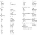
Table 1.
Laboratory data on admission
| Urinalysis | PG | 708 mg/dL | |
| pH | 7.5 | HbA1c | 13.4% |
| Protein | – | TSH | 0.52 μIU/mL |
| Glucose | >2,000 mg/dL | F‐T3 | 2.02 pg/mL |
| Blood | – | F‐T4 | 1.03 ng/dL |
| Ketone | – | Venous blood gases | |
| CBC | pH | 7.396 | |
| WBC | 2,700/μL | pCO2 | 36.9 mmHg |
| RBC | 540 × 104/μL | HCO3– | 22.1 mM/L |
| Hb | 11.1 g/dL | BE | −2.3 mM/L |
| Ht | 33.8% | Time course of serum C‐peptide | |
| Plt | 19.3 × 10⁴/μL | Day 1 | 0.97 ng/mL |
| Biochemistry | Day 2 | 0.54 ng/mL | |
| TP | 8.1 g/dL | Day 3 | 0.26 ng/mL |
| Alb | 3 g/dL | Day 7 | 0.49 ng/mL |
| BUN | 14 mg/dL | Time‐course of urinary C‐peptide | |
| Cre | 0.93 mg/dL | Day 2 | 4.02 μg/daily |
| Na | 127 mEq/L | Day 3 | 4.26 μg/daily |
| K | 3.7 mEq/L | Glucagon tolerance test C‐peptide | |
| Cl | 97 mEq/L | 0 min | 0.49 ng/mL |
| ALP | 290 IU/L | 6 min | 0.83 ng/mL |
| γ‐GT | 12 IU/L | HLA genotype | |
| AST | 17 IU/L | DRB1*09:03‐DQB1*03:03 | |
| ALT | 13 IU/L | DRB1*01:01‐DQB1*05:01 | |
| LDH | 260 IU/L | ||
| CK | 94 IU/L | ||
| T‐bil | 0.6 mg/dL | ||
| CRP | 1.8 mg/dL | ||
γ‐GT, γ‐glutamyl transpeptidase; AST, aspartate aminotransferase; Alb, albumin; ALP, alkaline phosphatase; ALT, alanine transaminase; BE, base excess; BUN, blood urea nitrogen; CBC, complete blood cell count; CK, creatine kinase; Cre, creatine; CRP, C‐reactive protein; F‐T3, free triiodothyronine; F‐T4, free thyroxine; Hb, hemoglobin; HbA1c, glycated hemoglobin; HLA, human leukocyte antigen; Ht, hematocrit; LDH, lactate dehydrogenase; PG, plasma glucose; Plt, platelets; RBC, red blood cells; T‐bil, total bilirubin; TSH, thyroid‐stimulating hormone; WBC, white blood cells.
Pneumonitis was first noted in the left lower lung lobe on computed tomography carried out on the first day of the seventh administration of nivolumab. Exacerbation of the pneumonitis was observed on computed tomography carried out 1 week after the 11th administration of nivolumab, which led to the termination of the scheduled 12th administration of nivolumab. Around the same time, the patient developed vitiligo on his forehead, forearm and back of the hand.
Hughes et al 2. reported that the time to the development of severe hyperglycemia or ketoacidosis spanned 1 week to 5 months after initiation of antiprogrammed death‐1 immunotherapy. In the present case, PG increased to 285 mg/dL 22 weeks after administration of nivolumab, and the patient developed symptoms associated with hyperglycemia with HbA1c of 9.4% 25 weeks after starting nivolumab. Considering the duration of hyperglycemia of more than 3 weeks, the increased HbA1c to more than 8.7% and the absence of ketoacidosis or ketonuria, the patient was diagnosed as having acute‐onset type 1 diabetes mellitus rather than fulminant type 1 diabetes mellitus3. DRB1*09:01‐DQB1*03:03 has been reported to confer strong susceptibility to acute‐onset type 1 diabetes mellitus in cases with at least one of the islet‐related autoantibodies4. Although the present case also had DRB1*09:01‐DQB1*03:03, autoantibodies for glutamic acid decarboxylase, insulin, insulinoma‐associated antigen‐2 and zinc transporter 8 were negative.
In summary, a patient who developed acute‐onset type 1 diabetes mellitus 22 weeks after taking nivolumab, with simultaneous exacerbation of pneumonitis and development of vitiligo, was described. This report might provide some insight into the pathophysiology of type 1 diabetes mellitus caused by nivolumab.
Disclosure
The authors declare no conflict of interest.
References
- 1. Spain L, Diem S, Larkin J. Management of toxicities of immune checkpoint inhibitors. Cancer Treat Rev 2016; 44: 51–60. [DOI] [PubMed] [Google Scholar]
- 2. Hughes J, Vudattu N, Sznol M, et al Precipitation of autoimmune diabetes with anti‐PD‐1 immunotherapy. Diabetes Care 2015; 38: e55–e57. [DOI] [PMC free article] [PubMed] [Google Scholar]
- 3. Kawasaki E, Maruyama T, Imagawa A, et al Diagnostic criteria for acute‐onset type 1 diabetes mellitus (2012): report of the Committee of Japan Diabetes Society on the Research of Fulminant and Acute‐onset Type 1 Diabetes Mellitus. J Diabetes Investig 2014; 5: 115–118. [DOI] [PMC free article] [PubMed] [Google Scholar]
- 4. Kawabata Y, Ikegami H, Awata T, et al Differential association of HLA with three subtypes of type 1 diabetes: fulminant, slowly progressive and acute‐onset. Diabetologia 2009; 52: 2513–2521. [DOI] [PubMed] [Google Scholar]