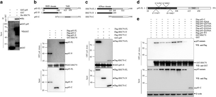
Figure 3.
RHD domain of p65 carries HSC70 recognition motifs. (a) Bacterially expressed GST-p65 was incubated with His-HSC70, followed by pull down with GSH beads. The precipitates were detected for proteins as indicated. (b) Upper panel: schematic illustrations of p65 fragments. Lower panel: Flag-p65-full-length (FL), N-terminus (p65-N) or C-terminus (p65-C) was transfected into HEK293T cells. And cell lysates were incubated with recombinant GST-HSC70 followed by pull down with GSH beads. (c) Upper panel: schematic illustrations of HSC70 fragments. Lower panel: Flag-HSC70-FL/N/C was transfected into HEK293T cells. Cell lysates were incubated with recombinant GST-p65-N followed by GST-pull down and immunoblots analysis. (d) Identification for putative KFERQ motifs within the RHD domain of p65 protein sequence. (e) Flag-p65-C, Flag-p65-FL-WT and Flag-p65-mutants were expressed in HEK293T cells. Cells lysates were incubated with GST-HSC70 or GST alone, followed by GST-pull down and immunoblotting analysis as indicated.

