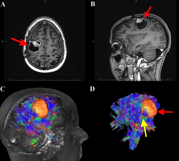
Figure 1. MRI of the brain and automated whole brain tractography (AWBT) for Case 1.
A) Axial MRI of the brain with contrast showing a right-sided mass, denoted by a red arrow. B) Sagittal MRI of the brain with contrast showing a right-sided mass, denoted by a red arrow. C) AWBT image showing the tumor to be sitting within the corticospinal tract. D) AWBT image showing the tumor (denoted by red arrow), sitting within the corticospinal tract (denoted by yellow arrow).
AWBT: automated whole brain tractography; MRI: magnetic resonance imaging

