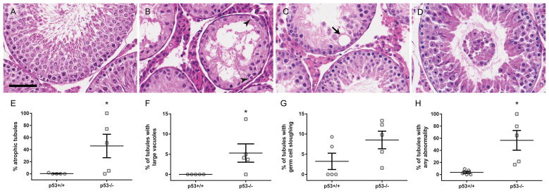
Figure 2.
p53−/− rats have higher rates of atrophy and other testicular pathology than p53+/+. p53+/+ and p53−/− rat testis sections were stained with H&E. Histological images show a normal seminiferous tubule in a p53+/+ testis (A), as well as examples of atrophy (B), a large vacuole (C, arrow), and germ cell sloughing (D) in p53−/− testes. Scale bar = 60 μm. Arrowheads in B identify spermatogonia in the atrophic tubule. p53−/− testes display significantly increased levels of seminiferous tubule atrophy (E) and large vacuoles (F), but not sloughing (G), compared to the wild-type. When all abnormalities were considered together, p53−/− rats also had a significantly higher rate than wild-type (H). Mean ± SEM reported in E–H. Scored 51–73 tubules per left testis from each animal (mean = 57.3 tubules). *p < 0.05 by the Mann-Whitney U test (atrophy, vacuoles, all) or t-test (sloughing) (n = 5 rats).

