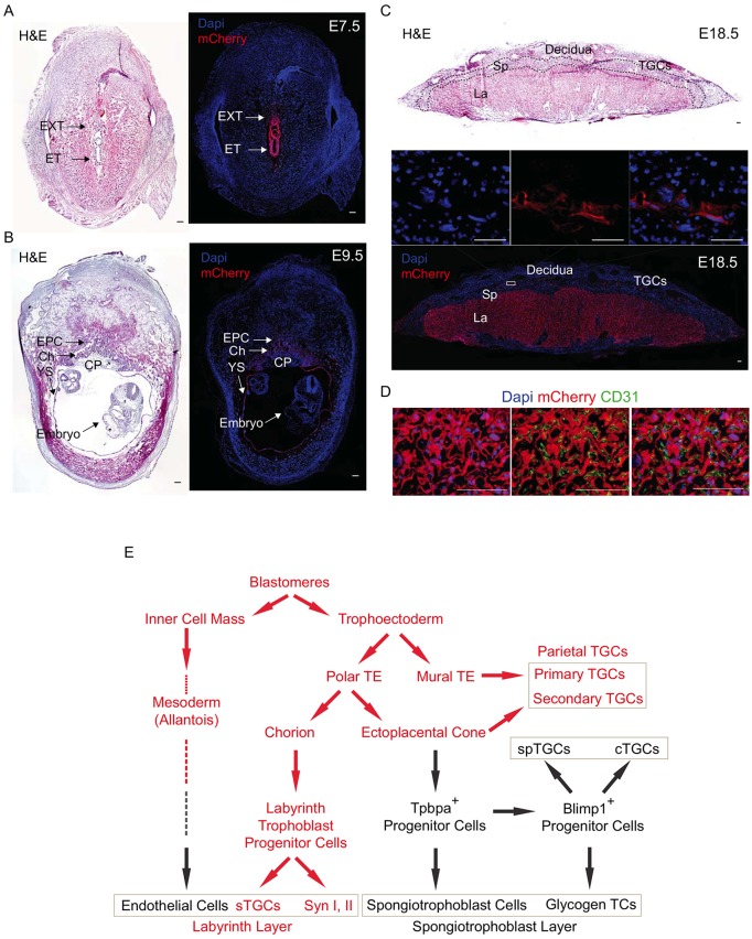
Fig. 2.
miR-290 cluster expression becomes localized to the trophoblast cells of the labyrinth and parietal TGC layers of the placenta. (A,B) H&E and immunofluorescent staining for miR-290-mCherry reporter at E7.5 and E9.5. At E7.5, the reporter is expressed strongly in extraembryonic tissue and also in embryonic tissue, whereas at E9.5 it is only expressed in extraembryonic tissues. (C) H&E and immunofluorescent staining for mCherry reporter of fully mature E18.5 placenta. It is expressed in labyrinth and parietal TGCs but not in the spongiotrophoblast cell layer. The boxed region is magnified above, showing single channels and merge. (D) The miR-290 cluster is expressed in trophoblast-derived cells of the labyrinth, whereas allantois-derived CD31+ endothelial cells are negative miR-290 cluster expression. DAPI (left), CD31 (middle) and merge (right) are shown with mCherry. (E) Ontogeny of cells expressing the miR-290 cluster during extraembryonic tissue formation. Red denotes expression of the miR-290 cluster in that cellular compartment. EXT, extraembryonic tissue; ET, embryonic tissue; EPC, ectoplacental cone; Ch, chorion; YS, yolk sac; CP, chorionic plate; La, labyrinth; Sp, spongiotrophoblast layer; TGCs, trophoblast giant cells; TE, trophectoderm; spTGCs, spiral artery TGCs; cTGCs, canal TGC; TCs, trophoblast cells; sTGCs, sinusoidal TGCs; Syn, syncytiotrophoblast cells. Scale bars: 100 μm.

