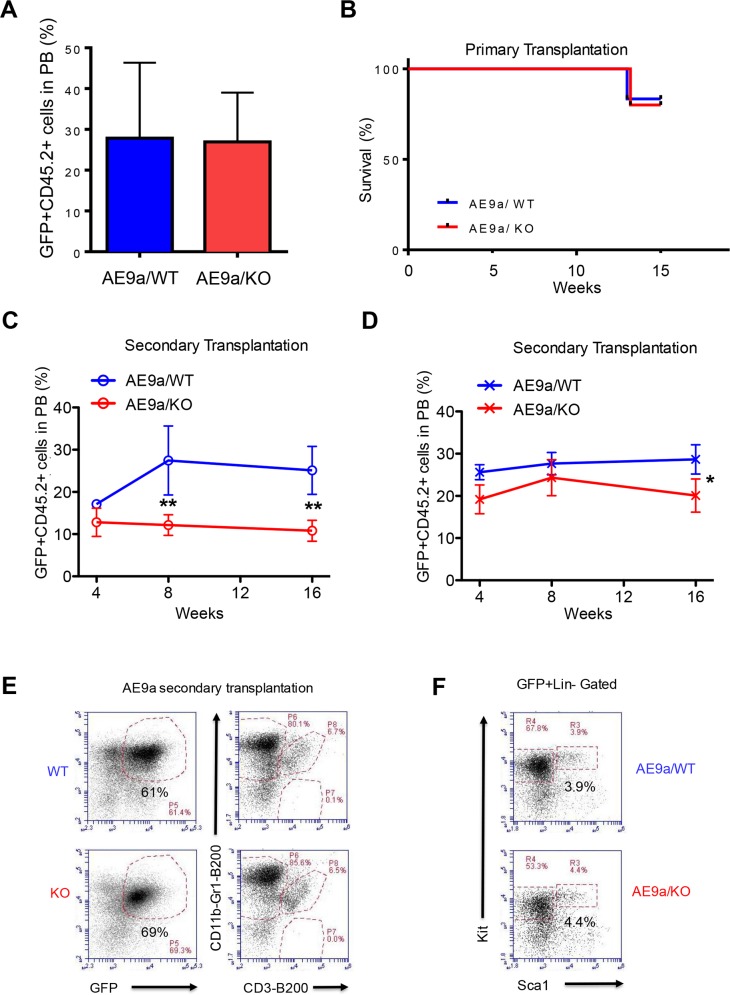
Figure 5. Necdin deficiency decreases the repopulating potential of hematopoietic stem and progenitor cells expressing AML1-ETO9a.
(A) Primary transplantation of fetal liver cells expressing AML1-ETO9a. The frequency of donor-derived cells (CD45.2+GFP+) in the peripheral blood (PB) of recipient mice at 8 weeks following transplantation was determined by flow cytometry analysis (p=0.2, n=5-6). (B) Survival curve of animals transplanted with WT or Necdin null fetal liver cells expressing AML1-ETO9a (P=0.2, n=5-6). (C) and (D) Secondary transplantation assays using 3 × 10⁵ (C) or 3 ×10⁶ (D) bone marrow cells from mice repopulated with WT or Necdin null fetal liver cell expressing AML1-ETO9a. The frequency of donor-derived cells (CD45.2+GFP+) in peripheral blood was measured by flow cytometry analysis every four weeks for 16 weeks. (*p<0.05, **p<0.01, n=5). (E) The frequency of donor-derived (CD45.2+GFP+) myeloid cells (CD11b+Gr1+), B cells (B220+), and T cells (CD3+) in the bone marrow of secondary recipient mice at 16 weeks following transplantation was determined by flow cytometry analysis. (F) The frequency of donor-derived (CD45.2+GFP+) Lin−Sca1−Kit+ and Lin−Sca1+Kit+ cells in the bone marrow of secondary recipient mice at 16 weeks following transplantation was determined by flow cytometry analysis.

