
Figure 4. The effects of emodin and rhein on the viability of MiaPaCa2 cells in vivo.
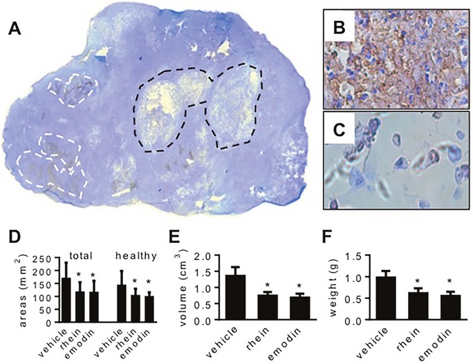
MiaPaCa2 cells grew as subcutaneous tumors in three groups of athymic mice (10 mice per group) for 8 weeks. In the last 4 weeks, the mice were treated with emodin, rhein, or vehicle, respectively. (A-C). A tumor whose host was treated with vehicle is used to show common histological features. (A). In the whole-section image (original magnification: 50x), white lines surround typical apoptotic regions and black lines surround central necrosis. (B). A region full of apoptosis (original magnification: 400x). (C). Central necrosis (original magnification: 400x). (D-F). Data shown in these panels are derived from all tumors. (D). Total section area and the section area that was occupied by healthy tissue. (E). Tumor volume. (F). Tumor weight. * P<0.05.