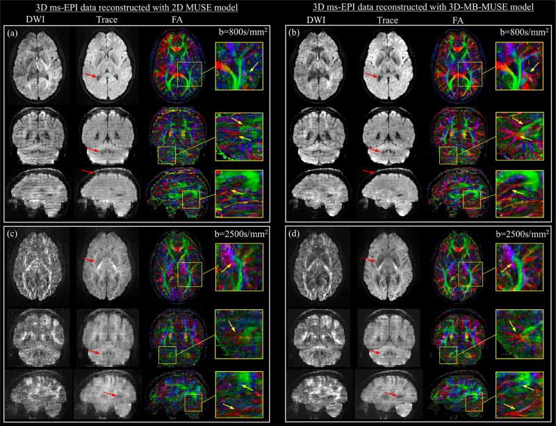
Fig. 7.
3D ms-EPI diffusion data reconstructed with 2D MUSE and 3D-MB-MUSE. Axial, coronal, and sagittal images with 1.0mm isotropic voxels are presented for diffusion weighted (DWI), trace weighted and color fractional anisotropy (FA) volumes generated from 3D ms-EPI dMRI data reconstructed by the 2D MUSE model (a) and 3D-MB-MUSE (b) with b=800s/mm2, and by the respective models with b=2500s/mm2 in (c) and (d). Differences between the 2D MUSE and 3D-MB-MUSE reconstructions are highlighted in trace weighted images with red arrows, while regions exhibiting differences in FA between the two models are highlighted by yellow arrows in enlarged images.

