Abstract
Background:
The oral cavity being a natural moist environment, topical application reduces its potency and effectiveness, but the nanoparticles in the Nano-Bio Fusion (NBF) gel are efficient in rapidly penetrating the cells. The purpose of the study aimed to evaluate clinically and microbiologically the effectiveness of locally delivered NBF technology gel as an adjunctive therapy to scaling and root planing (SRP) in the treatment of chronic periodontitis.
Materials and Methods:
Six chronic periodontitis patients with 76 sites and probing pocket depth (PD) between 5 and 7 mm were selected in a randomized controlled clinical trial. SRP was performed in both control and test group followed by NBF gel application in 38 sites. The plaque index, gingival index, papillary bleeding index, probing PD, and clinical attachment level (CAL) were recorded at baseline, 6 weeks, and 3 months. Supragingival microbial plaque analysis was done at baseline and 6 weeks interval. The statistical analysis with paired t-test was used to compare the test and control sites.
Results:
From baseline to a period of 3 months, a statistically significant difference was seen between both groups for PD, CAL, P value being PD (P = 0.001) and CAL (P = 0.01) along with the significant reduction of colony-forming units of aerobic periodontopathogens.
Conclusions:
Locally delivered NBF gel exhibited a significant improvement compared with SRP alone in chronic periodontitis.
Keywords: Local drug delivery, Nano-Bio Fusion gel, periodontal pockets
INTRODUCTION
Periodontal disease is an infectious inflammatory disease of multifactorial etiology. Bacteria present in the dental plaque are known to be one of the major causes of periodontitis. Periodontal disease progression occurs due to the shift in the composition of microorganism from supragingival plaque to subgingival plaque. Bacteria in the dental plaque grow apically and gradually forms periodontal pocket and eventually destroys the bone. In the periodontal pocket, the bacteria present apically forms a highly structured and complex biofilm leading to accomplish effective oral hygiene efforts.
It is, therefore, mandatory to treat the periodontal pockets by mechanical removal of local factors and disruption of subgingival plaque biofilm itself. The development of subgingivally placed controlled delivery systems has provided the possibility an effective intrapocket concentration levels of antibacterial agents for extended period of time, resulting in an altered subgingival flora and enhanced the healing of the attachment apparatus.[1]
Various chemical agents such as nonsteroidal anti-inflammatory drugs and antimicrobial agents, chlorhexidine and cetylpyridinium chloride, have gained popularity but simultaneously lead to conditions such as drug resistance and drug allergy. Thus, an emphasis on usage of herbal agents such as propolis, Aloe vera, green tea extracts, neem, and curcumin have gained popularity in recent times.
Propolis produced by honeybees is a resinous mixture collected from parts of plants, buds, and exudates. In 1908, the first scientific work with propolis, illustrating its composition and pleiotropic property, was published and was first patented in 1968. Nowadays, propolis is a natural remedy in the different formulation in the field of medicine and dentistry.[2] Vitamin C, a key processor of cell growth, healing, and repair of tissue, influences the metabolism of collagen within the periodontium. It also maintains the epithelial barrier function to bacterial products and integrity of periodontal microvasculature. Vitamin E serves as an antioxidant to limit free radical and to protect cells from lipid peroxidation. Vitamin E works in synergistic with Vitamin C and maintains the integrity of the cell membranes.[1]
The oral cavity being a natural moist environment, the potency and effectiveness of the topical application of various agents to prevent progression of disease are markedly reduced. Nano-Bio Fusion (NBF) gingival gel used in the study is a patented scientifically formulated, bioadhesive antioxidant gel harvesting naturally occurring antioxidants for targeted action. It mainly acts based on the NBF technology which allows the ultrafine antioxidants to surpass the moist intraoral environment, enter the cells and rejuvenate, revitalize, support, protect, and optimize gum and soft oral tissue.
The natural antioxidant power of propolis, vitamin C, and vitamin E is amplified with Nano Bio-Fusion technology. For example, nano Vitamin C is ten times more potent in hundred times smaller quantities, than Vitamin C on its own. Once applied, NBF gel creates nano-bioactive protective film which results in increased absorption, resulting in improved clinical effectiveness and visible results after application.
Thus, the present study aimed to evaluate the clinico-microbiological effectiveness of locally delivered NBF gel as an adjunctive therapy to scaling and root planing (SRP) in the treatment of periodontitis.
MATERIALS AND METHODS
The clinical trial was carried out in the Periodontology Department, Oxford Dental College, Bengaluru, Karnataka, in a randomized controlled design. Six chronic periodontitis patients comprising 76 sites were recruited in the study. Informed consent was taken from all the patients before the start of the study. The inclusion criteria were systemically healthy participants visiting the department within the age group of 30–60 years, with a minimum of twenty teeth present and a probing depth of 5–7 mm classified as localized/generalized chronic periodontitis. The exclusion criteria included the patients on chemical/herbal drugs for the past 3 months, endodontically treated teeth, teeth with furcation involvement, periodontal therapy in past 6 months, habit of tobacco chewing and smoking, and pregnant and lactating women.
The patients were randomly divided into two groups satisfying the above-mentioned criteria based on randomization software.
Group A: SRP only [Figure 1]
Group B: SRP followed by NBF gel application in periodontal pockets [Figure 2].
Figure 1.
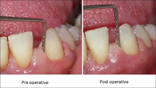
Group A (Control) - Scaling and root planing alone
Figure 2.
Group B (Test) - Scaling and root planing along with Nano-Bio Fusion gel application
Treatment protocol
The initial examination recorded plaque index (PI) 1963,[3] gingival index (GI) 1964,[4] papillary bleeding index 1977,[5] and probing pocket depth (PD) were measured from the gingival margin to the depth of the pocket using William graduated probe, clinical attachment level was measured from cementoenamel junction to base of pocket using William graduated probe at baseline.
Following the initial examination, NBF gel [Figure 3] was filled in the pockets through a blunt cannula until it was detected at the gingival margin in group B. To ensure retention of the gel for long duration to be effective in the pocket, a periodontal dressing (Coe-Pak) was placed.[6] Postoperative home care instructions including brushing two times daily with a soft brush were given. The patients in both the groups were evaluated at baseline, 6 weeks, and 3 months interval. Supragingival plaque sample was collected from tooth surface from two patients before scaling and 6 weeks after application of NBF gel for microbiological analysis using nutrient agar culture media [Figure 4].
Figure 3.
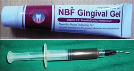
Nano-Bio Fusion gel and blunt cannula for application in periodontal pockets
Figure 4.
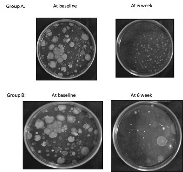
Colony-forming unit analysis at baseline and 6 weeks interval
Statistical analysis
Statistical analysis was conducted for the results obtained. Statistical analysis was done using SPSS Inc. Released 2009. PASW Statistics for Windows, Version 18.0. Chicago. The comparison of test and control sites was done using paired t-test. P < 0.05 was considered statistically significant.
RESULTS
Table 1 observed the comparison of mean PI which showed a statistically significant result at 6 weeks period (P = 0.002) from baseline, but no statistically significant difference was observed at 3 months interval as observed from [Figure 5]. Table 2 depicts the mean GI scores after application of NBF gel alone at various intervals. At 6 months, a statistically significant result was obtained (P = 0.031), but the P value at 3 months interval was 0.822, hence no statistically significant results were obtained as reflected in [Figure 6]. Table 3 explains the mean value of sulcus bleeding index (SBI) at various levels. At 6 weeks (P = 0.001) and 3 months (P = 0.422) interval, a statistically significant results were obtained as observed in [Figure 7]. Table 4 describes the periodontal pocket depth, which at 6 weeks (P = 0.050) and 3 months (0.001) interval in the test group has shown statistically significant difference as also depicted in [Figure 8]. Table 5 shows the means of clinical attachment level (CAL) at baseline, 6 weeks, and 3 months interval with a statistically significant difference in both the groups at 6 weeks and 3 months with P= 0.188 and P= 0.001, respectively as seen in [Figure 9]. Table 6 shows a statistically significant result in a mean reduction of colony-forming unit (CFU) at 6 weeks interval in test group from baseline unit (P = 0.001) as compared to control group as depicted in [Figure 10].
Table 1.
Plaque index - mean values at various intervals

Figure 5.
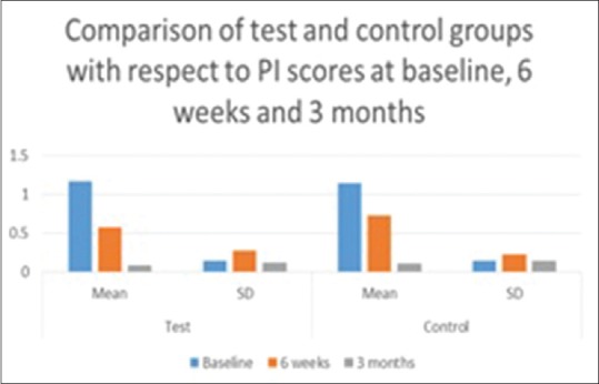
Mean values of plaque index at various intervals
Table 2.
Mean values of gingival index at various intervals

Figure 6.
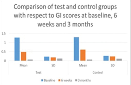
Mean values of gingival index at baseline, 6 weeks, and 3 months interval
Table 3.
Sulcus bleeding index - mean values at various intervals

Figure 7.
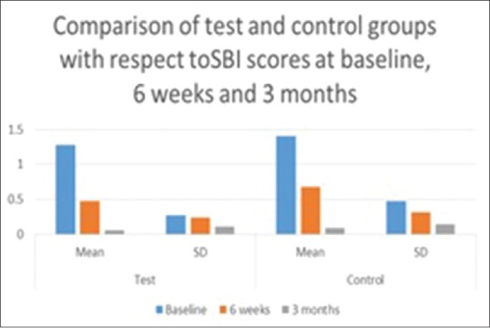
Mean values of sulcus bleeding index at baseline, 6 weeks, and 3 months interval
Table 4.
Periodontal pocket depth - mean values at various intervals

Figure 8.
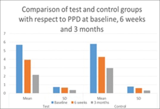
Mean values of periodontal pocket depth at baseline, 6 weeks, and 3 months interval
Table 5.
Mean values of clinical attachment level at various interval

Figure 9.
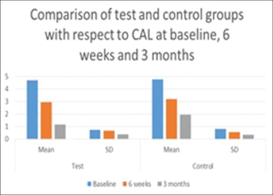
Mean values of clinical attachment level at baseline, 6 weeks, and 3 months interval
Table 6.
Colony forming unit/ml at baseline, 6 weeks (mean values)

Figure 10.
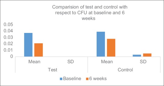
Mean values of colony forming unit (CFU) at baseline and 6 weeks interval
DISCUSSIONS
Local delivery agents placed in periodontal pockets have authenticated the reduction in microbial load and improvements in clinical signs of periodontitis.
Propolis, nature's powerful antibiotic and healing substantial, is natural resinous product extracted by honeybees (Apis mellifera) from botanical provenance to protect the hive from invasion and infection. Propolis is rich in a wide range of bioflavonoids and has been used since ancient times for its pleiotropic properties. Test tube and in vivo studies have shown various antioxidant, anti-inflammatory, and anticancer properties of propolis.[7,8,9] Antibiotic properties of propolis are retained in commercial formulations, as 1 mg/ml has exhibited the minimum inhibitory concentration of propolis extract across various periodontopathogens.[10]
The present study evaluated the various parameters in the treatment of chronic periodontitis with NBF gel. The PI score obtained at various intervals is in consistent with meticulous oral hygiene procedure, so at 3 months interval, intergroup analysis showed no significant results. With single application of NBF gel, GI and SBI showed statistically significant difference at 6 weeks interval and clinically significant difference at 3 months period which can be due to the diverse characteristic property of propolis.
Propolis as an anti-inflammatory agent has shown to inhibit synthesis of prostaglandins, activate the immune system by promoting phagocytic activity, stimulate cellular immunity, and facilitate healing effects on epithelial tissues. Propolis is a potent antioxidant and leads to reduction in reactive oxygen species (ROS) and limit lipid peroxidation activity. Propolis enhance synthesis of collagen due to the presence of iron and zinc elements.[11]
The once application of NBF gel with an appropriate drug carrier agent at baseline has shown a statistically significant reduction in PD, CAL, which could be attributed to the antimicrobial property of propolis and could also be confirmed by the reduction in cfu/mL as seen after 6-week interval after NBF gel application. The total viable counts between 105 and 106 cfu/mL are related to low number of periodontopathogenic organisms as per Wolff et al., 1994.[12] The variation in composition of propolis led to a complex mechanism of antimicrobial action which is not understood completely.
Hence, with an appropriate drug carrier agent, it is possible to administer the effect of this drug to their target site in the oral cavity. A suitable drug carrier agent could be best described through nanotechnology which uses the modern technology to manufacture a material or structure purposefully with dimensions between 1 and 100 nm to deliver the unique properties it has at that size. The sole intent is the targeted delivery of active ingredients into periodontium. Encapsulating the ingredients within a film protects from the moist environment of oral cavity and enhances activation and better penetrability at target site. However, along with all these beneficial attributes come various controversies with nanoparticles. It includes the poor understanding of depth of a nanoparticles penetration, the ability, and the extent at which it can enter the bloodstream. Some authors believe using nanoparticles reduce the efficacy in terms of function and others are concerned about whether the use of nanoparticles is harmful.
The effect of nanoemulsion as a base to carry the drug was studied by Chang and Park,[13] on treatment of gingival inflammation, exhibited its effectiveness in protection of gingiva and treatment of gingival diseases. At a specific concentration, the anti-inflammatory and antimicrobial association was established.
The results obtained in the present study are in accordance with previous studies where various formulation of propolis at different time frame was evaluated.
Koo et al.[14] evaluated propolis in mouth rinse formulation and a significant reduction in PI was obtained at 4th day of the study. Coutinho[15] used propolis as subgingival irrigation at 6 weeks interval period which showed a significant improvement in clinical and microbiological parameters. A study to treat gingivitis patients with topical application of NBF showed promising improvements in clinical parameters.[16]
In NBF gel, the NBF of propolis along with Vitamin C and Vitamin E has brought about the tremendous improvement in periodontal health of the patients. Vitamin C has many metabolic functions relevant to the health of the periodontium including collagen synthesis, and immune function. Vitamin C, a powerful antioxidant, causes a reduction in the protective antioxidant effect on its marginal shortfalls increasing the pathogenesis of periodontitis. The systemic administration of Vitamin C could be clinically beneficial in downregulating 2-fold, the gene expression encoding inflammation, including interleukin-1 alpha and interleukin-1 beta, in improving periodontitis-induced oxidative stress.[17] ROS is universally aimed at bacterial targets and is also released into the host extracellular environment, causing damage to DNA, cell proteins, and membrane lipid peroxidation. ROS also stimulates pro-inflammatory cytokine release by monocytes and macrophages,[18] and macrophages may also stimulate osteoclast activation.[19] Patients with gingivitis and periodontitis have high levels of many ROS in gingival fluid,[20] and thus a correlation between ROS and periodontal inflammation has been reported. Research suggests that powerful antioxidant properties of Vitamin E protect periodontal tissue from oxidative damage.
Although propolis in various formulations has been investigated, the results obtained from various trials are not comparable, due to the difference in composition of propolis, formulations, and the different study design used for the evaluation of its diverse activities. The present study with the usage of NBF gel in chronic periodontitis is the first of its own kind. The periodontal heath of the patients improved significantly in all the sites irrespective of the treatment rendered. However, the sites which were treated with a combination of scaling and NBF gel application showed better results (P < 0.001) as compared to control sites. Thus, nanoemulsion base along with active ingredient of propolis, Vitamin C, and Vitamin E significantly improved the clinical parameters of the patients which could be alluded to activation of the nano-sized propolis and vitamins.
CONCLUSIONS
The present study influenced the beneficial outcome of propolis along with Vitamin C and E and the nanotechnology amplified this effect in preventing disease progression. Although the results indicate anti-inflammatory, antibacterial, and antioxidant property of the NBF gel, SRP still dwells to be the gold standard treatment protocol, and NBF gel can be used as an adjunct for improving the periodontal status of an individual. Further research with a larger sample size is warranted to have a better understanding of the effectiveness of NBF gel in the protection of periodontium.
Financial support and sponsorship
Nil.
Conflicts of interest
There are no conflicts of interest.
REFERENCES
- 1.Newman MG, Takei HH, Klokkevold PR, Carranza FA. Carranza's Clinical Periodontology. 11th ed. St. Louis, MO: Elsevier/Saunders; c2012. p. 1938. [Google Scholar]
- 2.Ikeno K, Ikeno T, Miyazawa C. Effects of propolis on dental caries in rats. Caries Res. 1991;25:347–51. doi: 10.1159/000261390. [DOI] [PubMed] [Google Scholar]
- 3.Loe H, Silness J. Periodontal disease in pregnancy. I. Prevalence and severity. Acta Odontol Scand. 1963;21:533–51. doi: 10.3109/00016356309011240. [DOI] [PubMed] [Google Scholar]
- 4.Silness J, Loe H. Periodontal disease in pregnancy. II. Correlation between oral hygiene and periodontal condtion. Acta Odontol Scand. 1964;22:121–35. doi: 10.3109/00016356408993968. [DOI] [PubMed] [Google Scholar]
- 5.Mühlemann HR. Psychological and chemical mediators of gingival health. J Prev Dent. 1977;4:6–17. [PubMed] [Google Scholar]
- 6.Bhat G, Kudva P, Dodwad V. Aloe vera: Nature's soothing healer to periodontal disease. J Indian Soc Periodontol. 2011;15:205–9. doi: 10.4103/0972-124X.85661. [DOI] [PMC free article] [PubMed] [Google Scholar]
- 7.Pascual C, Gonzalez R, Torricella RG. Scavenging action of propolis extract against oxygen radicals. J Ethnopharmacol. 1994;41:9–13. doi: 10.1016/0378-8741(94)90052-3. [DOI] [PubMed] [Google Scholar]
- 8.Dobrowolski JW, Vohora SB, Sharma K, Shah SA, Naqvi SA, Dandiya PC. Antibacterial, antifungal, antiamoebic, antiinflammatory and antipyretic studies on propolis bee products. J Ethnopharmacol. 1991;35:77–82. doi: 10.1016/0378-8741(91)90135-z. [DOI] [PubMed] [Google Scholar]
- 9.Choi YH, Lee WY, Nam SY, Choi KC, Park YE. Apoptosis induced by propolis in human hepatocellular carcinoma cell line. Int J Mol Med. 1999;4:29–32. doi: 10.3892/ijmm.4.1.29. [DOI] [PubMed] [Google Scholar]
- 10.Gebara EC, Lima LA, Mayer MP. Propolis antimicrobial activity against periodontopathic bacteria. Braz J Microbiol. 2002;33:365–9. [Google Scholar]
- 11.Ozan F, Sümer Z, Polat ZA, Er K, Ozan U, Deger O. Effect of mouthrinse containing propolis on oral microorganisms and human gingival fibroblasts. Eur J Dent. 2007;1:195–201. [PMC free article] [PubMed] [Google Scholar]
- 12.Wolff L, Dahlén G, Aeppli D. Bacteria as risk markers for periodontitis. J Periodontol. 1994;65(5 Suppl):498–510. doi: 10.1902/jop.1994.65.5s.498. [DOI] [PubMed] [Google Scholar]
- 13.Chang CH, Park JW. The study on the effect of nanaoemulsion for the prevention and treatment of gingival inflammation. J Korean Oral Maxillofac Surg. 2007;33:1–6. [Google Scholar]
- 14.Koo H, Cury JA, Rosalen PL, Ambrosano GM, Ikegaki M, Park YK. Effect of a mouthrinse containing selected propolis on 3-day dental plaque accumulation and polysaccharide formation. Caries Res. 2002;36:445–8. doi: 10.1159/000066535. [DOI] [PubMed] [Google Scholar]
- 15.Coutinho A. Honeybee propolis extract in periodontal treatment: A clinical and microbiological study of propolis in periodontal treatment. Indian J Dent Res. 2012;23:294. doi: 10.4103/0970-9290.100449. [DOI] [PubMed] [Google Scholar]
- 16.Sneha V, Chatterjee A. Evaluate the efficacy of NBF gel as an adjunct to scaling in gingivitis – A clinical study. Guident. 2014;7:86–8. [Google Scholar]
- 17.Tomofuji T, Ekuni D, Sanbe T, Irie K, Azuma T, Maruyama T, et al. Effects of Vitamin C intake on gingival oxidative stress in rat periodontitis. Free Radic Biol Med. 2009;46:163–8. doi: 10.1016/j.freeradbiomed.2008.09.040. [DOI] [PubMed] [Google Scholar]
- 18.Chapple IL. Reactive oxygen species and antioxidants in inflammatory diseases. J Clin Periodontol. 1997;24:287–96. doi: 10.1111/j.1600-051x.1997.tb00760.x. [DOI] [PubMed] [Google Scholar]
- 19.Hall TJ, Schaeublin M, Jeker H, Fuller K, Chambers TJ. The role of reactive oxygen intermediates in osteoclastic bone resorption. Biochem Biophys Res Commun. 1995;207:280–7. doi: 10.1006/bbrc.1995.1184. [DOI] [PubMed] [Google Scholar]
- 20.Diab-Ladki R, Pellat B, Chahine R. Decrease in the total antioxidant activity of saliva in patients with periodontal diseases. Clin Oral Investig. 2003;7:103–7. doi: 10.1007/s00784-003-0208-5. [DOI] [PubMed] [Google Scholar]


