

Abstract
In this report, we describe the management of a child with bilateral cataract, nystagmus, and comitant sensory esotropia. Routine ultrasonography done before cataract surgery revealed bilateral disc edema confirmed as idiopathic intracranial hypertension by a pediatric neurologist. The primary intervention for cataract surgery was followed by nonresolving papilledema, despite maximum medical therapy. To salvage the optic nerve function in a nonverbal child, bilateral optic nerve sheath decompression was planned with simultaneous medial rectus recessions for the persistent esotropia with the satisfactory postoperative outcome.
Keywords: Congenital cataract, idiopathic intracranial hypertension, optic nerve sheath decompression in infants, sensory esotropia
Idiopathic intracranial hypertension (IIH) though classically described in obese, middle-aged females, may be seen in any age group in both sexes. However, it remains a rarity among infants. Here, we describe an incidental detection of IIH with comitant esotropia in an infant with bilateral congenital cataract and its successful management by surgery. It is possibly the first report of optic nerve sheath decompression (ONSD) at such a young age combined with rectus muscle surgery for simultaneous correction of esotropia following cataract surgery in both eyes.
Case Report
An 11-month-old infant was brought to the clinic by her parents with complaints of white opacity in both eyes for 3 months of age, inward deviation and shaking of the eyes [Fig. 1a] for 7 months. She was a full-term baby, born of cesarean delivery with an uneventful prenatal, perinatal, and postnatal course. Parents were nonconsanguineous with no significant family history.
Figure 1.
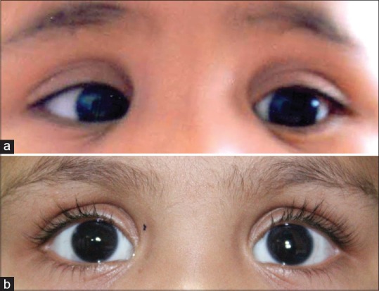
Clinical photograph of child; (a) preoperatively showing right esotropia with psuedophakia in both eyes, (b) postoperatively, photograph showing orthotropia and pseudophakia in both eyes
On examination, she was able to fix and follow light. There was a comitant right esotropia with good alternation and full abduction. She had a horizontal pendular nystagmus of moderate amplitude and high frequency in all gazes. Anterior segment examination revealed bilateral nuclear cataract with grossly normal pupillary reactions and no view of the fundus. A routine B-scan ultrasonography done to evaluate the posterior segment revealed increased optic nerve diameter (diameters more than 3.5 mm), with subarachnoid fluid[1,2] around the nerves [Fig. 2a]. This was confirmed on magnetic resonance imaging (MRI) as prominent perioptic subarachnoid spaces bilaterally and posterior scleral pole flattening at optic nerve head confirming papilledema. No hydrocephalus, TORCH imaging markers (intracranial calcifications, etc.,) cerebral sinovenous thrombosis or space-occupying lesions were seen on the MRI [Fig. 3]. Pediatric neurological evaluation recorded cerebrospinal fluid (CSF) manometric opening pressure of 29 cm of water pressure (normal CSF pressure <20 cm water pressure) with unremarkable CSF analysis, ruling out sepsis. Endocrinological causes for raised intracranial pressure (ICP) were excluded. Baseline hematological, biochemical, and vasculitic investigations including TORCH profile were normal. She was not on any regular medications that could have attributed to raised ICP. IIH was diagnosed after ruling out secondary causes of raised ICP as per modified Dandy's criteria[3] and was then started on oral acetazolamide.
Figure 2.

B-scan right (left image) and left eye (right image); (a) at presentation showing optic nerve head (thick arrow) with size of 5 mm in right eye and 5.30 mm in left eye and an echolucent crescent due to the sub-arachnoid fluid around the optic nerve (thin arrow), intraoperative image showing, (b) distended optic nerve sheath (arrowhead) and short posterior ciliary vessels (thin arrow) over optic nerve, (c) B-scan right (left image) and left eye (right image); postoptic nerve sheath decompression showing decrease in optic nerve head size (thick arrow) with resolution of sub-arachnoid fluid around optic nerve
Figure 3.

Magnetic resonance imaging of brain (T2 sagittal scan) showing distended optic nerve sheaths indicated by white arrows in both eyes
The child underwent cataract surgery with primary posterior chamber intraocular lens implantation in both eyes. Postoperative fundus evaluation confirmed papilledema in both eyes. Her vision started improving in both eyes following cataract surgery and her nystagmus amplitude reduced with moderate frequency. Following surgery, the esotropia measured to 35 prism diopter base out with good alternation for distance and near.
In our patient, monitoring the effect of IIH on the function of the optic nerve was challenging. Visual acuity measurements were influenced by congenital cataract in both eyes. The young age, limited assessment of formal visual fields, and view of dense amblyopia secondary to congenital cataract with nystagmus, visual evoked potentials would not be reliable in assessing the optic nerve status, hence fundus examination and serial B-scan measurements were done to evaluate response to treatment.[1] During this period, maximal therapy at a dose of 30 mg/kg/day was given in four divided doses for 3 months. As the child was otherwise asymptomatic for raised ICP features (no emesis, seizures, encephalopathy, focal lateralizing deficits, or developmental regression), medication was tapered over the next 2 months. Since the disc edema showed no resolution, which was confirmed on three serial B-scans and the child was systemically stable, no systemic shunt procedure was indicated, and bilateral ONSD was planned in conjunction with the pediatric neurologist to salvage optic nerve function. It was performed through transconjunctival approach in both eyes combined with bilateral medial rectus recession (5.5 mm) to tackle her esotropia simultaneously. Intraoperatively, the optic nerve sheaths were found to be distended with fluid [Fig. 2b]. The postoperative course was uneventful with a resolution of fluid on B-scan [Fig. 2c]. Thirty months after ONSD, she remained orthotropic [Fig. 1b] with small amplitude and frequency nystagmus with the acceptable vision of 20/50 with binocular viewing. She has achieved age-appropriate development with no new neurological or ocular features of IIH at follow-up.
Discussion
IIH can occur at any age but is rare in infants.[4,5] The response to medical management or lumbar puncture is good and few of them need surgical intervention,[5] which includes lumboperitoneal shunt, ventriculoperitoneal shunt, or ONSD.[6] A delay in the treatment of IIH can lead to optic atrophy and blindness.[4] Since assessing visual parameters reliably in children is difficult, intervention needs to be done earlier as a preventive strategy as opposed to waiting for objective testing. Furthermore, it can be very difficult to monitor optic nerve function in children. Hence, when the fluid around the optic nerve head did not show any resolution on serial B-scans, ONSD was planned to prevent permanent optic nerve damage. Bilateral ONSD was performed since the procedure was to be undertaken under general anesthesia as unilateral ONSD has eventually required other eye decompression in pediatric cases[7] and we had planned simultaneous recession of bilateral medial rectus muscles.
Conclusion
This case report highlights the importance of detailed ophthalmic evaluation and the role of B-scan ultrasonography in diagnosis and follow-up of IIH, even in a child. It emphasizes the role of ONSD in a refractory case of IIH to prevent permanent visual deficits along with simultaneous correction of the strabismus for the comitant esotropia associated with the congenital cataract with satisfactory results.
Financial support and sponsorship
Nil.
Conflicts of interest
There are no conflicts of interest.
References
- 1.Matalia J, Shirke S, Kekatpure M. An alternate technique for assessing optic nerve in papilledema by ultrasound B scan. Am J Emerg Med. 2015;33:971–3. doi: 10.1016/j.ajem.2015.03.027. [DOI] [PubMed] [Google Scholar]
- 2.Stone MB. Ultrasound diagnosis of papilledema and increased intracranial pressure in pseudotumor cerebri. Am J Emerg Med. 2009;27:376.e1–376.e2. doi: 10.1016/j.ajem.2008.08.007. [DOI] [PubMed] [Google Scholar]
- 3.Wall M. Idiopathic intracranial hypertension. Neurol Clin. 1991;9:73–95. [PubMed] [Google Scholar]
- 4.Ko MW, Liu GT. Pediatric idiopathic intracranial hypertension (pseudotumor cerebri) Horm Res Paediatr. 2010;74:381–9. doi: 10.1159/000321180. [DOI] [PubMed] [Google Scholar]
- 5.Phillips PH. Pediatric pseudotumor cerebri. Int Ophthalmol Clin. 2012;52:51–9, xii. doi: 10.1097/IIO.0b013e31825a12f6. [DOI] [PubMed] [Google Scholar]
- 6.Lee AG, Patrinely JR, Edmond JC. Optic nerve sheath decompression in pediatric pseudotumor cerebri. Ophthalmic Surg Lasers. 1998;29:514–7. [PubMed] [Google Scholar]
- 7.Thuente DD, Buckley EG. Pediatric optic nerve sheath decompression. Ophthalmology. 2005;112:724–7. doi: 10.1016/j.ophtha.2004.11.049. [DOI] [PubMed] [Google Scholar]