Abstract
Background
Chondrotoxic effects of local anaesthetics are well reported in humans and some animal species but knowledge on their toxic effects on synoviocytes or equine chondrocytes or the effects on cellular production of inflammatory cytokines is limited. The purpose of this study was to evaluate the in vitro effects of local anaesthetics, morphine, magnesium sulphate (MgSO4) or their combinations on cell viability and pro-inflammatory cytokine gene expression of equine synoviocytes and chondrocytes.
Equine synoviocytes and cartilage explants harvested from normal joints in a co-culture system were exposed to mepivacaine (4.4 mg/ml), bupivacaine (2.2 mg/ml), morphine (2.85 mg/ml) and MgSO4 (37 mg/ml) alone or each local anaesthetic plus morphine or MgSO4 or both together. Chondrocyte and synoviocyte cell viability was assessed by CellTiter-Glo Luminescent Cell Viability Assay. Synoviocyte gene expression of IL-1β, IL-6 or TNF-α was measured and compared using the ∆∆CT method.
Results
Morphine alone, MgSO4 alone or their combination did not alter cell viability or the expression of IL-1β, IL-6 or TNF-α. However, local anaesthetics alone or in combination with morphine and/or MgSO4 reduced cell viability and increased the gene expression of IL-1β, IL-6 or TNF-α. Single short exposure to local anaesthetics is toxic to both chondrocytes and synoviocytes and their combination with morphine and/or MgSO4 enhanced the cytotoxic effects.
Conclusions
This in vitro study gives further evidence of the absence of cytotoxic effects of morphine alone, MgSO4 alone or their combination on normal articular tissues. However, local anaesthetics alone or in combination with morphine and/or MgSO4 have cytotoxic effects on equine articular tissues.
Electronic supplementary material
The online version of this article (10.1186/s12917-017-1244-8) contains supplementary material, which is available to authorized users.
Keywords: Local anaesthetic, Morphine, Magnesium sulphate, Chondrocyte, Synoviocyte, Equine
Background
Intra-articular injections of local anaesthetics are commonly performed in humans and horses to determine sources of pain and as perioperative pain control [1]. Despite their widespread use, there is growing concern over the potential toxicity of these substances and their long-term effects on articular tissue [2, 3]. Chondrotoxic properties of local anaesthetic agents have been reported in humans and animals [2, 4–6], but knowledge of their effect on equine chondrocytes is limited [7, 8]. The majority of these studies have investigated their effects on chondrocyte viability, but the effects of local anaesthetics on synoviocytes are still largely unknown. The synovium contributes to nociceptive, inflammatory and degradative responses and therefore it is vital that the effects of intra-articular injections are also studied on the synovium. Recent studies on rabbits and dogs suggest that the toxic effects of local anaesthetic on synoviocytes may affect the onset of chondrolysis associated with intra-articular use of local anaesthetics [9–11].
Because of the local anaesthetic related chondrotoxic effects, alternatives for articular analgesia are being sought in humans [3]. Morphine is an opioid that provides excellent articular analgesic and anti-inflammatory effects when administered intra-articularly in humans [12, 13] with apparently minimal toxic effects on human and canine chondrocytes [2, 14]. Intra-articular administration of morphine causes analgesia, and reduces swelling and synovial inflammatory markers in horses [15–18], although it was associated with release of large molecular weight proteoglycans into the synovial fluid [19]. Magnesium sulphate (MgSO4) is routinely administered intra-articularly to human patients for peri-operative analgesia [20] and does not cause a significant reduction in human chondrocyte viability [21]. Moreover, addition of MgSO4 to local anaesthetics reduced the toxic effects of the latter on human chondrocytes in vitro [22]; and intra-articular administration of MgSO4 attenuated the development of osteoarthritis (OA) in a rat model [23].
We hypothesised that local anaesthetics but not morphine or MgSO4, would produce deleterious effects on chondrocyte and synoviocyte viability and increase the expression of pro-inflammatory cytokines. We further hypothesised that morphine or MgSO4 in combination with a local anaesthetic would prevent the negative effects exerted by local anaesthetics alone.
Methods
The aim of this study was to evaluate the in vitro effects of clinically-relevant doses of local anaesthetics, morphine, MgSO4 or their combinations on equine chondrocyte and synoviocyte viability and gene expression of pro-inflammatory cytokines in a co-culture in vitro model. We hypothesised that local anaesthetics would produce deleterious effects on chondrocyte and synoviocyte viability and increase the expression of pro-inflammatory cytokines. We further hypothesised that morphine and/or MgSO4 in combination with a local anaesthetic would reduce the impact of the negative effects exerted by local anaesthetics alone on cell viability and gene expression of pro-inflammatory cytokines.
Study design
An experimental in vitro study was performed on equine synoviocytes and cartilage explants harvested from normal joints and using a co-culture system.
Tissue sample collection
Tissue samples were obtained from metacarpophalangeal joints of 10 skeletally mature horses (6–10 years old) from an abattoir within 5 h of slaughter. Synoviocytes were obtained from three horses and articular cartilage samples obtained from seven different horses. Only horses with grossly normal joints were included in the study.
Under sterile conditions, synovial membrane was harvested from the entire synovial surface of the joint and synoviocytes isolated as previously described [24]. Synoviocytes were cultured in Dulbecco’s Modified Eagle Medium (DMEM) (Sigma-Aldrich, Darmstadt, Germany) supplemented with 10% foetal calf serum (FCS), 100 units/ml penicillin, 100 μg/ml streptomycin, L-glutamine 4 mM (all from Invitrogen, Paisley, UK) and 500 ng/ml amphotericin B (Bio Whittaker, Lonza, San Diego, California, USA), in routine laboratory conditions (37 °C, 5% O2) until 90% confluent. After treatment with 0.05% w/v trypsin, synoviocytes were passaged and cultured until 90% confluent. Synoviocytes (passage 2) were then frozen in 10% DMSO in DMEM complete (DMEM supplemented as above) and stored in liquid nitrogen. Full thickness cartilage was collected from the entire surface of the distal condylar area of the third metacarpus. Cartilage was diced into explants of approximately 2 mm x 2 mm, mixed and placed in complete DMEM complete at 37 °C, 5% O2 for 18 h to equilibrate.
Synoviocytes from three horses were thawed in a water bath at 37 °C for 1 min, mixed and suspended in DMEM complete to a final concentration of 50,000 live cells per ml. Synoviocytes were plated in 24-well plates at a concentration of 50,000 cells per well (2 wells per group). After equilibration at 37 °C, 5% O2 for 24 h, trans-well inserts (0.45 μm pore, 12 mm; Millicell Cell Culture Inserts PIHA01250; Sigma-Aldrich, Darmstadt, Germany) were located into the wells and cartilage explants from 7 horses were placed separately into the inserts (5 explants per insert) in duplicate. The co-culture system was maintained in an incubator at 37 °C, 5% O2 for 24 h at which time co-cultures were exposed to different treatments for 2 h (Table 1). Drugs were diluted in DMEM complete to a total volume of 2 ml per well to achieve the following final concentrations: mepivacaine 4.4 mg/ml, bupivacaine 2.2 mg/ml, morphine 2.85 mg/ml, magnesium sulphate 37 mg/ml. Concentrations were based on the average volume of synovial fluid in the equine metacarpophalangeal joint (12.5 mL) [25] and the doses commonly used in clinical practice [16, 17, 26–29]. Exposure time (2 h) was based on the reported half-life for the local anaesthetics in horses [26, 30]. After exposure, the medium containing the different treatments was removed and replaced by DMEM complete and cultured for additional 48 h. At this time synoviocytes and explants were harvested. Cartilage explants were snap-frozen in liquid nitrogen and stored at -80 °C. Synoviocytes from the same group were released from the wells using 250 μL Tri-reagent (Sigma-Aldrich, Darmstadt, Germany) per well for 30 min and immediately stored at -80 °C
Table 1.
Treatment groups
| Group | Treatment |
|---|---|
| 1 | Control |
| 2 | Mepivacaine only |
| 3 | Mepivacaine + morphine |
| 4 | Mepivacaine + MgSO4 |
| 5 | Mepivacaine + morphine + MgSO4 |
| 6 | Morphine only |
| 7 | MgSO4 only |
| 8 | Morphine + MgSO4 |
| 9 | Bupivacaine only |
| 10 | Bupivacaine + morphine |
| 11 | Bupivacaine + MgSO4 |
| 12 | Bupivacaine + morphine + MgSO4 |
Treatment groups to which synoviocyte-articular cartilage explant co-cultures were exposed. Concentrations used were mepivacaine 4.4 mg/ml, bupivacaine 2.2 mg/ml, morphine 2.85 mg/ml and magnesium sulphate 37 mg/ml diluted in DMEM complete to a total volume of 2 ml per well
Cell viability
Cell viability was performed on tissues from one well per group. Chondrocytes were recovered after digesting cartilage explants with 1 mg/ml collagenase II (Worthington Biochemical Corporation, Lakewood, US). Cell viability analysis was performed on synoviocytes and chondrocytes with CellTiter-Glo Luminescent Cell Viability Assay using a Glomax Detection System as per manufacturer instructions (Promega, Wisconsin, US). This assay determines the number of viable cells in culture based on quantification of adenosine triphosphate by luminescence, which signals the presence of metabolically active cells [31, 32].
RNA extraction and Real-time Quantitative Polymerase Chain Reaction (qPCR) analysis
RNA was extracted from cartilage explants and synoviocytes as previously described [33]. Total RNA concentration was determined using a Nanodrop-2000c (Thermo Fisher Scientific). Complimentary DNA (cDNA) was prepared [33]. Real-time qPCR was used to measure relative gene expression for markers of three pro-inflammatory cytokines: Interleukin 1 beta (IL1β), Interleukin 6 (IL6) and Tumour Necrosis Factor alpha (TNFα) compared to the reference gene glyceraldehydes-3-phosphatedhyrogenase (GAPDH) as described previously using SYBR Green technology [33]. Primer sequences are listed in Table 2.
Table 2.
Primer sequences of the reference gene (GAPDH) and pro-inflammatory cytokines IL1β, IL6 and TNFα used in the study
| Gene | GeneBank Accession | Direction | Primer Sequence (5′-3′) |
|---|---|---|---|
| GAPDH | NM_001163856 | Forward | GCATCGTGGAGGGACTCA |
| Reverse | GCCACATCTTCCCAGAGG | ||
| IL-1β | NM_001317261 | Forward | GCCTAAGAATACTACATCCAGAGA |
| Reverse | GGCATTGATTAGACAACAGTGAA | ||
| IL-6 | NM_001082496 | Forward | CTGCTCCTCGTGATGGCTAC |
| Reverse | CCGAGGATGTACTTAATGTGCTG | ||
| TNF-α | NM_001081819 | Forward | CCTTCCACTCAATCAACCCTCT |
| Reverse | CACGCCCACTCAGCCACT |
Primer sequences of the reference gene (GAPDH) and proinflammatory cytokines IL1β, IL6 and TNFα used in the study
Statistical analysis
Data were analysed using commercially available statistical software (SPSS, version 22.0, 2013, Chicago, USA). Graphical displays and Anderson-Darling test were used to check for departures from assumptions of normality. Log transformations were applied when data were non-normally distributed. Gene expression data were normalised to the reference gene using the ∆∆CT method [34] and statistical analysis applied to the 2^-∆CT values. General linear models with Dunnet’s comparisons with control group were performed. Significance was considered when p < 0.05. Gene expression results were normalised to the control group.
Results
Cell viability
Raw data of cell vaiability are provided in Additional file 1. The effects of different treatments on synoviocyte and chondrocyte viability are presented in Figs. 1 and 2 respectively. Compared with control group, chondrocyte or synoviocyte viability did not change in groups exposed to morphine alone, MgSO4 alone or morphine and MgSO4 in combination. Chondrocyte viability was significantly reduced in chondrocytes exposed mepivacaine alone or in combination with morphine and/or MgSO4 (groups 2, 3, 4 and 5). Exposure to bupivacaine alone did not reduce chondrocyte viability in comparison with control group; however, chondrocyte viability was significantly reduced in groups exposed to combinations of bupivacaine with either morphine or MgSO4 or both combines (groups 10, 11 and 12). Synoviocyte viability was reduced in all groups exposed to either local anaesthetic (mepivacaine or bupivacaine), and any of their combinations with morphine and/or MgSO4. (groups 2, 3, 4, 5, 9, 10, 11 and 12).
Fig. 1.
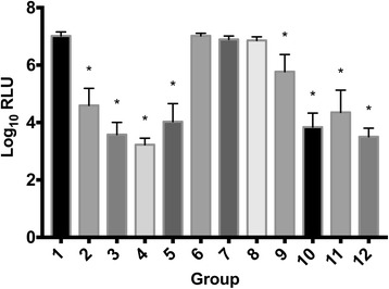
Synoviocyte viability Cell viability measured as luminescence signal (shown as log10 transformed Relative Light Units [RLU]) for synoviocytes exposed to different treatments (for further information on treatment groups refer to Table 1). Error bars indicate 95% confidence interval. Asterisk (*) indicates significant difference (p < 0.05) with group 1 (control). Groups included are: control (group 1); mepivacaine (group 2); mepivacaine + morphine (group 3); mepivacaine + MgSO4 (group 4); mepivacaine + morphine + MgSO4 (group 5); morphine (group 6); MgSO4 (group 7); morphine + MgSO4 (group 8); bupivacaine (group 9); bupivacaine + morphine (group 10); bupivacaine + MgSO4 (group 11); and bupivacaine + morphine + MgSO4 (group 12)
Fig. 2.
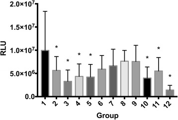
Chondrocyte viability Cell viability measured as luminescence signal (mean values of Relative Light Units [RLU]) for chondrocytes exposed to different treatment groups (for further information on treatment groups refer to Table 1). Error bars indicate 95% confidence interval. Asterisk (*) indicates significant difference (p < 0.05) with group 1 (control). Groups included are: control (group 1); mepivacaine (group 2); mepivacaine + morphine (group 3); mepivacaine + MgSO4 (group 4); mepivacaine + morphine + MgSO4 (group 5); morphine (group 6); MgSO4 (group 7); morphine + MgSO4 (group 8); bupivacaine (group 9); bupivacaine + morphine (group 10); bupivacaine + MgSO4 (group 11); and bupivacaine + morphine + MgSO4 (group 12)
Real-time qPCR
Raw data on gene expression are provided in Additional file 1. Treatment with morphine, MgSO4 or their combination did not have an effect on synoviocyte gene expression of IL-1β, IL-6 or TNF-α. Exposure to mepivacaine alone (group 2) increased expression of IL-1 β, IL6 and TNF-α, while exposure to bupivacaine alone (group 9) only significantly increased expression of IL6. Combination of either mepivacaine or bupivacaine with morphine, MgSO4, or both (groups 3, 4, 5, 10, 11 and 12) increased expression of IL-1β, IL-6 and TNF-α (Figs. 3, 4, 5). Gene expression from cartilage explants did not yield consistent amount of RNA for downstream qRT-PCR.
Fig. 3.
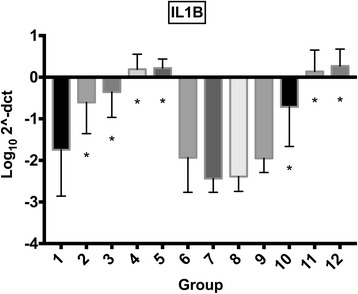
Synoviocyte IL1β gene expression IL1β gene expression (mean log102^-ΔCT) for synoviocytes exposed to different treatment groups (for further information on treatment groups refer to Table 1). Error bars indicate 95% confidence interval. Asterisk (*) indicates significant difference (p < 0.05) with group 1 (control). Groups included are: control (group 1); mepivacaine (group 2); mepivacaine + morphine (group 3); mepivacaine + MgSO4 (group 4); mepivacaine + morphine + MgSO4 (group 5); morphine (group 6); MgSO4 (group 7); morphine + MgSO4 (group 8); bupivacaine (group 9); bupivacaine + morphine (group 10); bupivacaine + MgSO4 (group 11); and bupivacaine + morphine + MgSO4 (group 12).
Fig. 4.
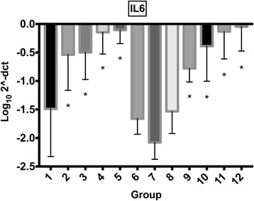
Synoviocyte IL6 gene expression IL6 gene expression (mean log102^-ΔCT) for synoviocytes exposed to different treatment groups (for further information on treatment groups refer to Table 1). Error bars indicate 95% confidence interval. Asterisk (*) indicates significant difference (p < 0.05) with group 1 (control). Groups included are: control (group 1); mepivacaine (group 2); mepivacaine + morphine (group 3); mepivacaine + MgSO4 (group 4); mepivacaine + morphine + MgSO4 (group 5); morphine (group 6); MgSO4 (group 7); morphine + MgSO4 (group 8); bupivacaine (group 9); bupivacaine + morphine (group 10); bupivacaine + MgSO4 (group 11); and bupivacaine + morphine + MgSO4 (group 12).
Fig. 5.
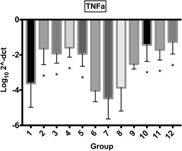
Synoviocyte TNFα gene expression (mean log102^-ΔCT) for synoviocytes exposed to different treatment groups (for further information on treatment groups refer to Table 1). Error bars indicate 95% confidence interval. Asterisk (*) indicates significant difference (p < 0.05) with group 1 (control). Groups included are: control (group 1); mepivacaine (group 2); mepivacaine + morphine (group 3); mepivacaine + MgSO4 (group 4); mepivacaine + morphine + MgSO4 (group 5); morphine (group 6); MgSO4 (group 7); morphine + MgSO4 (group 8); bupivacaine (group 9); bupivacaine + morphine (group 10); bupivacaine + MgSO4 (group 11); and bupivacaine + morphine + MgSO4 (group 12)
Discussion
Chondrotoxic effects of local anaesthetics are reported in the literature and results of this study confirm that a single short exposure to local anaesthetics causes significant articular cytotoxic effects characterised by decreased viability of both equine synoviocytes and chondrocytes. These effects were not counteracted by the addition of morphine and/or MgSO4. Except for the mepivacaine effect on TNF-α expression, local anaesthetics alone, as well as morphine alone, MgSO4 alone, or morphine and MgSO4 combined, did not increase the expression of pro-inflammatory cytokines by synoviocytes; however, the pro-inflammatory cytokines expression were increased when local anaesthetics were combined with morphine and/or MgSO4.
The present study demonstrates that bupivacaine at a concentration of 0.22% is harmful for articular equine synoviocytes but its negative effects are less pronounced on chondrocytes. A protective effect from the existence of intact extracellular matrix on chondrocytes from the cartilage explant has been previously suggested [6, 14], although this was recently questioned on an experimental canine model [10]. Local anaesthetics have drug-, dose- and time-dependent cytotoxic effects [5, 10, 35–37]. Bupivacaine 0.0625% did not cause cell death in canine cartilage and synovium explants [10] and varying toxicity has been observed for bupivacaine concentrations in the range of 0.125–0.25% [2, 5, 10, 35]. Bupivacaine concentrations ≥0.5% have consistently been reported to exert toxic effects on human and animal chondrocytes [5, 35], including horses [7] but interestingly did not affect the viability of rabbit type B synoviocytes in monolayer [11]. Different species sensitivities, methodologies or cell lines may explain different results between studies. Mepivacaine is less frequently administered as an intra-articular analgesic in people and there are fewer studies investigating its effects on articular cells. In contrast to our study, mepivacaine was less toxic than bupivacaine on human [35] and equine [7] chondrocytes on monolayer cultures. However, the bupivacaine concentration used in the present study was lower than in previous studies and has been previously associated with chondrotoxic effects on human cartilage [35] and equine chondrocytes [38]. Mepivacaine is commonly administered intra-articularly in horses at doses similar to that in the present study and temporary synovitis has occasionally been reported [27].
Few studies have investigated the effects of local anaesthetics on the production of inflammatory molecules by articular tissues. Following exposure to bupivacaine a decrease in nitric oxide and PGE2 concentrations was observed in an IL-1-treated co-culture explant model; and that was suggested to be the result of the lack of viable cells remaining after the exposure [2]. However, in our study increased expression of IL1-β, IL6 or TNF-α was observed despite decreased viability
Intra-articular administration of morphine or MgSO4 produces analgesia without clinical evidence of adverse effects in people [12, 13, 20, 39–41]. The present study corroborates the absence of deleterious effects of morphine or MgSO4 on canine human and canine chondrocyte viability in vitro [2, 14, 21, 22], and extends this absence of toxicity to synoviocytes. Furthermore, the combination of morphine and MgSO4 did not have negative effects on cell viability, which warrants investigation of the potential analgesic effect of the combination in vivo. In addition, no effects on expression of pro-inflammatory cytokines were observed when tissues were exposed to morphine and/or MgSO4. Anti-inflammatory effects of opioids have been reported after systemic or local administration. Morphine exposure of IL-1-treated cartilage explants decreased nitric oxide and PGE2 production [2]. Intra-articular administration of morphine reduced nucleated cell count in synovial fluid of chronic arthritis patients [42] and serum amyloid A and total protein levels in horses with experimentally-induced synovitis [15]. Anti-inflammatory effects of magnesium have also been recognised. Low magnesium promotes inflammation and up-regulates IL1α and IL6 production in endothelial cells [43] while magnesium supplementation attenuated the development of OA [23] and rheumatoid arthritis (RA) [44]. MgSO4 acts on N-methyl-D-aspartate receptors and although the present study did not include an OA or RA model, N-methyl-D-aspartate receptors involved in the development of OA and RA are also present in normal human and mouse articular chondrocytes [45]. Results of this study support the clinical use of morphine and warrant further investigation on MgSO4 as intra-articular analgesic drugs in horses and other species.
Addition of MgSO4 to local anaesthetics counteracted harmful effects on human chondrocyte viability in vitro [22] and we aimed to determine if the same was true in a synoviocyte and cartilage explant co-culture model. However, the addition of morphine, MgSO4 or both to mepivacaine or bupivacaine did not counteract cell death caused by mepivacaine and furthermore, decreased chondrocyte viability in comparison with the control group when combined with bupivacaine. Combination of either local anaesthetic with morphine and/or MgSO4 also enhanced the synoviocyte expression of IL1β, IL6 and TNFα. The reasons for the enhanced toxicity of the drug combinations are unclear. The mechanisms of action of local anaesthetics, morphine and MgSO4 differ. The cytotoxic effects of local anaesthetics are related to a concentration-dependent mitochondrial depolarization with subsequent alteration in transmembrane potential and cell death [46]. Osmolality-related cytotoxicity has been observed with high concentrations of magnesium (500 mg/ml) [22] but the concentration used in the present study was 37 mg/ml. Interaction between molecules or their combination with the culture medium or changes in the pH of the environment may be involved [47]. A time-dependent increased chondrotoxicity has been observed for the combination of corticosteroid and local anaesthetics [48]. The pH of the treatment solutions in the present study were nor determined and buffer solutions were not used; however, low pH was not the cause for decreased viability of bovine chondrocytes [1, 49] and addition of buffering solution can increase chondrotoxicity [47]. Chemical incompatibility between the anaesthetic solution and culture medium leading to crystal formation and high cell death rates has been suggested by some authors [1, 7]; however, precipitation was not observed in the present or other studies [2, 10, 38]. Although combination of local anaesthetics and MgSO4 are being administered clinically into joints without reported clinical side effects [20, 50] further research into the chemical compatibility or interactions between drugs is warranted.
The present study has a number of limitations. A co-culture equine model was used to allow evaluations of both cartilage and synovium components as many inflammatory mediators and analgesic receptors are found in the synovium, and synoviocytes have important roles in maintaining articular health and participating in pathophysiological processes [51, 52]. However, results from this in vitro study may not be the same in vivo. Use of synovial explants [2, 10] could have enhanced the system although recent studies support the comparability of explants and monolayer cultures to assess cytotoxicity [2, 10]. The preferential type of synoviocyte present in the culture is uncertain and a mixture of both macrophage-like and fibroblast-like synoviocytes is suspected in this study as they were frozen at passage 2 [52]. Drug concentrations and duration of exposure were based on clinically used doses, joint volume and pharmacokinetics in horses. However, the drug concentrations remained constant during the 2 h exposure, which could be interpreted as a relative overdose as clearance of the drug is expected to occur already within this time in the live animal. The horse is an accepted translational models of naturally-occurring osteoarthritis [53] but drug effects can differ between species. Normal articular tissues were used and more severe chondrotoxic effects are expected on degenerative versus normal cartilage [35]. The reasons for the poor RNA extraction from cartilage explants are uncertain. This methodology has been used successfully in other studies but equine mature articular cartilage is relatively acellular and we used small size explants. Previous studies in our lab have yielded 1 μg of RNA from the cartilage harvested from both third metacarpal condyles in adult horses (Peffers M.J. Unpublished data). The use of isolated chondrocytes, larger size or higher number of cartilage explants, or use of a different methodology may have yielded gene expression data from the cartilage explants. Quantification of adenosine triphosphate by luminescence has been shown reliable method to assess cell viability [14, 31].
Conclusions
The present study corroborate the toxic effects of single short exposure to local anaesthetics on both equine chondrocytes and synoviocytes using an in vitro co-culture model, with more severe effects observed for mepivacaine than bupivacaine. It also provides evidence that synoviocytes respond to drugs commonly administered intra-articularly, which warrants further investigation. The present study indicates that morphine and MgSO4 are not toxic to synoviocytes when exposed either alone or in combination, which supports the clinical use of morphine and warrants further investigation of MgSO4 as intra-articular analgesic drugs in horses and other species. However, further investigation into drug incompatibilities is required as combinations of either mepivacaine or bupivacaine with morphine, MgSO4 or both did not alleviate any detrimental effects from the local anaesthetics, but actually caused cell death and increased expression of pro-inflammatory enzymes.
Acknowledgements
We wish to thank Ian Richards for his contribution in the acquisition of data. None of the authors have conflicts of interests.
Funding
This study was funded by the Horserace Betting Levy Board (SPrj015). Mandy Peffers is funded through a Wellcome Trust Clinical Intermediate Fellowship (107471). Funding bodies did not take part in the study design or conclusion of the study.
Availability of data and materials
All data generated and analysed during this study are included in this published article and its supplementary information files.
Abbreviations
- cDNA
Complimentary deoxyribonucleic acid
- DMSO
Dimethyl sulfoxide
- FCS
Foetal calf Serum
- GAPDH
Glyceraldehyde-3-phosphatedhyrogenase
- IL-1β
Interleukin 1β
- IL-6
Interleukin 6
- MgSO4
Magnesium sulphate
- OA
Osteoarthritis
- qRT-PCR
Real-time quantitative polymerase chain reaction
- TNF-α
Tumour necrosis factor α
Additional file
Raw data on chondrocyte and synoviocyte viability and synoviocyte gene expression. (XLSX 18 kb)
Authors’ contributions
LMRM, MJP, ER and PC participated in the design of the study and the experiments and interpretation of the data. LRM, MCM and SW participated in the design of the study, performing the majority of the experiments and interpretation of data. LRM and ER performed statistical analysis and interpretation of the data. MJP and PC supervised all the experiments. All authors read and approved the final manuscript.
Ethics approval and consent to participate
No samples were obtained from equine patients. Tissue samples were collected from the abattoir which are considered a by-product of the agricultural industry. Specifically, the Animal (Scientific Procedures) Act 1986, Schedule 2, does not define collection from these sources as scientific procedures. Ethical approval was therefore not required for this study.
Consent for publication
Not applicable.
Competing interests
All authors declare that they have no competing interests.
Publisher’s Note
Springer Nature remains neutral with regard to jurisdictional claims in published maps and institutional affiliations.
Footnotes
Electronic supplementary material
The online version of this article (10.1186/s12917-017-1244-8) contains supplementary material, which is available to authorized users.
Contributor Information
L. M. Rubio-Martínez, Email: luis.rubiomartinez@hotmail.com
E. Rioja, Email: evarioja@hotmail.com
M. Castro Martins, Email: mcm@liverpool.ac.uk
S. Wipawee, Email: wipawee.s@rmutsv.ac.th
P. Clegg, Email: p.d.clegg@liverpool.ac.uk
M. J. Peffers, Email: peffs@liverpool.ac.uk
References
- 1.Bogatch MT, Ferachi DG, Kyle B, Popinchalk S, Howell MH, Ge D, You Z, Savoie FH. Is chemical incompatibility responsible for chondrocyte death induced by local anesthetics? Am J Sports Med. 2010;38(3):520–526. doi: 10.1177/0363546509349799. [DOI] [PubMed] [Google Scholar]
- 2.Anz A, Smith MJ, Stoker A, Linville C, Markway H, Branson K, Cook JL. The effect of bupivacaine and morphine in a coculture model of diarthrodial joints. Arthroscopy. 2009;25(3):225–231. doi: 10.1016/j.arthro.2008.12.003. [DOI] [PubMed] [Google Scholar]
- 3.Gulihar A, Robati S, Twaij H, Salih A, Taylor GJ. Articular cartilage and local anaesthetic: A systematic review of the current literature. J Orthop. 2015;12(Suppl 2):S200–S210. doi: 10.1016/j.jor.2015.10.005. [DOI] [PMC free article] [PubMed] [Google Scholar]
- 4.Dragoo JL, Braun HJ, Kim HJ, Phan HD, Golish SR. The in vitro chondrotoxicity of single-dose local anesthetics. Am J Sports Med. 2012;40(4):794–799. doi: 10.1177/0363546511434571. [DOI] [PubMed] [Google Scholar]
- 5.Chu CR, Izzo NJ, Coyle CH, Papas NE, Logar A. The in vitro effects of bupivacaine on articular chondrocytes. J Bone Joint Surg Br. 2008;90(6):814–820. doi: 10.1302/0301-620X.90B6.20079. [DOI] [PMC free article] [PubMed] [Google Scholar]
- 6.Chu CR, Izzo NJ, Papas NE, Fu FH. In vitro exposure to 0.5% bupivacaine is cytotoxic to bovine articular chondrocytes. Arthroscopy. 2006;22(7):693–699. doi: 10.1016/j.arthro.2006.05.006. [DOI] [PubMed] [Google Scholar]
- 7.Park J, Sutradhar BC, Hong G, Choi SH, Kim G. Comparison of the cytotoxic effects of bupivacaine, lidocaine, and mepivacaine in equine articular chondrocytes. Vet Anaesth Analg. 2011;38(2):127–133. doi: 10.1111/j.1467-2995.2010.00590.x. [DOI] [PubMed] [Google Scholar]
- 8.Piat P, Richard H, Beauchamp G, Laverty S. In vivo effects of a single intra-articular injection of 2% lidocaine or 0.5% bupivacaine on articular cartilage of normal horses. Vet Surg. 2012;41(8):1002–1010. doi: 10.1111/j.1532-950X.2012.01039.x. [DOI] [PubMed] [Google Scholar]
- 9.Sherman SL, James C, Stoker AM, Cook CR, Khazai RS, Flood DL, Cook JL. In Vivo Toxicity of Local Anesthetics and Corticosteroids on Chondrocyte and Synoviocyte Viability and Metabolism. Cartilage. 2015;6(2):106–112. doi: 10.1177/1947603515571001. [DOI] [PMC free article] [PubMed] [Google Scholar]
- 10.Sherman SL, Khazai RS, James CH, Stoker AM, Flood DL, Cook JL. In Vitro Toxicity of Local Anesthetics and Corticosteroids on Chondrocyte and Synoviocyte Viability and Metabolism. Cartilage. 2015;6(4):233–240. doi: 10.1177/1947603515594453. [DOI] [PMC free article] [PubMed] [Google Scholar]
- 11.Braun HJ, Busfield BT, Kim HJ, Scuderi GJ, Dragoo JL. The effect of local anaesthetics on synoviocytes: a possible indirect mechanism of chondrolysis. Knee Surg Sports Traumatol Arthrosc. 2013;21(6):1468–1474. doi: 10.1007/s00167-012-2104-5. [DOI] [PubMed] [Google Scholar]
- 12.Grabowska-Gawel A, Gawel K, Hagner W, Bilinski PJ. Morphine or bupivacaine in controlling postoperative pain in patients subjected to knee joint arthroscopy. Ortop Traumatol Rehabil. 2003;5(6):758–762. [PubMed] [Google Scholar]
- 13.Kalso E, Smith L, McQuay HJ, Andrew Moore R. No pain, no gain: clinical excellence and scientific rigour--lessons learned from IA morphine. Pain. 2002;98(3):269–275. doi: 10.1016/S0304-3959(02)00019-2. [DOI] [PubMed] [Google Scholar]
- 14.Ickert I, Herten M, Vogl M, Ziskoven C, Zilkens C, Krauspe R, Kircher J. Opioids as an alternative to amide-type local anaesthetics for intra-articular application. Knee Surg Sports Traumatol Arthrosc. 2015;23(9):2674–2681. doi: 10.1007/s00167-014-2989-2. [DOI] [PubMed] [Google Scholar]
- 15.Lindegaard C, Gleerup KB, Thomsen MH, Martinussen T, Jacobsen S, Andersen PH. Anti-inflammatory effects of intra-articular administration of morphine in horses with experimentally induced synovitis. Am J Vet Res. 2010;71(1):69–75. doi: 10.2460/ajvr.71.1.69. [DOI] [PubMed] [Google Scholar]
- 16.Lindegaard C, Thomsen MH, Larsen S, Andersen PH. Analgesic efficacy of intra-articular morphine in experimentally induced radiocarpal synovitis in horses. Vet Anaesth Analg. 2010;37(2):171–185. doi: 10.1111/j.1467-2995.2009.00521.x. [DOI] [PubMed] [Google Scholar]
- 17.Santos LC, de Moraes AN, Saito ME. Effects of intraarticular ropivacaine and morphine on lipopolysaccharide-induced synovitis in horses. Vet Anaesth Analg. 2009;36(3):280–286. doi: 10.1111/j.1467-2995.2009.00452.x. [DOI] [PubMed] [Google Scholar]
- 18.van Loon JP, de Grauw JC, van Dierendonck M, L’Ami JJ, Back W, van Weeren PR. Intra-articular opioid analgesia is effective in reducing pain and inflammation in an equine LPS induced synovitis model. Equine Vet J. 2010;42(5):412–419. doi: 10.1111/j.2042-3306.2010.00077.x. [DOI] [PubMed] [Google Scholar]
- 19.Tulamo RM, Raekallio M, Taylor P, Johnson CB, Salonen M. Intra-articular morphine and saline injections induce release of large molecular weight proteoglycans into equine synovial fluid. Zentralbl Veterinarmed A. 1996;43(3):147–153. doi: 10.1111/j.1439-0442.1996.tb00439.x. [DOI] [PubMed] [Google Scholar]
- 20.Elsharnouby NM, Eid HE, Abou Elezz NF, Moharram AN: Intraarticular injection of magnesium sulphate and/or bupivacaine for postoperative analgesia after arthroscopic knee surgery. Anesth Analg 2008, 106(5):1548–1552, table of contents. [DOI] [PubMed]
- 21.Baker JF, Walsh PM, Byrne DP, Mulhall KJ. In vitro assessment of human chondrocyte viability after treatment with local anaesthetic, magnesium sulphate or normal saline. Knee Surg Sports Traumatol Arthrosc. 2011;19(6):1043–1046. doi: 10.1007/s00167-011-1437-9. [DOI] [PubMed] [Google Scholar]
- 22.Baker JF, Byrne DP, Walsh PM, Mulhall KJ. Human chondrocyte viability after treatment with local anesthetic and/or magnesium: results from an in vitro study. Arthroscopy. 2011;27(2):213–217. doi: 10.1016/j.arthro.2010.06.029. [DOI] [PubMed] [Google Scholar]
- 23.Lee CH, Wen ZH, Chang YC, Huang SY, Tang CC, Chen WF, Hsieh SP, Hsieh CS, Jean YH. Intra-articular magnesium sulfate (MgSO4) reduces experimental osteoarthritis and nociception: association with attenuation of N-methyl-D-aspartate (NMDA) receptor subunit 1 phosphorylation and apoptosis in rat chondrocytes. Osteoarthritis Cartilage. 2009;17(11):1485–1493. doi: 10.1016/j.joca.2009.05.006. [DOI] [PubMed] [Google Scholar]
- 24.Rosengren S, Boyle DL, Firestein GS. Acquisition, culture, and phenotyping of synovial fibroblasts. Methods Mol Med. 2007;135:365–375. doi: 10.1007/978-1-59745-401-8_24. [DOI] [PubMed] [Google Scholar]
- 25.Ekman L, Nilsson G, Persson L, Lumsden JH. Volume of the synovia in certain joint cavities in the horse. Acta Vet Scand. 1981;22(1):23–31. doi: 10.1186/BF03547202. [DOI] [PMC free article] [PubMed] [Google Scholar]
- 26.Lindegaard C, Frost AB, Thomsen MH, Larsen C, Hansen SH, Andersen PH. Pharmacokinetics of intra-articular morphine in horses with lipopolysaccharide-induced synovitis. Vet Anaesth Analg. 2010;37(2):186–195. doi: 10.1111/j.1467-2995.2010.00526.x. [DOI] [PubMed] [Google Scholar]
- 27.Bassage LH, Ross MW. Diagnostic analgesia. In: Ross MW, Dyson SJ, editors. Diagnosis and management of lameness in the horse. 2. St. Louis, Missouri: Elsevier Saunders; 2011. pp. 100–134. [Google Scholar]
- 28.Koltka K, Koknel-Talu G, Asik M, Ozyalcin S. Comparison of efficacy of intraarticular application of magnesium, levobupivacaine and lornoxicam with placebo in arthroscopic surgery. Knee Surg Sports Traumatol Arthrosc. 2011;19(11):1884–1889. doi: 10.1007/s00167-011-1497-x. [DOI] [PubMed] [Google Scholar]
- 29.Zeng C, Gao SG, Cheng L, Luo W, Li YS, Tu M, Tian J, Xu M, Zhang FJ, Jiang W, et al. Single-Dose Intra-Articular Morphine After Arthroscopic Knee Surgery: A Meta-Analysis of Randomized Placebo-Controlled Studies. Arthroscopy. 2013;29(8):1450–1458. doi: 10.1016/j.arthro.2013.04.005. [DOI] [PubMed] [Google Scholar]
- 30.Wintzer HJ, Fitzek A, Frey HH. Pharmacokinetics of procaine injected into the hock joint of the horse. Equine Vet J. 1981;13(1):68–69. doi: 10.1111/j.2042-3306.1981.tb03460.x. [DOI] [PubMed] [Google Scholar]
- 31.Niles AL, Moravec RA, Riss TL. In vitro viability and cytotoxicity testing and same-well multi-parametric combinations for high throughput screening. Curr Chem Genomics. 2009;3:33–41. doi: 10.2174/1875397300903010033. [DOI] [PMC free article] [PubMed] [Google Scholar]
- 32.Riss TL, Moravec RA, Niles AL, Benink HA, Worzella TJ, Minor L: Cell Viability Assays. In: Assay Guidance Manual. edn. Edited by Sittampalam GS, Coussens NP, Nelson H, Arkin M, Auld D, Austin C, Bejcek B, Glicksman M, Inglese J, Iversen PW et al. Bethesda (MD); 2004.
- 33.Peffers M, Liu X, Clegg P. Transcriptomic signatures in cartilage ageing. Arthritis Res Ther. 2013;15(4):R98. doi: 10.1186/ar4278. [DOI] [PMC free article] [PubMed] [Google Scholar]
- 34.Livak KJ, Schmittgen TD. Analysis of relative gene expression data using real-time quantitative PCR and the 2(−Delta Delta C(T)) Method. Methods. 2001;25(4):402–408. doi: 10.1006/meth.2001.1262. [DOI] [PubMed] [Google Scholar]
- 35.Breu A, Rosenmeier K, Kujat R, Angele P, Zink W. The cytotoxicity of bupivacaine, ropivacaine, and mepivacaine on human chondrocytes and cartilage. Anesth Analg. 2013;117(2):514–522. doi: 10.1213/ANE.0b013e31829481ed. [DOI] [PubMed] [Google Scholar]
- 36.Grishko V, Xu M, Wilson G, Pearsall AW. Apoptosis and mitochondrial dysfunction in human chondrocytes following exposure to lidocaine, bupivacaine, and ropivacaine. J Bone Joint Surg Am. 2010;92(3):609–618. doi: 10.2106/JBJS.H.01847. [DOI] [PubMed] [Google Scholar]
- 37.Miyazaki T, Kobayashi S, Takeno K, Yayama T, Meir A, Baba H. Lidocaine cytotoxicity to the bovine articular chondrocytes in vitro: changes in cell viability and proteoglycan metabolism. Knee Surg Sports Traumatol Arthrosc. 2011;19(7):1198–1205. doi: 10.1007/s00167-010-1369-9. [DOI] [PubMed] [Google Scholar]
- 38.Bolt DM, Ishihara A, Weisbrode SE, Bertone AL. Effects of triamcinolone acetonide, sodium hyaluronate, amikacin sulfate, and mepivacaine hydrochloride, alone and in combination, on morphology and matrix composition of lipopolysaccharide-challenged and unchallenged equine articular cartilage explants. Am J Vet Res. 2008;69(7):861–867. doi: 10.2460/ajvr.69.7.861. [DOI] [PubMed] [Google Scholar]
- 39.Eroglu A, Saracoglu S, Erturk E, Kosucu M, Kerimoglu S. A comparison of intraarticular morphine and bupivacaine for pain control and outpatient status after an arthroscopic knee surgery under a low dose of spinal anaesthesia. Knee Surg Sports Traumatol Arthrosc. 2010;18(11):1487–1495. doi: 10.1007/s00167-010-1061-0. [DOI] [PubMed] [Google Scholar]
- 40.Elkousy H, Kannan V, Calder CT, Zumwalt J, O'Connor DP, Woods GW. Intra-articular morphine versus bupivacaine for postoperative pain management. Orthopedics. 2013;36(9):e1121–e1127. doi: 10.3928/01477447-20130821-12. [DOI] [PubMed] [Google Scholar]
- 41.Bondok RS, Abd El-Hady AM. Intra-articular magnesium is effective for postoperative analgesia in arthroscopic knee surgery. Br J Anaesth. 2006;97(3):389–392. doi: 10.1093/bja/ael176. [DOI] [PubMed] [Google Scholar]
- 42.Stein A, Yassouridis A, Szopko C, Helmke K, Stein C. Intraarticular morphine versus dexamethasone in chronic arthritis. Pain. 1999;83(3):525–532. doi: 10.1016/S0304-3959(99)00156-6. [DOI] [PubMed] [Google Scholar]
- 43.Bernardini D, Nasulewic A, Mazur A, Maier JA. Magnesium and microvascular endothelial cells: a role in inflammation and angiogenesis. Front Biosci. 2005;10:1177–1182. doi: 10.2741/1610. [DOI] [PubMed] [Google Scholar]
- 44.Nagai N, Fukuhata T, Ito Y, Tai H, Hataguchi Y, Nakagawa K. Preventive effect of water containing magnesium ion on paw edema in adjuvant-induced arthritis rat. Biol Pharm Bull. 2007;30(10):1934–1937. doi: 10.1248/bpb.30.1934. [DOI] [PubMed] [Google Scholar]
- 45.Salter DM, Wright MO, Millward-Sadler SJ. NMDA receptor expression and roles in human articular chondrocyte mechanotransduction. Biorheology. 2004;41(3–4):273–281. [PubMed] [Google Scholar]
- 46.Irwin W, Fontaine E, Agnolucci L, Penzo D, Betto R, Bortolotto S, Reggiani C, Salviati G, Bernardi P. Bupivacaine myotoxicity is mediated by mitochondria. J Biol Chem. 2002;277(14):12221–12227. doi: 10.1074/jbc.M108938200. [DOI] [PubMed] [Google Scholar]
- 47.Syed HM, Green L, Bianski B, Jobe CM, Wongworawat MD. Bupivacaine and triamcinolone may be toxic to human chondrocytes: a pilot study. Clin Orthop Relat Res. 2011;469(10):2941–2947. doi: 10.1007/s11999-011-1834-x. [DOI] [PMC free article] [PubMed] [Google Scholar]
- 48.Farkas B, Kvell K, Czompoly T, Illes T, Bardos T. Increased chondrocyte death after steroid and local anesthetic combination. Clin Orthop Relat Res. 2010;468(11):3112–3120. doi: 10.1007/s11999-010-1443-0. [DOI] [PMC free article] [PubMed] [Google Scholar]
- 49.Karpie JC, Chu CR. Lidocaine exhibits dose- and time-dependent cytotoxic effects on bovine articular chondrocytes in vitro. Am J Sports Med. 2007;35(10):1621–1627. doi: 10.1177/0363546507304719. [DOI] [PubMed] [Google Scholar]
- 50.Chen Y, Zhang Y, Zhu YL, Fu PL. Efficacy and safety of an intra-operative intra-articular magnesium/ropivacaine injection for pain control following total knee arthroplasty. J Int Med Res. 2012;40(5):2032–2040. doi: 10.1177/030006051204000548. [DOI] [PubMed] [Google Scholar]
- 51.Chang X, Shen J, Yang H, Xu Y, Gao W, Wang J, Zhang H, He S. Upregulated expression of CCR3 in osteoarthritis and CCR3 mediated activation of fibroblast-like synoviocytes. Cytokine. 2016;77:211–219. doi: 10.1016/j.cyto.2015.09.012. [DOI] [PubMed] [Google Scholar]
- 52.Bartok B, Firestein GS. Fibroblast-like synoviocytes: key effector cells in rheumatoid arthritis. Immunol Rev. 2010;233(1):233–255. doi: 10.1111/j.0105-2896.2009.00859.x. [DOI] [PMC free article] [PubMed] [Google Scholar]
- 53.Gregory MH, Capito N, Kuroki K, Stoker AM, Cook JL, Sherman SL. A review of translational animal models for knee osteoarthritis. Arthritis. 2012;2012:764621. doi: 10.1155/2012/764621. [DOI] [PMC free article] [PubMed] [Google Scholar]
Associated Data
This section collects any data citations, data availability statements, or supplementary materials included in this article.
Data Availability Statement
All data generated and analysed during this study are included in this published article and its supplementary information files.


