Abstract
We report on a 74-year-old man with seropositive rheumatoid arthritis (RA) and radiographic osteoarthritis (OA) who underwent dual-phase high-resolution 99mTc-MDP SPECT/CT. Early radiotracer enhancement was noted in two RA joints of the right hand, both presenting with a ring-like uptake pattern around the joint, consistent with synovitis. Insignificant early enhancement was noted at the first carpometacarpal joint, despite presentation of CT features of OA. The delayed-phase enhancement patterns were distinct, showing asymmetry in RA joints, but a symmetric, centered pattern for the OA joint.
A 74-year-old man with seropositive rheumatoid arthritis (RA) (Disease Activity Score 28 [DAS-28] of 3.61) presented prior to starting therapy. The patient underwent dual-phase SPECT/CT bone scan, conducted 15 min (early soft tissue phase) and 220 min (delayed osseous phase) respectively, after a single, 925 MBq, intravenous injection of 99mTechnetium-methylene diphosphonate (99mTc-MDP) radiotracer.
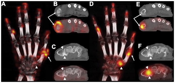
Images of the right hand are presented. The soft-tissue phase images (A–C) showed markedly increased ring-like radiotracer distribution around the first metacarpophalangeal (MP) joint (arrow, A & B). That pattern is consistent with hyperemia and synovitis. Hyperemia at the fifth proximal interphalangeal (PIP) joint was also noted (A, not shown in cross sections). Corresponding CT images showed joint space narrowing with no evidence of bone erosion. On blinded examination, the patient was found to have swelling and tenderness at both these joints, consistent with RA. For the first carpometacarpal (CMC) joint, CT images showed joint space narrowing with increased sclerosis and two < 2 mm osteophytes, consistent with Eaton stage II osteoarthritis (OA). Insignificant elevation in radiotracer uptake was, however, noted for this joint in the soft-tissue phase (arrowhead, C).
In the osseous phase images (D–F), both first MP and fifth PIP joints demonstrated focal increased tracer activity, more on the proximal aspect of the joints (arrow, D & E), while the first CMC joint showed a rather symmetrically-increased joint-centered radiotracer uptake pattern (arrowhead, F).
This case illustrates the potential of 99mTc-MDP high-resolution SPECT/CT imaging for assessing inflammatory activity and bone turnover in RA and OA via a single radiotracer injection. The early enhancement of 99mTc-MDP activity in soft tissues is suggestive of hypervascularity and inflammatory synovitis, a hallmark of RA, and less commonly of OA1, 2. The tracer distribution on delayed images, which displays considerably different patterns is suggestive of osteoblastic activity3 which could be seen with both RA and OA4–6. In the absence of erosive changes, bone remodeling in RA could be attributed to osteitis from the overlying inflamed synovium (outside-in hypothesis)7, 8.
Figure 1.
Acknowledgments
Funding: This study was funded in part by research grants from Philips Healthcare (Andover, Massachusetts, United States) and the National Institutes of Health grants 2K12 HD051958 and R03 EB015099. The views expressed in this article are the authors’ own and do not necessarily represent the views of Philips Healthcare, or the National Institutes of Health.
The authors thank Drs. Piotr Maniawski and Angela Da Silva from Philips Healthcare for insightful discussions regarding imaging protocol development and data interpretation.
References
- 1.Kim JY, Choi YY, Kim CW, et al. Bone Scintigraphy in the Diagnosis of Rheumatoid Arthritis: Is There Additional Value of Bone Scintigraphy with Blood Pool Phase over Conventional Bone Scintigraphy? J Korean Med Sci. 2016;31:502–509. doi: 10.3346/jkms.2016.31.4.502. [DOI] [PMC free article] [PubMed] [Google Scholar]
- 2.Klett R, Grau K, Puille M, et al. Comparison of HIG scintigraphy and bloodpool scintigraphy using HDP in arthritic joint disease. Nuklearmedizin. 2000;39:33–37. [PubMed] [Google Scholar]
- 3.Ostendorf B, Scherer A, Wirrwar A, et al. High-resolution multipinhole single-photon-emission computed tomography in experimental and human arthritis. Arthritis Rheum. 2006;54:1096–1104. doi: 10.1002/art.21732. [DOI] [PubMed] [Google Scholar]
- 4.Buchbender C, Sewerin P, Mattes-Gyorgy K, et al. Utility of combined high-resolution bone SPECT and MRI for the identification of rheumatoid arthritis patients with high-risk for erosive progression. Eur J Radiol. 2013;82:374–379. doi: 10.1016/j.ejrad.2012.10.011. [DOI] [PubMed] [Google Scholar]
- 5.Schleich FS, Schurch M, Huellner MW, et al. Diagnostic and therapeutic impact of SPECT/CT in patients with unspecific pain of the hand and wrist. EJNMMI Res. 2012;2:53. doi: 10.1186/2191-219X-2-53. [DOI] [PMC free article] [PubMed] [Google Scholar]
- 6.Ostendorf B, Wirrwar A, Mattes-Gyorgy K, et al. High-resolution SPECT imaging of bony pathology in early arthritis of finger joints. Rheumatology (Oxford) 2009;48:853–854. doi: 10.1093/rheumatology/kep115. [DOI] [PubMed] [Google Scholar]
- 7.Schett G, Firestein GS. Mr Outside and Mr Inside: classic and alternative views on the pathogenesis of rheumatoid arthritis. Ann Rheum Dis. 2010;69:787–789. doi: 10.1136/ard.2009.121657. [DOI] [PubMed] [Google Scholar]
- 8.Ostendorf B, Mattes-Gyorgy K, Reichelt DC, et al. Early detection of bony alterations in rheumatoid and erosive arthritis of finger joints with high-resolution single photon emission computed tomography, and differentiation between them. Skeletal Radiol. 2010;39:55–61. doi: 10.1007/s00256-009-0761-3. [DOI] [PubMed] [Google Scholar]


