Summary
Mapping hundreds of genetic variants through genome wide association studies provided an opportunity to gain insights into the pathobiology of immune‐mediated diseases. However, as most of the disease variants fall outside the gene coding sequences the functional interpretation of the exact role of the associated variants remains to be determined. The integration of disease‐associated variants with large scale genomic maps of cell‐type‐specific gene regulation at both chromatin and transcript levels deliver examples of functionally prioritized causal variants and genes. In particular, the enrichment of disease variants with histone marks can point towards the cell types most relevant to disease development. Furthermore, chromatin contact maps that link enhancers to promoter regions in a direct way allow the identification of genes that can be regulated by the disease variants. Candidate genes implicated with such approaches can be further examined through the correlation of gene expression with genotypes. Additionally, in the context of immune‐mediated diseases it is important to combine genomics with immunology approaches. Genotype correlations with the immune system as a whole, as well as with cellular responses to different stimuli, provide a valuable platform for understanding the functional impact of disease‐associated variants. The intersection of immunogenomic resources with disease‐associated variants paints a detailed picture of disease causal mechanisms. Here, we provide an overview of recent studies that combine these approaches to identify disease vulnerable pathways.
Keywords: activation, autoimmunity, cell activation, genomics
Glossary
- Chromatin accessibility
a term describing regions of the genome free of nucleosomes, which might be a result of the sequence occupation by a DNA binding protein, such as transcription factors. The open chromatin regions, which can be assayed with ATAC‐seq (assay for transposase accessible chromatin followed by sequencing) or DNase‐seq, often have a functional role in regulating transcription and are enriched at promoters and enhancers.
- Chromatin immunoprecipitation followed by sequencing (ChIP‐seq)
an assay in which antibodies against an epitope of interest, such as a transcription factor or a histone modification, are used to pull down the protein of interest along with the genomic sequences interacting with it. The proteins are then washed off and the DNA is sequenced. The sequencing results in pile ups of reads concentrated at the positions of DNA ‐protein interaction, referred to as peaks.
- Genome‐wide association studies (GWAS)
a method that leverages the information from genetic variants spread through the whole genome and compares allele frequencies between a group of patients and a group of healthy controls, within the same population. If the frequency difference is statistically significant between the two groups (P‐value < 5 x 10‐8), the variant is reported as disease‐associated. GWAS can also be applied within a single group with quantitative measurements, such as a group of patients for which a response to a treatment is measured, or a group of healthy individuals for which blood cell counts have been recorded.
- Hi‐C
a genome‐wide method that enables identification of chromatin conformation in three‐dimensional space.
- Linkage disequilibrium (LD)
a correlation between genetic variants, resulting from the non‐random inheritance of genetic regions.
- Quantitative trait locus (QTL)
a region of the genome that is correlated with a quantitative phenotype. Typically, genetic variants, such as single nucleotide polymorphisms (SNPs), are correlated with the gene expression levels (eQTL).
Introduction
The primary role of the immune system is to protect the host from infection by a variety of pathogens constantly present in the environment. As such, genetic defects that cause loss of the immune system's activity result in recurrent infections and severe immunodeficiencies that are often life threatening. However, uncontrolled activation of immune cells may result in the response being targeted towards healthy cells causing tissue destruction and consequently autoimmunity.
It is estimated that one in five people suffer from at least one of the 81 documented autoimmune diseases in the USA.1 Despite the high prevalence, the molecular mechanisms that predispose to autoimmunity are not well understood. The clustering of autoimmune diseases in families has indicated a strong genetic component underlying pathological processes driving many complex immune‐mediated diseases. Early studies identified several loci with large effect sizes, including HLA‐DQB1 associated with type 1 diabetes (T1D),2 HLA‐DQ2 and HLA‐DQ8 associated with coeliac disease (CeD),3 and HLA‐DR4 associated with rheumatoid arthritis (RA).4 It is now well appreciated that most autoimmune diseases share a strong association with the MHC region.5
The susceptible genetic background of the HLA alone is often not sufficient to lead to the development of an immune‐mediated disease. For example, T1D, RA, multiple sclerosis (MS) and CeD, result from the combination of the risk genotypes of both the HLA and non‐HLA genes, as well as an environmental trigger. Genome‐wide association studies (GWAS) revealed that complex immune traits develop as a consequence of the interplay between hundreds to thousands of common variants6 with individually small effects on the overall disease phenotype. More than 497 susceptibility loci for autoimmune disorders have been identified,7 however, despite the large number of mapped variants, the heritability explained by the non‐HLA loci remains moderate. Even for the most successful examples such as MS, T1D or RA, where over a hundred risk variants have been mapped, the explained heritability varies between 20% for MS,8 10% for T1D9 and about 5% for RA.10
A clear picture emerging from GWAS is that immune‐mediated diseases to some extent result from the dysregulation of the same biological pathways. For example, a locus encoding genes for receptors that control T‐cell activation, CD28, ICOS and CTLA4, is associated to CeD, RA and T1D. The sharing of genetic regions associated with immune diseases is widespread; of the 90 risk loci associated with T1D, RA, CeD and MS, 33 (37%) overlap between two or more diseases.11 However, the molecular mechanisms by which genetic variants hinder control of the immune system and cause autoimmunity have been determined for only a small number of variants. For example, missense mutations in the exon of the key immune regulator, CTLA4, induce severe immunodeficiency and a spectrum of autoimmune and autoinflammatory diseases.12 These rare monogenic disorders provide an insight into the control of the immune system in the presence of the dysfunctional genes. However, the same genotype‐to‐function logic cannot be easily applied to complex traits. First, associated loci map to regions of the genome with extended linkage disequilibrium (LD). The LD blocks often comprise tens to hundreds of highly correlated single nucleotide polymorphisms (SNPs). Therefore, in a statistical test, the high correlation between SNPs results in equivalent strength of the association signal spread throughout all of the variants in LD. Practically, this renders the variants indistinguishable from one another and hinders the prioritization of the causal variants based on the association statistics alone. Second, the majority of the associated variants localize to the non‐coding regions of the genome, implicating that the disease variants are likely to act through the dysregulation of gene expression. This poses a challenge because gene expression regulation can be cell‐type‐specific and therefore functional follow‐up studies have to be carried out in the cell types most relevant to the disease. However, for many immune diseases the exact pathological cell type is unknown. The final challenge is linking the associated SNPs to effector genes, as the causal variants may not necessarily affect the closest genes but instead act through long‐range genomic interactions.
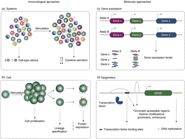
Here, we provide an overview of the recent advances in the genetics of immune‐mediated diseases. We discuss how the immunogenomic cross‐disciplinary toolkit can be used as a platform for inferring correlations between genotypes and individual genes, cellular traits, as well as the immune system as a whole (Fig. 1). Finally, we discuss the challenges of mapping associated variants to the molecular mechanisms through which they act.
Figure 1.

Immunogenomic approaches to infer the role of disease variants. Variants associated to immune diseases can be functionally annotated using immunological or a genomic approaches. Immunological approaches include the (a) systemic analysis of immune function, such as measuring cell type ratios and cytokine levels, and (b) cellular analysis, such as proliferation assays of a specific cell type in response to a stimulus, the lineage specification markers or protein expression levels. Genomic assays, include (c) gene expression and (d) epigenetics; transcription factor binding sites, chromatin accessibility, histone modifications and DNA methylation.
Correlating disease variants with molecular traits
Context‐specific effects of disease variants on gene expression
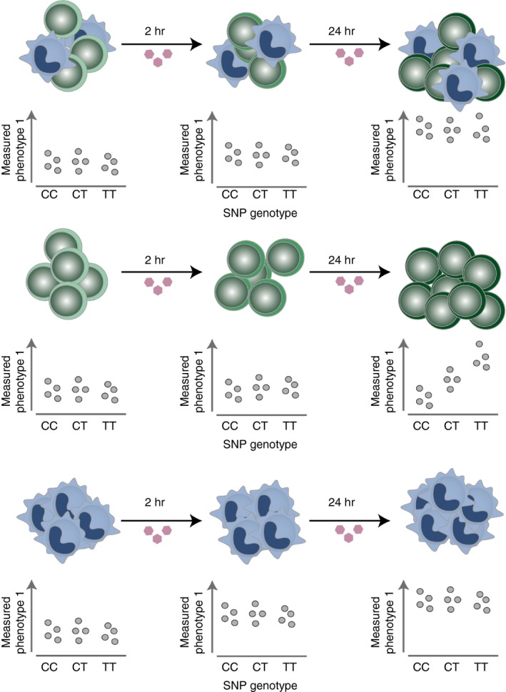
The effects of genetic variants on gene expression are often assessed through genotype correlation with gene expression levels measured across tens to thousands of individuals (expression quantitative trait loci, eQTL; Fig. 2). Disease‐associated variants are enriched for eQTLs, suggesting that a large proportion of disease variants are likely to function through gene expression regulation.13, 14 A relevant and easily accessible tissue for immune diseases is blood. Early studies demonstrated that immune disease SNPs also affect gene expression in the whole blood or peripheral blood mononuclear cells. For example, over 50% of CeD variants also expressed an eQTL effect.15 Disease‐associated variants affecting gene expression can point towards a dysregulated specific pathway, e.g. five of the inflammatory bowel disease (IBD) risk variants were also eQTLs that increased the expression levels of ITGA, ITGAL, ICAM and ITGB8 genes, encoding integrins, the pro‐inflammatory cell surface proteins that mediate leucocyte accumulation at the site of inflammation.16
Figure 2.

Cell‐type‐specific and cell‐state‐specific expression quantitative trait loci. Cellular phenotypes, such as gene expression, cytokine secretion or chromatin accessibility, might be affected by a genetic variant only in a specific cell type and under specific conditions, e.g. at a specific time‐point following a stimulation. Here, a bulk of cell types (upper panel), as well as each cell type individually, were stimulated for 2 and 24 hr. In all scenarios, the measured phenotype, e.g. gene expression, increased upon stimulation, but only in the green cell type (middle panel) was the effect correlated with the genotypes. The effect was missed when measured in the sample containing the mixed cell population (upper panel). The blue cell type (bottom panel) expresses the strongest up‐regulation in expression upon stimulation and largely drives the observed increase in expression in the bulk sample.
As the gene expression studies increased in sample size,17 and expanded across different tissues18 and cell states,19 it became evident that the eQTL effects are widespread. For example, the study of whole blood gene expression from over 8000 individuals identified that nearly 6500 genes (44% of all tested genes) were under genetic control.17 However, importantly, recent studies have highlighted that the initially observed over‐representation of GWAS SNPs among eQTLs may have been overestimated. This is because of the confounding effects of the LD and cell‐type‐specific gene expression. The LD results in long‐distance correlations between tens to hundreds of variants. The GWAS and eQTL signals can overlap in genetic location; however, it is critical to determine whether the overlap is coincidental or driven by the same functional variants. Therefore, simple overlap between the eQTL and the GWAS SNPs is not sufficient, instead other more stringent methods that co‐localize the LD variants between the two signals need to be applied.20, 21, 22 Guo et al.20 assessed the co‐localization of 595 variants from 154 non‐overlapping regions associated with ten different immune‐mediated diseases with gene expression variants from primary resting and stimulated monocytes as well as resting B cells. Of the 1414 genes mapping to these regions 125 showed an eQTL effect that also overlapped with a disease SNP. However, for only six genes was there a strong support of co‐localizing signals.
The very early eQTL mapping studies have already recognized the importance of cell‐type‐specific gene regulation. Dimas et al.13 used lymphoblastoid cell lines, primary fibroblasts and T cells from umbilical cords and reported that 69–80% of regulatory variants affected gene expression in a specific cell context. Indeed, a recent study found that only a small proportion of IBD variants overlap with whole blood eQTL SNPs (8 of 76 IBD loci).23 The lack of enrichment could be due to the presence of heterogeneous populations of immune cells in the whole blood. In fact, a higher enrichment was observed with eQTLs from CD4+, ileum and CD14+ cells, underscoring the importance of the cell‐type‐specific context when assessing the function of GWAS variants. Kasela et al.24 assessed gene expression in CD4+ and CD8+ T cells from 313 individuals and observed that the protective allele associated with T1D, a missense variant in IL27, decreased expression levels of IRF1, STAT1 and REC8 specifically in CD4+ T cells. Additionally, the expression levels of IL27RA and IL6ST genes, which together comprise the IL27 receptor, were higher in CD4+ T cells in comparison to CD8+ T cells. This implies that a decreased level of interleukin‐27 (IL‐27) receptor in CD4+ T‐cell pathways could play a protective role for T1D.
Regulatory variants can exhibit opposite effects across different cell types.25 For example, of over 7000 eQTLs that were shared between monocytes and T cells, Raj et al.26 identified 42 eQTLs as having inverse effects between the two cell types. One of these eQTLs affected the expression of CD52, a target for antibody therapy used in MS treatment.27 Furthermore, even closely related cell types, such as CD4+ and CD8+ T cells, showed a limited overlap between eQTLs and GWAS SNPs (21 SNPs affecting the expression of 133 genes) based on a cohort of 313 healthy individuals.24 Better understanding of disease‐associated variants in a cell‐type‐specific context can therefore inform future drug development strategies, for instance by ensuring that a drug is targeting a protein within a specific, disease pathological cell type.
However, even if the relevant cell type is identified, the functional effect of a variant may not be detected unless the cell is challenged in an appropriate environment. Fairfax et al.,19 stimulated monocytes with lipopolysaccharide and interferon‐γ and demonstrated that 467 eQTLs overlapped with disease‐associated GWAS SNPs, 53% of which were stimulation specific. One of the eQTL‐GWAS SNPs was an MS variant that affects IRF8 expression following 2 hr stimulation with lipopolysaccharide. Earlier studies that investigated the expression levels of IRF8 in peripheral blood mononuclear cells using microarrays failed to identify a variant controlling the expression of this gene,28 probably due to the cell‐type‐specific effect. The same locus is also associated with SLE but with an opposite effect. IRF8 is a regulatory factor of type‐1 interferons, which are elevated in patients with SLE, whereas patients with MS present low interferon levels.29 Importantly, the authors observed eQTL effects that differed between early and late stimulatory responses. Similarly, the dynamic nature of gene expression regulation was also observed in dendritic cells stimulated with interferon‐γ,30 and in CD4+ T cells stimulated with anti‐CD3/anti‐CD28 beads.31 Ye et al. identified 157 GWAS SNPs that overlapped with genetic variants that affected gene expression in CD4+ T cells in a cohort of 348 healthy individuals.31 Notably, an ulcerative colitis variant nearby IL23R and a variant nearby IL2RA associated with T1D, MS and vitiligo, only presented an effect on gene expression 48 hr following stimulation. Given that most effector functions of immune cells are performed following stimulation, it is not surprising that the majority of immune disease variants are functional in activated cells. Further studies are necessary to investigate different stimulation contexts in more detail.
Co‐localization of SNPs with chromatin marks points to disease causal cell types
Gene expression regulation results from the tight interplay between gene enhancers and promoters. Chromatin immunoprecipitation followed by sequencing (ChIP‐seq), assesses DNA–protein interactions by pulling down regions of the genome that are bound by the protein of interest. Recently, this method has been used to annotate the activity of non‐coding regions of the genome through the presence of different post‐translational histone modifications. Histone marks highlight genomic regions that regulate gene expression, they reflect change in chromatin structure that is often coupled with DNA accessibility for binding by different proteins, such as transcription factors (TFs). International consortia have worked together to comprehensively annotate the non‐coding sequences of the genome across a wide range of cell lines and primary cell types.32, 33, 34
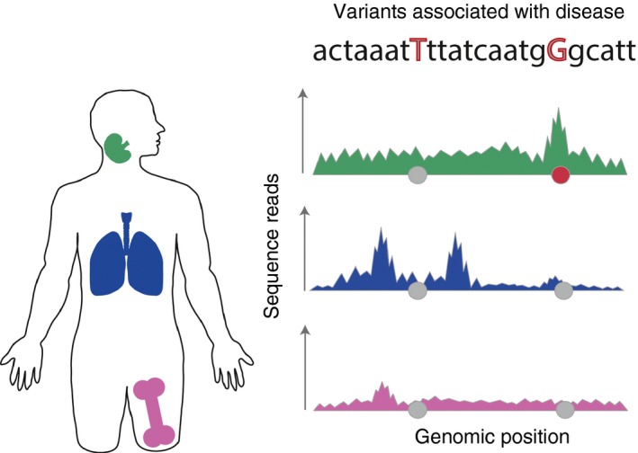
Comprehensive annotation of the regulatory chromatin provides an opportunity to interpret the role of non‐coding disease variants. Indeed, development of statistical approaches that integrate GWAS SNPs with histone marks proved to be a valuable approach to prioritize disease‐relevant cell types35, 36, 37 (Fig. 3). Enrichment of GWAS variants in cell‐type‐specific promoters marked by histone‐3 lysine‐4 trimethylation (H3K4me3), confirmed the importance of CD4+ T‐cell subsets in a number of autoimmune disorders, including T1D and RA. CD4+ memory T cells and regulatory T (Treg) cells showed high enrichment for variants associated with a number of diseases, including RA, IBD and CeD.38 This observation converges with the previous immune studies pointing towards impaired function of Treg cells in autoimmune diseases.39 Notably, a study by the Todd group has demonstrated that Treg cells from patients with SLE have lower levels of CD25, the α‐chain of IL‐2 receptor, which affects their phenotype and decreases their survival.40
Figure 3.

Causal disease variants overlap cell‐type‐specific chromatin marks. Chromatin immunoprecipitation followed by sequencing and chromatin accessibility assays produce pile ups of reads that form ‘peaks’. Such genomic annotations generated from different tissues [lymph nodes (green), lungs (blue), femur (pink)] provide a valuable roadmap of cell‐type‐specific genome activity. The genome annotations can be overlapped with genetic variants associated with a phenotype of interest, such as an autoimmune disease, represented as grey circles. If a statistically significant proportion of associated variants overlaps with peaks specific to a cell type it can point towards disease‐relevant tissue and prioritize the causal variants. Here, we illustrate a single‐associated locus where only one single nucleotide polymorphism (red circle) overlaps with a peak specific to the lymph nodes and absent from the other two tissues.
Leveraging ChIP‐seq data allowed the mapping of a proportion of GWAS variants in active enhancer regions of CD4+ T‐cell subsets marked by histone‐3 lysine‐27 acetylation (H3K27ac).37 Consistent with eQTL data, stimulation‐induced enhancers had high enrichment for autoimmune disease GWAS SNPs, whereas no enrichment was observed in enhancers specific to resting cells.37 If performed across tens or hundreds of individuals, histone marks can be directly correlated with genotypes.41 That is because ChIP‐seq assays result in pile ups of sequence reads at genomic regions that map the interaction with proteins. In that respect, they produce a quantitative measurement and, just like gene expression, can be analysed in light of correlations with different alleles, as QTLs. Regulatory annotations are not only a good predictor of gene activity but can also suggest a mechanism of action, e.g. by implicating a specific TF binding site,42 or when overlapping with eQTLs.43 For example, histone acetylation QTLs in human lymphoblastoid cell lines are highly enriched for immune disease GWAS variants, particularly from MS. In a study of three major immune cell types; neutrophils, monocytes and CD4+ T cells, from 200 individuals, Chen et al.44 mapped coordinated genetic effects on the epigenome and transcriptome.
In addition to ChIP‐seq, genome activity can also be measured by chromatin accessibility, either through mapping regions that undergo cutting by DNase (DNase hypersensitive sites)45 or by incorporation of the Tn5 transposase (ATAC‐seq).46 Analysis of 349 tissues and cell types generated using DNase‐seq found that > 75% of non‐coding GWAS‐associated variants lie within the chromatin accessible sites. This implies that assaying chromatin activity provides a valuable approach for understanding molecular mechanisms underlying the disease variants. For example, around 25% (n = 262) of GWAS SNPs associated with autoimmune diseases residing within the DNase hypersensitive sites in immune cells altered the TF binding motif for the IRF9 pathway.45 The IRF9 network along with the Jak/Stat cascade are initiated in the presence of interferon‐γ, pointing towards a potentially important role for this pathway in the development of autoimmunity.
Despite the observed enrichment of disease variants in regulatory non‐coding sequences it is estimated that only 10–20% of 823 disease variants lie within transcription factor binding motifs, suggesting other mechanisms of gene expression regulation,37 for instance by affecting spatial genomic interactions. To test this, a promising approach includes an integration of GWAS variants with Hi‐C assay47 that infers chromosome conformation by mapping interactions between genomic regions located nearby in the three‐dimensional space. The technique allows both the high level mapping of chromatin loops as well as identification of more granular interactions between enhancers and gene promoters. The latter can be directly used in mapping non‐coding disease variants to target genes. For example, using Hi‐C, a recent study demonstrated that the non‐coding region on chromosome 6 containing a variant that has been associated with RA and psoriasis, interacts not only with the promoter of TNFAIP3, the closest gene, but also with IL20RA.48 Furthermore, a comprehensive examination of 17 human primary blood cell types found an enrichment of autoimmune disease SNPs in Hi‐C domains in lymphoid cells compared with myeloid cells.49 The authors identified that the majority of identified genes (76%) had not been previously linked with immune‐mediated diseases. Notably, five of the identified genes associated with RA and SLE were also eQTLs, e.g. RASGRP1, a gene that activates the extracellular signal‐regulated kinase/mitogen‐activated protein kinase cascade and regulates T‐cell and B‐cell development and differentiation.
Gene expression regulation is also reflected through DNA methylation. By combining DNA methylation with RNA sequencing from 3841 Dutch individuals, Bonder et al. observed that trans methylation QTLs, where genetic variants affect distant rather than local methylation status, were enriched for immune‐associated GWAS traits.50 Although methylation has typically been linked to repressed regions, the authors found two distinct functions for methylation depending on its location. To investigate these findings further, the authors carried out TF ChIP‐seq and identified 13 trans‐methylation QTLs that influenced the TF binding sites, including two SNPs on chromosome 4 associated with ulcerative colitis. They prioritized one of them as the causal SNP based on its association with lower methylation and the higher gene expression of NFKB1. By incorporating Hi‐C assay with DNA methylation and RNA sequencing, Bonder et al., showed that inter‐chromosomal contacts provide a mechanism by which some trans‐methylation QTLs can act. The 402 identified CpG islands overlapped with CTCF, RAD21 and SMC3 TF binding sites.
Correlating genetic variation with immunological readouts
To gain comprehensive insights into the effects of genetic variants on the immune system, genomic assays have to be complemented with traditional immunological approaches. The network of cellular interactions can be assessed at a global scale, for instance by looking at mechanisms that control cell frequency and cell proliferation, as well as at the cellular level, by measuring protein expression and identifying genes that drive the phenotype of interest. To accurately infer the causal relationships between genetic variants and cellular traits it is important to carry out the experiments in cells isolated from healthy individuals, limiting the effects of active disease or ongoing treatment.
Linking GWAS variants to immune cell counts and function
The ratio between different immune cell subsets is heritable and could partly account for one of the pathobiological disease mechanisms.51, 52 Through an association study of genome‐wide SNPs with cell counts, two independent genetic signals were identified to control the CD4+ : CD8+ T‐cell ratio. Both signals mapped to the MHC region, explaining 8% of the observed variance. Interestingly, one of these associations overlapped with a T1D variant where the T1D risk allele increases the number of CD4+ cells by decreasing their apoptosis rate.53 Past years have seen many systematic, large‐scale efforts aiming to associate genetic variants with cell counts. Astle et al.54 identified over 2500 variants associated with 36 different haematopoietic traits. They observed an overlap between asthma‐associated variants and the eosinophil counts, highlighting that the established positive association between eosinophil counts and asthma is genetically controlled. Additionally, variants within the MHC locus and nearby COG6, SPRED2, RUNX1 and ATXN2/SH2B3/BRAP genes pointed towards a novel link between eosinophil function and RA. Study of immune cell frequencies and surface protein expression levels of 170 dizygotic and 75 monozygotic pairs of twins using seven distinct 14‐plex antibody panels identified 151 independent heritable immune traits, including, for example, the proportion of CD10+ immature B cells associated with the genotypes of the MME gene, which encodes for a metallo‐peptidase.55 This study reported that one of the most heritable traits is the frequency of CD39+ Treg cells. The authors identified a SNP that increases the level of CD39, and so alters the proportion of CD39+ Treg cells. CD39 along with CD73 are enzymes that degrade the pro‐inflammatory ATP molecule to an anti‐inflammatory adenosine.56 Dysregulation of this machinery has been observed in MS, RA and IBD patients, with multiple drugs targeting these two proteins. Additionally, the same variant had previously been identified in a study that measured counts of 95 different cell types from a cohort of 1629 individuals from Sardinia.57
In another study, the authors demonstrated that genes located near RA‐associated variants were highly expressed in CD4+ effector memory T cells.58 Following this observation, Hu et al.59 isolated CD4+ T effector memory cells and measured relative cell abundance, as well as gene expression of 215 genes located in RA loci, and cell proliferation capacity upon in vitro stimulation with anti‐CD28/anti‐CD3. The authors identified a group of genes whose basal level could predict the proliferative potential of effector memory cells, along with a non‐coding genetic variant that increased cell division capacity. Although, this study has not linked RA risk loci with the effect on CD4+ memory cell proliferation, it exemplifies how to link genotype to immune cell function.
Genetic control of cytokine level in response to stimuli
The effector function of immune cells is to communicate with each other through expression of receptors and receptor ligands, combined with the secretion of specific molecules, such as cytokines. Alteration of these cellular functions results in an impaired immune response. Interestingly, the cytokine levels in the blood have been shown to be highly heritable.52 Systematic characterization of the cell response to different bacterial and fungal infections identified six cytokine QTLs (cQTLs) that explained the IL‐6, IL‐8, IL‐10 and tumour necrosis factor‐α levels.60 The genetic control of cytokine secretion in response to pathogens is relevant, as some of the identified cytokines have also been associated with autoimmune diseases and are being targeted by drugs, e.g. IL‐6 with RA and psoriasis.61 Interleukin‐6 pathways, along with IL‐1β were identified as being mostly driven by genetics, compared with environmental factors and the microbiome.62 Assessment of the responses of peripheral blood mononuclear cells, whole blood and monocyte‐derived macrophages to pathogenic and non‐microbial stimuli in 500 individuals identified cell‐type‐specific cQTLs, with monocyte‐specific cQTLs being associated with susceptibility to infectious diseases and T‐cell‐specific cQTLs being associated with autoimmune diseases. Separately, co‐localization of genetic variants that control cytokine levels with those that contribute to susceptibility to autoimmune diseases linked the vascular endothelial growth factor cascade with IBD and expression of the α subunit of the IL‐2 receptor with Crohn's disease and MS.63 By standardizing the blood culture of healthy volunteers and using flow cytometry analysis, Duffy et al., de‐convoluted complex immune response signatures to a range of stimuli.64, 65 They built a detailed reference of cytokine levels from healthy individuals and identified a group of donors that do not release IL‐1α upon stimulation, despite the presence of IL‐1β, suggesting a genetic component underlying this effect.64 Together these data demonstrated that the levels of a number of pro‐ and anti‐inflammatory cytokines are under genetic control. Future large‐scale characterization of the molecular mechanisms that control cytokine levels in health and disease will provide further insights into disease pathology.
Discussion and future perspectives
Over the last decade, hundreds of immune disease loci have been successfully mapped. Recent advances in genomics have enabled functional follow‐up studies that provide insights into the molecular mechanisms by which risk variants drive disease pathology.
In this review, we have provided recent examples where the correlations of quantitative immunogenomic phenotypes with disease‐associated loci prioritized causal variants and suggested disrupted gene pathways and cellular functions. Although the majority of individual allelic effects have a miniscule effect on the overall phenotype, the effects can be higher on the molecular or cellular levels. Hence, future efforts have to be focused on identifying the critical disease cell types in which the associated variants are functional. These studies are challenging to conduct, as certain cell populations only represent a small proportion of the whole blood and need to be isolated using reliable markers and cell sorting. Importantly, isolated cell populations are often difficult to culture in vitro. This process has to be repeated a number of times on blood drawn from individuals of the same population to reach a cohort size large enough that it would allow for reliable identification of QTL effects. QTL effects have been annotated mostly in the context of expression assays, however, a proportion of GWAS variants may not act through gene regulation measured by bulk gene expression assays, calling for further development of genomic tools.
A challenge in GWAS follow‐up studies is that often the causal cell types are unknown. Typically, cell types have been identified through immunology studies where a limited set of specific cell markers, both intracellular and extracellular, have been used to characterize cell populations. A promising new method, that is unbiased to a predefined set of markers, is single‐cell RNA sequencing (scRNA‐seq). This approach can identify previously unknown heterogeneity in a sample based on gene expression measured at the individual cell level. There is now a major international effort that aims to use scRNA‐seq to characterize all human cells, the Human Cell Atlas.66 This resource will provide the most comprehensive annotation of gene expression, and when integrated with GWAS variants it will present a tremendous opportunity for the improved understanding of immune‐mediated diseases.
Disclosure
The authors have no competing interests.
Acknowledgements
We thank the Wellcome Trust for their funding supporting our research (grant WT206194).
References
- 1. Hayter SM, Cook MC. Updated assessment of the prevalence, spectrum and case definition of autoimmune disease. Autoimmun Rev 2012; 11:754–65. [DOI] [PubMed] [Google Scholar]
- 2. Todd JA, Bell JI, McDevitt HO. HLA‐DQ β gene contributes to susceptibility and resistance to insulin‐dependent diabetes mellitus. Nature 1987; 329:599–604. [DOI] [PubMed] [Google Scholar]
- 3. Sollid LM, Markussen G, Ek J, Gjerde H, Vartdal F, Thorsby E. Evidence for a primary association of celiac disease to a particular HLA‐DQ α/β heterodimer. J Exp Med 1989; 169:345–50. [DOI] [PMC free article] [PubMed] [Google Scholar]
- 4. Nepom GT. Major histocompatibility complex‐directed susceptibility to rheumatoid arthritis. Adv Immunol 1998; 68:315–32. [DOI] [PubMed] [Google Scholar]
- 5. Lenz TL, Deutsch AJ, Han B, Hu X, Okada Y, Eyre S et al Widespread non‐additive and interaction effects within HLA loci modulate the risk of autoimmune diseases. Nat Genet 2015; 47:1085–90. [DOI] [PMC free article] [PubMed] [Google Scholar]
- 6. Stahl EA, Raychaudhuri S, Remmers EF, Xie G, Eyre S, Thomson BP et al Genome‐wide association study meta‐analysis identifies seven new rheumatoid arthritis risk loci. Nat Genet 2010; 42:508–14. [DOI] [PMC free article] [PubMed] [Google Scholar]
- 7. Gutierrez‐Arcelus M, Rich SS, Raychaudhuri S. Autoimmune diseases – connecting risk alleles with molecular traits of the immune system. Nat Rev Genet 2016; 17:160–74. [DOI] [PMC free article] [PubMed] [Google Scholar]
- 8. Consortium IMSG, Others . Analysis of immune‐related loci identifies 48 new susceptibility variants for multiple sclerosis. Nat Genet 2013; 45:1353–60. [DOI] [PMC free article] [PubMed] [Google Scholar]
- 9. Hu X, Deutsch AJ, Lenz TL, Onengut‐Gumuscu S, Han B, Chen W‐M et al Additive and interaction effects at three amino acid positions in HLA‐DQ and HLA‐DR molecules drive type 1 diabetes risk. Nat Genet 2015; 47:898–905. [DOI] [PMC free article] [PubMed] [Google Scholar]
- 10. Okada Y, Wu D, Trynka G, Raj T, Terao C, Ikari K et al Genetics of rheumatoid arthritis contributes to biology and drug discovery. Nature 2014; 506:376–81. [DOI] [PMC free article] [PubMed] [Google Scholar]
- 11. Fortune MD, Guo H, Burren O, Schofield E, Walker NM, Ban M et al Statistical colocalization of genetic risk variants for related autoimmune diseases in the context of common controls. Nat Genet 2015; 47:839–46. [DOI] [PMC free article] [PubMed] [Google Scholar]
- 12. Schubert D, Bode C, Kenefeck R, Hou TZ, Wing JB, Kennedy A et al Autosomal dominant immune dysregulation syndrome in humans with CTLA4 mutations. Nat Med 2014; 20:1410–6. [DOI] [PMC free article] [PubMed] [Google Scholar]
- 13. Dimas AS, Deutsch S, Stranger BE, Montgomery SB, Borel C, Attar‐Cohen H et al Common regulatory variation impacts gene expression in a cell type‐dependent manner. Science 2009; 325:1246–50. [DOI] [PMC free article] [PubMed] [Google Scholar]
- 14. Nicolae DL, Gamazon E, Zhang W, Duan S, Eileen Dolan M, Cox NJ. Trait‐associated SNPs are more likely to be eQTLs: annotation to enhance discovery from GWAS. PLoS Genet 2010; 6:e1000888. [DOI] [PMC free article] [PubMed] [Google Scholar]
- 15. Dubois PCA, Trynka G, Franke L, Hunt KA, Romanos J, Curtotti A et al Multiple common variants for celiac disease influencing immune gene expression. Nat Genet 2010; 42:295–302. [DOI] [PMC free article] [PubMed] [Google Scholar]
- 16. de Lange KM, Moutsianas L, Lee JC, Lamb CA, Luo Y, Kennedy NA et al Genome‐wide association study implicates immune activation of multiple integrin genes in inflammatory bowel disease. Nat Genet 2017; 49:256–61. [DOI] [PMC free article] [PubMed] [Google Scholar]
- 17. Westra H‐J, Peters MJ, Esko T, Yaghootkar H, Schurmann C, Kettunen J et al Systematic identification of trans eQTLs as putative drivers of known disease associations. Nat Genet 2013; 45:1238–43. [DOI] [PMC free article] [PubMed] [Google Scholar]
- 18. GTEx Consortium . Human genomics. The Genotype‐Tissue Expression (GTEx) pilot analysis: multitissue gene regulation in humans. Science 2015;348:648–60. [DOI] [PMC free article] [PubMed] [Google Scholar]
- 19. Fairfax BP, Humburg P, Makino S, Naranbhai V, Wong D, Lau E et al Innate immune activity conditions the effect of regulatory variants upon monocyte gene expression. Science 2014; 343:1246949. [DOI] [PMC free article] [PubMed] [Google Scholar]
- 20. Guo H, Fortune MD, Burren OS, Schofield E, Todd JA, Wallace C. Integration of disease association and eQTL data using a Bayesian colocalisation approach highlights six candidate causal genes in immune‐mediated diseases. Hum Mol Genet 2015; 24:3305–13. [DOI] [PMC free article] [PubMed] [Google Scholar]
- 21. Chick JM, Munger SC, Simecek P, Huttlin EL, Choi K, Gatti DM et al Defining the consequences of genetic variation on a proteome‐wide scale. Nature 2016; 534:500–5. [DOI] [PMC free article] [PubMed] [Google Scholar]
- 22. Chun S, Casparino A, Patsopoulos NA, Croteau‐Chonka DC, Raby BA, De Jager PL et al Limited statistical evidence for shared genetic effects of eQTLs and autoimmune‐disease‐associated loci in three major immune‐cell types. Nat Genet 2017; 49:600–5. [DOI] [PMC free article] [PubMed] [Google Scholar]
- 23. Huang H, Fang M, Jostins L, Mirkov MU, Boucher G, Anderson CA et al Fine‐mapping inflammatory bowel disease loci to single‐variant resolution. Nature 2017; 547:173–8. [DOI] [PMC free article] [PubMed] [Google Scholar]
- 24. Kasela S, Kisand K, Tserel L, Kaleviste E, Remm A, Fischer K et al Pathogenic implications for autoimmune mechanisms derived by comparative eQTL analysis of CD4+ versus CD8+ T cells. PLoS Genet 2017; 13:e1006643. [DOI] [PMC free article] [PubMed] [Google Scholar]
- 25. Solovieff N, Cotsapas C, Lee PH, Purcell SM, Smoller JW. Pleiotropy in complex traits: challenges and strategies. Nat Rev Genet 2013; 14:483–95. [DOI] [PMC free article] [PubMed] [Google Scholar]
- 26. Raj T, Rothamel K, Mostafavi S, Ye C, Lee MN, Replogle JM et al Polarization of the effects of autoimmune and neurodegenerative risk alleles in leukocytes. Science 2014; 344:519–23. [DOI] [PMC free article] [PubMed] [Google Scholar]
- 27. CAMMS223 Trial Investigators , Coles AJ, Compston DAS, Selmaj KW, Lake SL, Moran S et al Alemtuzumab vs. interferon beta‐1a in early multiple sclerosis. N Engl J Med 2008; 359:1786–801. [DOI] [PubMed] [Google Scholar]
- 28. De Jager PL, Jia X, Wang J, de Bakker PIW, Ottoboni L, Aggarwal NT et al Meta‐analysis of genome scans and replication identify CD6, IRF8 and TNFRSF1A as new multiple sclerosis susceptibility loci. Nat Genet 2009; 41:776–82. [DOI] [PMC free article] [PubMed] [Google Scholar]
- 29. Chrabot BS, Kariuki SN, Zervou MI, Feng X, Arrington J, Jolly M et al Genetic variation near IRF8 is associated with serologic and cytokine profiles in systemic lupus erythematosus and multiple sclerosis. Genes Immun 2013; 14:471–8. [DOI] [PMC free article] [PubMed] [Google Scholar]
- 30. Lee MN, Ye C, Villani A‐C, Raj T, Li W, Eisenhaure TM et al Common genetic variants modulate pathogen‐sensing responses in human dendritic cells. Science 2014; 343:1246980. [DOI] [PMC free article] [PubMed] [Google Scholar]
- 31. Ye CJ, Feng T, Kwon H‐K, Raj T, Wilson MT, Asinovski N et al Intersection of population variation and autoimmunity genetics in human T cell activation. Science 2014; 345:1254665. [DOI] [PMC free article] [PubMed] [Google Scholar]
- 32. ENCODE Project Consortium . An integrated encyclopedia of DNA elements in the human genome. Nature 2012; 489:57–74. [DOI] [PMC free article] [PubMed] [Google Scholar]
- 33. Roadmap Epigenomics Consortium , Kundaje A, Meuleman W, Ernst J, Bilenky M, Yen A et al Integrative analysis of 111 reference human epigenomes. Nature 2015; 518:317–30. [DOI] [PMC free article] [PubMed] [Google Scholar]
- 34. Stunnenberg HG, International Human Epigenome Consortium , Hirst M. The International Human Epigenome Consortium: a blueprint for scientific collaboration and discovery. Cell 2016; 167:1897. [DOI] [PubMed] [Google Scholar]
- 35. Trynka G, Sandor C, Han B, Xu H, Stranger BE, Liu XS et al Chromatin marks identify critical cell types for fine mapping complex trait variants. Nat Genet 2013; 45:124–30. [DOI] [PMC free article] [PubMed] [Google Scholar]
- 36. Pickrell JK. Joint analysis of functional genomic data and genome‐wide association studies of 18 human traits. Am J Hum Genet 2014; 94:559–73. [DOI] [PMC free article] [PubMed] [Google Scholar]
- 37. Farh KK‐H, Marson A, Zhu J, Kleinewietfeld M, Housley WJ, Beik S et al Genetic and epigenetic fine mapping of causal autoimmune disease variants. Nature 2015; 518:337–43. [DOI] [PMC free article] [PubMed] [Google Scholar]
- 38. Trynka G, Raychaudhuri S. Using chromatin marks to interpret and localize genetic associations to complex human traits and diseases. Curr Opin Genet Dev 2013; 23:635–41. [DOI] [PMC free article] [PubMed] [Google Scholar]
- 39. Buckner JH. Mechanisms of impaired regulation by CD4+ CD25+ FOXP3+ regulatory T cells in human autoimmune diseases. Nat Rev Immunol 2010; 10:849–59. [DOI] [PMC free article] [PubMed] [Google Scholar]
- 40. Ferreira RC, Rainbow DB, García AR, Pekalski ML, Porter L, Oliveira JJ et al Human IL‐6R hi TIGIT‐ CD4+ CD127 low CD25+ T cells display potent in vitro suppressive capacity and a distinct Th17 profile. Clin Immunol 2017; 179:25–39. [DOI] [PMC free article] [PubMed] [Google Scholar]
- 41. Kumasaka N, Knights AJ, Gaffney DJ. Fine‐mapping cellular QTLs with RASQUAL and ATAC‐seq. Nat Genet 2016; 48:206–13. [DOI] [PMC free article] [PubMed] [Google Scholar]
- 42. McVicker G, van de Geijn B, Degner JF, Cain CE, Banovich NE, Raj A et al Identification of genetic variants that affect histone modifications in human cells. Science 2013; 342:747–9. [DOI] [PMC free article] [PubMed] [Google Scholar]
- 43. del Rosario RC‐H, Poschmann J, Rouam SL, Png E, Khor CC, Hibberd ML et al Sensitive detection of chromatin‐altering polymorphisms reveals autoimmune disease mechanisms. Nat Methods 2015; 12:458–64. [DOI] [PubMed] [Google Scholar]
- 44. Chen L, Ge B, Casale FP, Vasquez L, Kwan T, Garrido‐Martín D et al Genetic drivers of epigenetic and transcriptional variation in human immune cells. Cell 2016; 167:1398–414.e24. [DOI] [PMC free article] [PubMed] [Google Scholar]
- 45. Maurano MT, Humbert R, Rynes E, Thurman RE, Haugen E, Wang H et al Systematic localization of common disease‐associated variation in regulatory DNA. Science 2012; 337:1190–5. [DOI] [PMC free article] [PubMed] [Google Scholar]
- 46. Buenrostro JD, Giresi PG, Zaba LC, Chang HY, Greenleaf WJ. Transposition of native chromatin for fast and sensitive epigenomic profiling of open chromatin, DNA‐binding proteins and nucleosome position. Nat Methods 2013; 10:1213–8. [DOI] [PMC free article] [PubMed] [Google Scholar]
- 47. Belton J‐M, McCord RP, Gibcus JH, Naumova N, Zhan Y, Dekker J. Hi‐C: a comprehensive technique to capture the conformation of genomes. Methods 2012; 58:268–76. [DOI] [PMC free article] [PubMed] [Google Scholar]
- 48. McGovern A, Schoenfelder S, Martin P, Massey J, Duffus K, Plant D et al Capture Hi‐C identifies a novel causal gene, IL20RA, in the pan‐autoimmune genetic susceptibility region 6q23. Genome Biol 2016; 17:212. [DOI] [PMC free article] [PubMed] [Google Scholar]
- 49. Javierre BM, Burren OS, Wilder SP, Kreuzhuber R, Hill SM, Sewitz S et al Lineage‐specific genome architecture links enhancers and non‐coding disease variants to target gene promoters. Cell 2016; 167:1369–84. [DOI] [PMC free article] [PubMed] [Google Scholar]
- 50. Bonder MJ, Luijk R, Zhernakova DV, Moed M, Deelen P, Vermaat M et al Disease variants alter transcription factor levels and methylation of their binding sites. Nat Genet 2017; 49:131–8. [DOI] [PubMed] [Google Scholar]
- 51. Hall MA, Ahmadi KR, Norman P, Snieder H, MacGregor AJ, Vaughan RW et al Genetic influence on peripheral blood T lymphocyte levels. Genes Immun 2000; 1:423–7. [DOI] [PubMed] [Google Scholar]
- 52. Brodin P, Jojic V, Gao T, Bhattacharya S, Angel CJL, Furman D et al Variation in the human immune system is largely driven by non‐heritable influences. Cell 2015; 160:37–47. [DOI] [PMC free article] [PubMed] [Google Scholar]
- 53. Ferreira MAR, Mangino M, Brumme CJ, Zhao ZZ, Medland SE, Wright MJ et al Quantitative trait loci for CD4 : CD8 lymphocyte ratio are associated with risk of type 1 diabetes and HIV‐1 immune control. Am J Hum Genet 2010; 86:88–92. [DOI] [PMC free article] [PubMed] [Google Scholar]
- 54. Astle WJ, Elding H, Jiang T, Allen D, Ruklisa D, Mann AL et al The allelic landscape of human blood cell trait variation and links to common complex disease. Cell 2016; 167:1415–29.e19. [DOI] [PMC free article] [PubMed] [Google Scholar]
- 55. Roederer M, Quaye L, Mangino M, Beddall MH, Mahnke Y, Chattopadhyay P et al The genetic architecture of the human immune system: a bioresource for autoimmunity and disease pathogenesis. Cell 2015; 161:387–403. [DOI] [PMC free article] [PubMed] [Google Scholar]
- 56. Antonioli L, Pacher P, Vizi ES, Haskó G. CD39 and CD73 in immunity and inflammation. Trends Mol Med 2013; 19:355–67. [DOI] [PMC free article] [PubMed] [Google Scholar]
- 57. Orrù V, Steri M, Sole G, Sidore C, Virdis F, Dei M et al Genetic variants regulating immune cell levels in health and disease. Cell 2013; 155:242–56. [DOI] [PMC free article] [PubMed] [Google Scholar]
- 58. Hu X, Kim H, Stahl E, Plenge R, Daly M, Raychaudhuri S. Integrating autoimmune risk loci with gene‐expression data identifies specific pathogenic immune cell subsets. Am J Hum Genet 2011; 89:496–506. [DOI] [PMC free article] [PubMed] [Google Scholar]
- 59. Hu X, Kim H, Raj T, Brennan PJ, Trynka G, Teslovich N et al Regulation of gene expression in autoimmune disease loci and the genetic basis of proliferation in CD4+ effector memory T cells. PLoS Genet 2014; 10:e1004404. [DOI] [PMC free article] [PubMed] [Google Scholar]
- 60. Li Y, Oosting M, Deelen P, Ricaño‐Ponce I, Smeekens S, Jaeger Ms et al Corrigendum: inter‐individual variability and genetic influences on cytokine responses to bacteria and fungi. Nat Med 2016; 22:1192. [DOI] [PubMed] [Google Scholar]
- 61. Ishihara K, Hirano T. IL‐6 in autoimmune disease and chronic inflammatory proliferative disease. Cytokine Growth Factor Rev 2002; 13:357–68. [DOI] [PubMed] [Google Scholar]
- 62. Li Y, Oosting M, Smeekens SP, Jaeger M, Aguirre‐Gamboa R, Le KTT et al A functional genomics approach to understand variation in cytokine production in humans. Cell 2016; 167:1099–110.e14. [DOI] [PubMed] [Google Scholar]
- 63. Ahola‐Olli AV, Würtz P, Havulinna AS, Aalto K, Pitkänen N, Lehtimäki T et al Genome‐wide association study identifies 27 loci influencing concentrations of circulating cytokines and growth factors. Am J Hum Genet 2017; 100:40–50. [DOI] [PMC free article] [PubMed] [Google Scholar]
- 64. Duffy D, Rouilly V, Libri V, Hasan M, Beitz B, David M et al Functional analysis via standardized whole‐blood stimulation systems defines the boundaries of a healthy immune response to complex stimuli. Immunity 2014; 40:436–50. [DOI] [PubMed] [Google Scholar]
- 65. Urrutia A, Duffy D, Rouilly V, Posseme C, Djebali R, Illanes G et al Standardized whole‐blood transcriptional profiling enables the deconvolution of complex induced immune responses. Cell Rep 2016; 16:2777–91. [DOI] [PubMed] [Google Scholar]
- 66. Regev A, Others. The Human Cell Atlas [Internet]. genome.gov; 2016. URL: https://www.genome.gov/multimedia/slides/gspfuture2014/10_regev.pdf Posted May 8, 2017.


