Abstract
Human parainfluenza virus type 3 (hPIV3) recognizes both α2,3- and α2,6-linked sialic acids, whereas human parainfluenza virus type 1 (hPIV1) recognizes only α2,3-linked sialic acids. To identify amino acid residues that confer α2,6-linked sialic acid recognition of hPIV3, amino acid residues in or neighboring the sialic acid binding pocket of the hPIV3 hemagglutinin–neuraminidase (HN) glycoprotein were substituted for the corresponding residues of hPIV1 HN. Hemadsorption assay with sialyl linkage-modified red blood cells indicated that amino acid residues at positions 275, 277, 372, and 426 contribute to α2,6-linked sialic acid recognition of the HN3 glycoprotein.
Keywords: Human parainfluenza virus, Hemagglutinin-neuraminidase, Sialyl linkage, Receptor binding, Sialic acid, HN
1. Introduction
Human parainfluenza virus type 3 (hPIV3) and type 1 (hPIV1) are respiratory pathogens that cause laryngotracheobronchitis and bronchiolitis in infants and young children [1]. hPIV1 and hPIV3, which belong to the genus Respirovirus in the family Paramyxoviridae, have two envelope glycoproteins, hemagglutinin–neuraminidase (HN) glycoprotein and fusion glycoprotein. Both hPIVs bind to terminal sialic acids of glycoconjugates on the host cell surface through the HN glycoprotein to initiate infection [2].
Sialic acids are typically found at the terminals of oligosaccharides on gangliosides and glycoproteins. They are mainly linked to the terminal galactose residue by α2,3- or α2,6-linkage [3]. Virus binding specificity for each sialyl linkage is believed to be one of major determinants of tissue tropism or virulence. Among paramyxoviruses, Sendai virus binds to both gangliosides with terminal α2,3- and α2,6-linked sialic acids [4]. Newcastle disease virus has been suggested to use both α2,3- and α2,6-linked sialic acids on N-linked glycoproteins as viral receptors [5]. However, specific amino acid residues of paramyxoviruses that determine the recognition for each sialyl linkage have not been identified.
Our previous studies showed that hPIV1 and hPIV3 have different recognition specificities for terminal sialyl linkages: hPIV1 and the hPIV1 HN (HN1) glycoprotein preferentially recognize α2,3-linked sialic acids, whereas hPIV3 and the hPIV3 HN (HN3) glycoprotein recognize both α2,3- and α2,6-linked sialic acids on the host cell surfaces [6,7]. Elucidation of the mechanism of sialyl linkage recognition is important for understanding cell and tissue tropism of hPIVs. In this study, we identified amino acid residues that confer the α2,6-linked sialic acid recognition of hPIV3. Amino acid residues in or neighboring the sialic acid binding pocket of the HN3 glycoprotein were substituted for the corresponding residues of the HN1 glycoprotein. Recognition activities for α2,6-linked sialic acid of these HN3 glycoproteins were evaluated by a hemadsorption (HAD) assay with sialyl linkage-modified red blood cells (RBCs).
2. Materials and methods
2.1. Cells, viruses, and antibody
Lewis lung carcinoma-monkey kidney (LLC-MK2) cells were maintained in Eagle’s minimum essential medium (MEM) supplemented with 5% heat-inactivated fetal bovine serum (FBS). African green monkey kidney (COS-7) cells were maintained in Dulbecco’s modified Eagle’s medium (DMEM) supplemented with 10% FBS. hPIV1 C35 strain and hPIV3 C243 strain were kindly provided by Allen Portner of St. Jude Children’s Research Hospital. Both hPIVs were propagated in LLC-MK2 cells. Rabbit anti-hPIV3 antibody was prepared by immunization with hPIV3 C243 strain as described previously and purified on a Protein G HiTrap™ Protein G HP (CE Healthcare Biosciences Corp., Piscataway, NJ, USA) [7].
2.2. Preparation of sialyl linkage-modified red blood cells
Human RBCs were collected from a healthy adult and washed 3 times with phosphate-buffered saline (PBS; pH 7.2, 131 mM NaCl, 14 mM Na2HPO4 and 2.7 mM KCl) (native RBCs). AUSD-RBCs were prepared from complete desialylation on native RBCs by incubation of 10% native RBCs with PBS containing 25 mU/ml of sialidase from Arthrobacter ureafaciens (AUSD; Nacalai Tesque, Kyoto, Japan) at 37 °C for 2 h and then washing with PBS 5 times. 23SD-RBCs were prepared from desialylation of α2,3-linked sialic acids on native RBCs by incubation of 10% native RBCs with PBS containing 100 U/ml of cloned α2,3 neuraminidase from Salmonella typhimurium LT2 (23SD; New England Biolabs Inc., Ipswich, MA, USA) and 5 mM CaCl2 at 37 °C for 30, 45, or 60 min and then washing with PBS 5 times. ST6-RBCs were prepared from resialylation of α2, 6-linked sialic acids on AUSD-RBCs by incubation of 10% AUSD-RBCs with PBS containing 1.5 mg/ml of cytidine-5′-monophospho-N-acetylneuraminic acid sodium salt (CMP-Sia; Nacalai Tesque, Kyoto, Japan) and 75 mU/ml of α2,6-sialyltransferase from Photobacterium damselae (ST6; CosmoBio Co., Ltd., Tokyo, Japan) at 37 °C for 17 h and then washing with PBS 5 times.
2.3. Quantitation of cell surface expression of HN3 glycoproteins
Expression vectors of mutated HN3 glycoprotein were constructed as described in Supplementary method 1. To quantify cell surface expression (CSE) of wild-type (WT) and mutated HN3 glycoproteins, a 70% confluent monolayer of COS-7 cells in a 24-well plate was transfected with 500 ng of pCAGGS expression plasmid vectors containing the WT or mutated HN3 genes using the transfection reagent TransIT-LT1 (Mirus, Madison, WI, USA) according to the manufacturer’s instructions. The cells were also mock-treated without plasmid vectors. After incubation at 37 °C for 6 h, the transfected cells were washed with PBS and then cultured in a serum-free medium (Hybridoma-SFM; SFM, Invitrogen, Corp., Carlsbad, CA, USA). After incubation at 37 °C for 48 h, the cells were harvested by treatment with 200 μl of 0.125% trypsin-PBS. The cells were washed with PBS, incubated with rabbit anti-hPIV3 antibody, and then incubated with tetramethylrhodamine (TRITC)-conjugated goat anti-rabbit IgG antibody (Sigma–Aldrich, St. Louis, MO, USA) for 30 min at 4 °C. Fluorescence for cells was measured with an excitation of the 488-nm line of an argon laser on a FACSCanto II flow cytometer (BD, Franklin Lakes, NJ, and USA). At least 1 × 10⁴ cells were analyzed for each sample. The background detected in non-transfected cells was subtracted.
2.4. Hemadsorption assay
To determine HAD activity of HN1 and HN3 glycoproteins, a 70% confluent monolayer of COS-7 cells in a 48-well plate was transfected with 300 ng of pCAGGS plasmid vectors containing WT HN1, WT HN3, or mutated HN3 genes. Cells were also mock-treated without plasmids vectors. After incubation at 37 °C for 48 h, the cells were washed with PBS and incubated with 0.5% native RBCs, 23SD-RBCs, AUSD-RBCs, or ST6-RBCs in SFM for 30 min on ice. The cells were washed 3 times with SFM and incubated with 100 μl of ultrapure water for 10 min on ice. Fifty microliters of the supernatant was applied in a 96-well plate, and the amount of hemoglobin was determined by measuring the absorbance at 415 nm [7]. The background detected in non-transfected cells was subtracted.
3. Results
3.1. Comparison of amino acid sequences of the sialic acid binding pockets in HN1 and HN3 glycoproteins
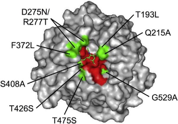
The HN1 glycoprotein recognizes only α2,3-linked sialic acid, but the HN3 glycoprotein recognizes both α2,3- and α2,6-linked sialic acids [6,7], meaning that the HN3 glycoprotein, but not the HN1 glycoprotein, has amino acid residues that interact with α2,6-linked sialic acids in the binding pocket. As another explanation for the lack of binding of the HN1 glycoprotein to α2,6-linked sialic acids, some amino acids in the binding pocket of the HN1 glycoprotein might interfere with the fit of the receptor moiety of α2,6-linked sialic acids. For Fig. 1 showing the structure of the HN3 glycoprotein [8], red amino acid residues in the receptor binding pocket directly interact with the sialic acid residue of the receptor through a hydrogen bond. These amino acid resides are conserved between the HN1 and NH3 glycoproteins. When amino acid sequences of the HN3 glycoprotein were compared with those of the HN1 glycoprotein, 9 amino acid differences between them were found in or neighboring the sialic acid binding pocket. These residues are colored in green in the structure of the HN3 glycoprotein shown in Fig. 1. These 9 amino acid residues of the HN3 glycoprotein were substituted for those of the HN1 glycoprotein (T193L, Q215A, D275N/R277T, F372L, S408A, T426S, T475S, and G529A, based on amino acid numbering of the HN3 glycoprotein) for changing sialic acid linkage binding specificity of the HN3 glycoprotein. Neighboring amino acid resides at positions 275 and 277 were simultaneously substituted.
Fig. 1.

Locations of amino acid substitutions and the sialic acid binding pocket in the HN3 glycoprotein. The three-dimensional structure of HN3 glycoprotein with sialic acid (PDB ID: 1V3C) was shown by PyMOL software. There were 9 amino acid residues that were different in sequences of the HN1 and HN3 glycoproteins in or neighboring the sialic acid binding pocket (green residues). These 9 residues of the HN3 glycoprotein (left of position numbers) were substituted for the corresponding residues of the HN1 glycoprotein (right of position numbers). Red residues mean that amino acids directly interacted with the sialic acid residue through a hydrogen bond. Sialic acid is shown by a stick model in green.
3.2. HAD activity and CSE of amino acid-substituted HN3 glycoproteins
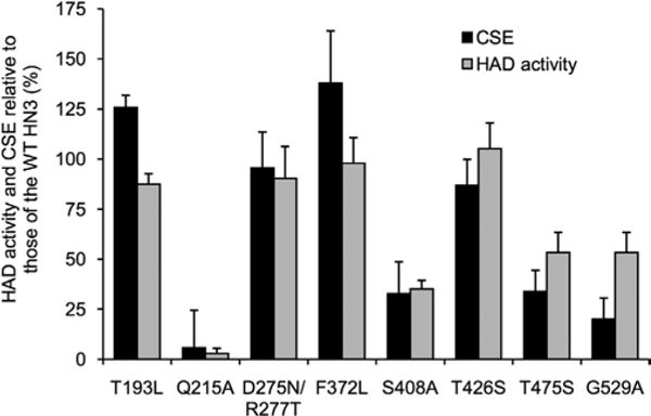
The effects of amino acid substitutions of HN3 glycoproteins were evaluated by HAD activity and CSE. The HN3 glycoproteins carrying Q215A, S408A, T475S, or G529A substitutions showed significant reduction of HAD activity and CSE (Fig. 2). These amino acid substitutions were probably needed to maintain normal CSE of the HN3 glycoprotein. T193L, D275N/R277T, F372L, or T426S substitutions of the HN3 glycoproteins resulted in HAD activity and CSE similar to those of the WT HN3 glycoprotein. These results indicated that T193L, D275N/R277T, F372L, and T426S substitutions had little effect on the sialic acid binding affinity of the HN3 glycoprotein.
Fig. 2.

HAD activity and CSE of amino acid-substituted HN3 glycoproteins. COS-7 cells were transfected with HN3 genes. CSE (filled column) was quantitated by flow cytometry with rabbit anti-hPIV3 antibody. HAD activity (gray column) was determined by the amount of native RBCs adsorbed on the HN3 glycoprotein-expressing cells. All data are represented as relative percentages to the WT HN3 glycoprotein.
3.3. Alpha2,6-linked sialic acid recognition of amino acid-substituted HN3 glycoproteins
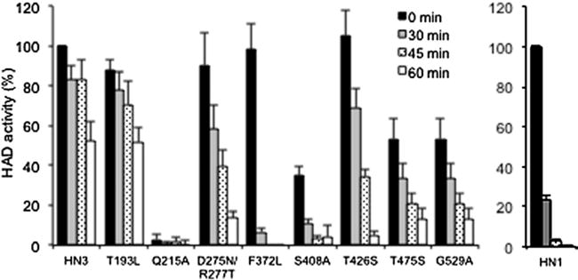
Alpha2,6-linked sialic acid recognition of the amino acid-substituted HN3 glycoproteins was estimated by HAD assays with α2,3-linked sialic acid-removed RBCs, 23SD-RBCs. Some 23SD-RBCs were prepared by treatment of native RBCs (expressing both α2,3-and α2,6-linked sialic acids) with 23SD that selectively removed α2,3-linked sialic acids, for 30, 45, and 60 min. While the WT HN1 glycoprotein hardly bound to 23SD-RBCs, the WT HN3 glycoprotein bound to 23SD-RBCs and retained 52% HAD activity even for 23SD-RBCs after 60-min incubation with 23SD relative to native RBCs. This result indicated that the WT HN3 glycoprotein recognized α2,6-linked sialic acids that remained on the surface 23SD-RBCs after 23SD treatment but that the HN1 glycoprotein did not (Fig. 3). This sialic acid recognition specificity coincides with results of our previous study [7]. The HN3 glycoprotein carrying the T193L substitution showed HAD activity similar to that of the WT HN3 glycoprotein (52% HAD activity even for 23SD-RBCs after 60-min incubation with 23SD). The HN3 glycoproteins with D275N/R277T, F372L, and T426S substitutions showed HAD activity similar to that of the WT HN3 glycoprotein for native RBCs, whereas these substitutions in the HN3 glycoproteins decreased HAD activity for 23SD-RBCs (14%, 0%, and 5% HAD activity for 23SD-RBCs after 60-min incubation with 23SD, respectively), indicating that these substitutions affected the α2,6-linked sialic acid recognition of the WT HN3 glycoprotein. S408A, T475S, and G529A substitutions also decreased HAD activity for native RBCs and 23SD-RBCs compared to the HAD activity of the WT HN3 glycoprotein, probably due to reduction of CSE of the HN3 glycoprotein.
Fig. 3.

HAD activities of WT HN3, WT HN1, and amino acid-substituted HN3 glycoproteins for 23SD-RBCs. 23SD-RBCs were prepared by 23SD treatment of native RBCs for 30 (gray column), 45 (hatched column), or 60 min (empty column) at 37 °C. HAD activities of HN3 and HN1 glycoproteins for native RBCs (filled column) and 23SD-RBCs are represented as relative percentages to those of WT HN3 glycoprotein and WT HN1 glycoprotein for native RBCs, respectively.
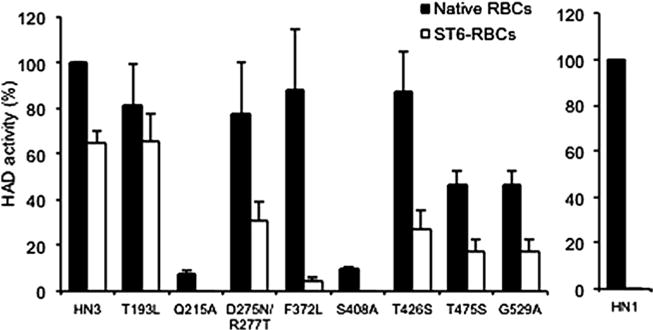
Alpha2,6-linked sialic acid recognition of amino acid-substituted HN3 glycoproteins was also estimated by HAD assays with α2,6-linked sialic acid-resialylated RBCs, ST6-RBCs. ST6-RBCs were prepared by treatment of native RBCs with AUSD, which removed all terminal sialic acids (AUSD-RBCs), and then by treatment of AUSD-RBCs with ST6, which specifically transferred α2,6-linked sialic acids to the terminal galactose residues. HAD activities of the HN3 glycoproteins for native RBCs and ST6-RBCs were expressed as relative activity to that of the WT HN3 glycoprotein for native RBCs. Again, the WT HN3 glycoprotein, but not the WT HN1 glycoprotein, showed 65% HAD activity for ST6-RBCs relative to that for native RBCs (Fig. 4). The HN3 glycoprotein carrying the T193L substitution showed HAD activity similar to that of the WT HN3 glycoprotein (65% HAD activity for ST6-RBCs). D275N/R277T, F372L, and T426S substitutions had little effect on HAD activity for native RBCs, whereas these substitutions resulted in reduction of HAD activity (31% HAD activity for D275N/R277T and 27% HAD activity for T426S) or loss of HAD activity (5% HAD activity for F372L) for ST6-RBCs. All of the HN glycoproteins showed no HAD activity for AUSD-RBCs (data not shown). These results indicated that amino acid residues at positions 275, 277, 372, and 426 in the sialic acid binding pocket were required for α2,6-linked sialic acid recognition of the HN3 glycoprotein, with little effect on α2,3-linked sialic acid recognition.
Fig. 4.

HAD activities of WT HN3, the WT HN1, and the amino acid-substituted HN3 glycoproteins for ST6-RBCs. ST6-RBCs were prepared by α2,6 linkage-specific resialylation of AUSD-RBCs with ST6. HAD activities of HN3 and HN1 glycoproteins for ST6-RBCs are represented as relative percentages to those of WT HN3 and WT HN1 glycoproteins for native RBCs, respectively.
4. Discussion
Sialyl linkage recognition of hPIVs determines cell infectivity [7]. Elucidation of the mechanism of sialyl linkage recognition is important for understanding cell and tissue tropism. To identify amino acid residues that confer α2,6-linked sialic acid recognition to the HN3 glycoprotein, amino acid residues in or neighboring the sialic acid binding pocket of the HN3 glycoprotein were substituted for the corresponding residues of the HN1 glycoprotein. HN3 glycoproteins with Q215A, S408A, T475S, and G529A substitutions significantly decreased CSE of the HN3 glycoprotein, meaning that these amino acid residues were needed to maintain normal CSE of the HN3 glycoprotein. HN3 glycoproteins with D275N/R277T, F372L, and T426S substitutions showed CSE and HAD activity for native RBCs similar to that of the WT HN3 glycoprotein, whereas these substitution resulted in reduction or loss of the binding activity for α2,6-linked sialic acids on the surface of RBCs. Based on these results, it is concluded that amino acid residues at positions 275, 277, 372, and 426 in the sialic acid binding pocket are required for α2,6-linked sialic acid recognition of the HN3 glycoprotein.
Simulations of docking of the disaccharides of α2,3-linked and α2,6-linked sialic acids with galactose to the HN3 glycoprotein were performed to elucidate the key interactions between the sialic acid and amino acid residues in the binding pocket of the HN3 glycoprotein. Our simulations predicted that α2,6-linked galactose could be located in the neighborhood of amino acid residues at positions 275, 277, and 372 (Supplementary Fig. 1) but that α2,3-linked galactose could not (Supplementary Fig. 2). The amino acid residues at positions 275, 277, and 372 may interact with or stabilize α2,6-linked galactose in and neighboring the binding pocket of the HN3 glycoprotein.
The amino acid residue at position 426 is located in the sialic acid binding pocket of the HN3 glycoprotein neighboring C-9 of sialic acid (Supplementary Figs. 1 and 2). In an influenza A virus study, it was shown that amino acid residues in the sialic acid binding pocket of the hemagglutinin glycoprotein regulate sialyl linkage recognition specificity in interacting with the C-8 or C-9 hydroxyl group of sialic acid [9]. Similar to the hemagglutinin glycoprotein, the amino acid residue of the HN3 glycoprotein at position 426 may regulate sialyl linkage recognition in interacting with the C-9 hydroxyl group of sialic acid.
Human trachea cells predominantly express α2,6-linked sialic acids [3]. Alpha 2,6-linked sialic acid recognition is thought to be related to the higher pathogenicity of hPIV3 than that of hPIV1. An amino acid sequence comparison of the HN3 glycoproteins deposited in Virus Pathogen Database and Analysis Resource [10] showed that there were no amino acid substitutions at positions 275, 277, 372, and 426 of the HN3 glycoproteins from 260 strains isolated over the past 10 years. This suggests that α2,6-linked sialic acid recognition continues to be conserved among hPIV3 strains. The present work is the first study to identify amino acid residues that determine sialyl linkage recognition in the paramyxovirus receptor binding protein. Our findings will be helpful to reveal the biological significance of tropism and pathogenesis of hPIVs.
Supplementary Material
Acknowledgments
We thank Allen Portner (St. Jude Children’s Research Hospital) for providing virus strains. Keijo Fukushima is a recipient of a scholarship by Honjo International Scholarship Foundation. This work was supported by JSPS KAKENHI, Series of single-year Grants, 2510690, and in part by MEXT/JSPS KAKENHI Grant (challenging Exploratory Research, 26670064; Young Scientists A, 15H05644). H.T. acknowledges Rikkyo SFR project, 2014–2016. T. Takimoto acknowledges National Institutes of Health grant, R01AI081779.
Appendix A. Supplementary data
Supplementary data associated with this article can be found, in the online version, at http://dx.doi.org/10.1016/j.febslet.2015.03.036.
Footnotes
Author Contributions
TS conceived and supervised the study; KF, TT and TS designed experiments; KF, HU, MT, SI and KO performed experiments; EI and HT performed simulation studies; KF, TT, AM, TOT and TS analyzed data; KF, TT and TS wrote the manuscript; KF, TT and TS made manuscript revisions.
References
- 1.Henrickson KJ, Chanock R. Parainfluenza viruses. Clin Microbiol Rev. 2003;16:242–264. doi: 10.1128/CMR.16.2.242-264.2003. [DOI] [PMC free article] [PubMed] [Google Scholar]
- 2.Scheid A, Caliguiri LA, Compans RW, Choppin PW. Isolation of paramyxovirus glycoproteins. Association of both hemagglutinating and neuraminidase activities with the larger SV5 glycoprotein. Virology. 1972;50:640–652. doi: 10.1016/0042-6822(72)90418-7. [DOI] [PubMed] [Google Scholar]
- 3.Suzuki Y. Sialobiology of influenza: molecular mechanism of host range variation of influenza viruses. Biol Pharm Bull. 2005;28:399–408. doi: 10.1248/bpb.28.399. [DOI] [PubMed] [Google Scholar]
- 4.Müthing J, Unland F. A comparative assessment of TLC overlay technique and microwell adsorption assay in the examination of influenza A and Sendai virus specificities towards oligosaccharides and sialic acid linkages of gangliosides. Glycoconj J. 1994;11:486–492. doi: 10.1007/BF00731285. [DOI] [PubMed] [Google Scholar]
- 5.Sánchez-Felipe L, Villar E, Muñoz-Barroso I. A2-3- and α2-6- N-linked sialic acids allow efficient interaction of Newcastle Disease Virus with target cells. Glycoconj J. 2012;29:539–549. doi: 10.1007/s10719-012-9431-0. [DOI] [PMC free article] [PubMed] [Google Scholar]
- 6.Suzuki T, Portner A, Scroggs RA, Uchikawa M, Koyama N, Matsuo K, Suzuki Y, Takimoto T. Receptor specificities of human respiroviruses. J Virol. 2001;75:4604–4613. doi: 10.1128/JVI.75.10.4604-4613.2001. [DOI] [PMC free article] [PubMed] [Google Scholar]
- 7.Fukushima K, Takahashi T, Ito S, Takaguchi M, Takano M, Kurebayashi Y, Oishi K, Minami A, Kato T, Park EY, Nishimura H, Takimoto T, Suzuki T. Terminal sialic acid linkages determine different cell infectivities of human parainfluenza virus type 1 and type 3. Virology. 2014;464–465:424–431. doi: 10.1016/j.virol.2014.07.033. [DOI] [PubMed] [Google Scholar]
- 8.Lawrence MC, Borg NA, Streltsov VA, Pilling PA, Epa VCC, Varghese JN, McKimm-Breschkin JL, Colman PM. Structure of the haemagglutinin–neuraminidase from human parainfluenza virus type III. J Mol Biol. 2004;335:1343–1357. doi: 10.1016/j.jmb.2003.11.032. [DOI] [PubMed] [Google Scholar]
- 9.Vines A, Wells K, Matrosovich M, Castrucci MR, Ito T, Kawaoka Y. The role of influenza A virus hemagglutinin residues 226 and 228 in receptor specificity and host range restriction. J Virol. 1998;72:7626–7631. doi: 10.1128/jvi.72.9.7626-7631.1998. [DOI] [PMC free article] [PubMed] [Google Scholar]
- 10.Pickett BE, Sadat EL, Zhang Y, Noronha JM, Squires RB, Hunt V, Liu M, Kumar S, Zaremba S, Gu Z, Zhou L, Larson CN, Dietrich J, Klem EB, Scheuermann RH. ViPR: an open bioinformatics database and analysis resource for virology research. Nucleic Acids Res. 2012;40:D593–D598. doi: 10.1093/nar/gkr859. [DOI] [PMC free article] [PubMed] [Google Scholar]
Associated Data
This section collects any data citations, data availability statements, or supplementary materials included in this article.


