Abstract
The ε4 allele of apolipoprotein E (APOE) is the dominant genetic risk factor for late-onset Alzheimer’s disease (AD). However, the reason APOE4 is associated with increased AD risk remains a source of debate. Neuronal hyperactivity is an early phenotype in both AD mouse models and in human AD, which may play a direct role in the pathogenesis of the disease. Here, we have identified an APOE4-associated hyperactivity phenotype in the brains of aged APOE mice using four complimentary techniques—fMRI, in vitro electrophysiology, in vivo electrophysiology, and metabolomics—with the most prominent hyperactivity occurring in the entorhinal cortex. Further analysis revealed that this neuronal hyperactivity is driven by decreased background inhibition caused by reduced responsiveness of excitatory neurons to GABAergic inhibitory inputs. Given the observations of neuronal hyperactivity in prodromal AD, we propose that this APOE4-driven hyperactivity may be a causative factor driving increased risk of AD among APOE4 carriers.
The APOE4 allele is the leading risk factor for late-onset Alzheimer’s disease, but how it might contribute to the disease is not clear. Here the authors show that a mouse expressing the human APOE4 allele displays hyperactivity in the entorhinal cortex due to a decreased inhibitory tone, which may in part explain accelerated Alzheimer’s pathology in APOE4 carriers.
Introduction
Carriers of the ε4 allele of apolipoprotein E (APOE) are at significantly greater risk of developing late-onset Alzheimer’s disease (AD)1. In normal physiology, the apoE protein plays a vital role in the transport of cholesterol and other lipids through the bloodstream, as well as within the brain2–4. However, the reason why APOE4 carriers have a higher incidence of AD compared to non-carriers is not understood. While much research has focused on the ability of apoE4 to increase the aggregation and decrease the clearance of Aβ5–11, possession of an APOE4 allele also affects a wide array of additional processes in the brain (see reviews by Huang12 and Wolf et al.13), and it is unclear if these other functions may play a role in the pathogenesis of AD among APOE4 carriers.
Recent studies have shown that numerous transgenic mouse models of AD manifest early and pronounced neuronal hyperactivity in AD-vulnerable brain regions such as the hippocampus14–17. In addition, functional magnetic resonance imaging (fMRI) studies have shown that humans with mild cognitive impairment (MCI)18–22, as well as presymptomatic carriers of familial AD (FAD) mutations23, 24, display increased activity in these same regions. Given the link between increased brain activity and accelerated AD pathology25–30, this has led to speculation that the observed increase in brain activity early in the pathogenic process may be a driving factor in the development of AD.
With this in mind, it is important to understand if neuronal hyperactivity may be relevant to the pathobiology of APOE4 carriers. To that end, several neuroimaging studies have examined the effects of APOE4 on human brain metabolism. In particular, several studies utilizing fMRI to measure task-based brain activity in APOE4 vs. non-carriers have reported increased blood oxygen level-dependent responses in APOE4 carriers31, 32. However, numerous other task-based fMRI studies have found contradictory findings, regardless of age or family history of AD (see review by Trachtenberg et al.33). Interestingly, two other studies that measured resting state brain activity found increased cerebral blood flow in the hippocampal region of cognitively normal, middle aged and elderly APOE4 carriers34, 35.
Since APOE4 is known to accelerate AD pathology, it is not clear if these neuroimaging findings are a direct result of APOE4 expression, or if they reflect an interaction with incipient disease, especially as it is now known that the pathophysiological process of AD starts decades before the onset of symptoms36. Therefore, to distinguish the APOE variant-specific effects on brain metabolism from those caused by AD pathology, we used a well-established mouse model that expresses the human APOE3 or APOE4 gene in place of the mouse Apoe gene, but which does not develop the plaques and tangles present in AD brain37, 38. In order to analyze the neuronal activation state in these mice, we utilized four complementary techniques—fMRI, in vitro electrophysiology, in vivo electrophysiology, and metabolomics. The results from each of these analyses reveal an increase in brain metabolism/activity in the hippocampal formation of aged APOE4 mice, most notably in the entorhinal cortex (EC). We also report a decrease in the inhibitory tone of excitatory neurons in the EC of aged APOE4 mice, which is likely to mediate the observed hyperactivity in these mice. We hypothesize that this APOE4-associated circuit imbalance may contribute to the pathogenesis of AD among APOE4 carriers.
Results
Hypermetabolism in the hippocampal formation of APOE4 mice
To test the effects of APOE4 on brain metabolism in the absence of AD pathology, we studied young (mean age = 8 months; 6 APOE3, and 8 APOE4 males) and old (mean age = 20 months; 7 APOE3, and 8 APOE4 males) APOE-targeted replacement mice, which express the human APOE gene in place of the endogenous mouse Apoe gene37, 38. We utilized a high-resolution variant of fMRI39, 40 that relies on basal cerebral blood volume (CBV), an established indicator of basal brain metabolism41–43. CBV possesses excellent spatial resolution, which is useful for visualizing specific regions of the brain, including the hippocampal formation (EC, dentate gyrus (DG), CA3 and CA1 subfields, and the subiculum), an early and important site of AD pathology44.
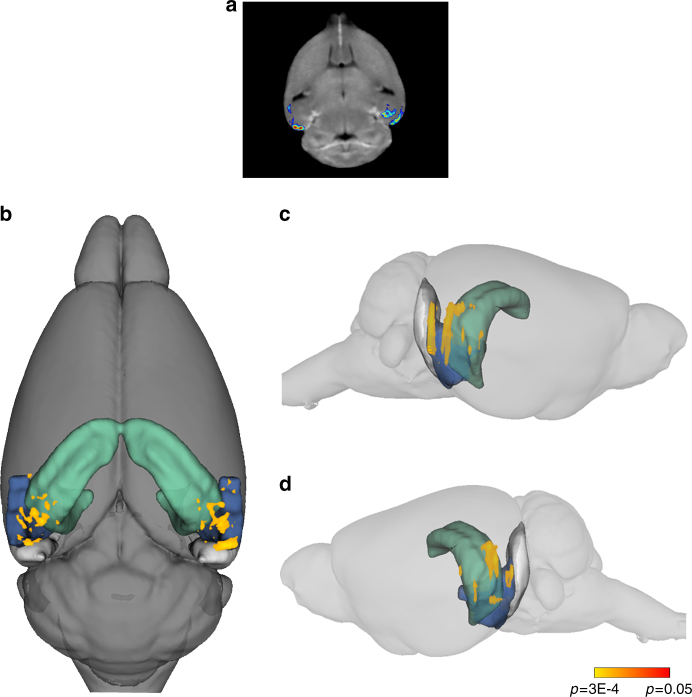
Whole-brain CBV maps were generated with the steady-state gadolinium-enhanced fMRI technique, as previously described40, followed by segmentation of the hippocampal formation region of interest (ROI) using operational criteria described previously45. An analysis of variance (ANOVA) revealed no significant differences for the younger mice, but a significant difference was detected in the hippocampal formation of the older mice. Compared to APOE3 mice, aged APOE4 mice were found to have higher CBV values in both the left and right hippocampal formations (Fig. 1), with the majority of the hypermetabolism centered around the lateral region of the EC, with additional hypermetabolism detected in the medial EC, the subiculum, and the CA1 region of the hippocampus (Fig. 1b, c).
Fig. 1.
APOE4 is associated with entorhinal cortex hypermetabolism in aged mice. fMRI analysis was performed on aged APOE mice (mean age = 20 months; 7 APOE3 and 8 APOE4 males), and a voxel-based analysis was performed to generate CBV maps of the whole brain of each mouse, followed by co-registration of each image, ROI selection of the hippocampal formation, and statistical comparison between genotypes by a Student’s t test, followed by multiple test correction. a Voxel-based analyses revealed that aged APOE4 mice had increased CBV in the hippocampal formation compared with APOE3 mice, primarily centered around the EC. b 3D superior view of CBV increase detected in aged APOE4 vs. APOE3 mice, as indicated using a heat scale by p value. c Right sagittal view of the same analysis. d Left sagittal view of the same analysis. For each panel, the colored regions depict the hippocampus and subiculum in green, the lateral entorhinal cortex (LEC) in blue, and the medial entorhinal cortex (MEC) in gray
EC hyperexcitability in awake freely moving APOE4 mice
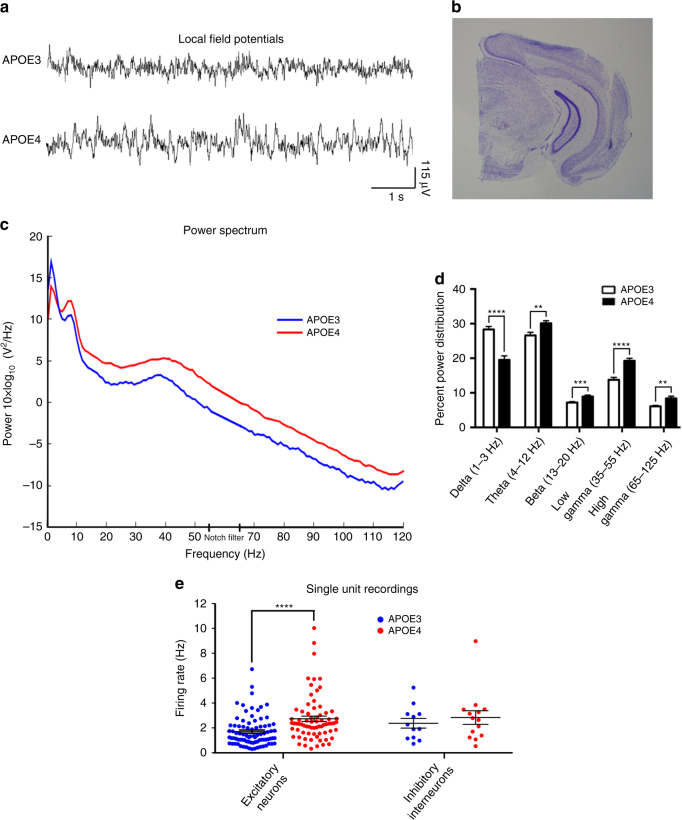
In order to confirm and expand upon the fMRI findings, in vivo electrophysiology was performed in awake, freely moving APOE mice using 16-channel microdrives, as previously described46 (Fig. 2). Briefly, tetrodes were inserted into the lateral region of the EC of aged APOE mice (mean age = 18 months; 5 APOE3 and 4 APOE4 males) using previously established coordinates (A/P: 3.4, L/M: 4.3, D/V: 2.5–3.5)47 that were independently verified in our laboratory. Recordings were performed twice daily for 10–20 min, and the depth of the tetrode was increased by 100 µm every 2–3 days. To determine the local field potential (LFP) and single unit activity in the EC, recordings from depths of 3.1–3.3 mm were pooled and quantified. For LFP measurements, a speed filter of 5–20 cm/s was applied. As shown in Fig. 2c, d, we observed APOE4-specific increases in theta, beta, and gamma oscillations of awake, freely moving mice, indicative of an increase in neuronal activity in this region.
Fig. 2.
APOE4 is associated with increased neuronal activity in awake freely moving mice. In vivo electrophysiology was performed on aged APOE mice (mean age = 18 months; 5 APOE3 and 4 APOE4 males) using a 16-channel microdrive placed directly into the LEC. a Representative local field potentials (LFPs) in awake, freely moving (5–20 cm/s) APOE3 and APOE4 mice, recorded at a depth of 3.3 mm from the brain surface. b A Nissl-stained brain section from a mouse used in this experiment, showing a track mark made by the implanted tetrode, which was descended to a depth of 3.3 mm from the brain surface when the mouse was killed. c Power spectra of LFPs recorded at a depth of 3.1–3.3 mm from the brain surface in freely moving (5–20 cm/s) APOE3 and APOE4 mice. (Frequencies between 55–65 Hz were notch-filtered to remove electrical noise and are extrapolated in the graph using straight lines.) d Percent power distributions comparing LFPs recorded at a depth of 3.1–3.3 mm from the brain surface in freely moving (5–20 cm/s) APOE3 and APOE4 mice (mean values and SEM). A Student’s t test with Welch’s correction was used for statistical comparison between genotypes (delta, p < 0.0001; theta, p = 0.0028; beta, p = 0.0008; low gamma, p < 0.0001; high gamma, p = 0.0015). e Single unit recordings detected at a depth of 3.1–3.3 mm from the brain surface in APOE3 and APOE4 mice, separated into putative excitatory neurons and interneurons based on peak size (black lines represent mean values and SEM). An increased firing rate of excitatory neurons (p < 0.0001) was observed using a Student’s t test with Welch’s correction. (**p < 0.01, ***p < 0.001, ****p < 0.0001)
In order to identify the source of these observed increases in theta, beta, and gamma oscillations, we analyzed single unit recordings from this same region in APOE3 and APOE4 mice. Putative excitatory neurons and inhibitory interneurons were distinguished based on spike width, as previously described48. As shown in Fig. 2e, we observed an increase in the firing rates of excitatory neurons in the EC of APOE4 mice (2.73 ± 0.22 Hz) vs. APOE3 mice (1.70 ± 0.13 Hz). This was based on the firing rates of 155 total excitatory neurons (83 from APOE3 and 72 from APOE4) detected at a depth of 3.1–3.3 mm. We did not observe a significant difference in the firing rates of inhibitory interneurons in APOE4 vs. APOE3 mice. However, we were only able to detect 26 total inhibitory interneurons (12 from APOE3 and 14 from APOE4) at the target depths, which is likely insufficient to accurately assess the activity of inhibitory interneurons in this region.
Increased energy-related metabolites in APOE4 EC
To investigate the neuromolecular basis of the observed hyperactivity in the EC, we performed untargeted and targeted metabolite profiling on brain tissues from aged APOE mice (14–15 months old; 8 APOE3/3, 9 APOE3/4, and 7 APOE4/4 males) using a previously described mass spectrometry (MS) platform49. The EC region was compared to the primary visual cortex (PVC), which is relatively resistant to AD pathology44, 50, 51. Small-molecule metabolites (50–1000 Da) from the EC and the PVC were extracted and analyzed by MS, and bioinformatics was performed to compare the levels of both untargeted (Supplementary Tables 1 and 2) and targeted (Tables 1 and 2) metabolites. An initial analysis of untargeted metabolites showed that these metabolites clustered independently of each other based on brain region and APOE genotype (Supplementary Fig. 2b, c). Differential expression analysis (false-positive discovery rate (FDR) <0.05) between the APOE3 and APOE4 genotypes revealed robust differences in a wide range of untargeted metabolites (Supplementary Fig. 2d).
Table 1.
Differentially expressed targeted metabolites from the EC of APOE4 vs. APOE3 mice
| Metabolite | Regulation in E4/4 | Fold change | P-value | FDR | Detection mode | CAS number |
|---|---|---|---|---|---|---|
| Fatty acids | ||||||
| Gamma-linolenic acid | Down | 1.31 | 1.19E−03 | 0.093 | Positive | 506-26-3 |
| Myristic acid | Down | 3.92 | 0.004 | 0.118 | Positive | 544-63-8 |
| Docosahexaenoic acid | Up | 1.36 | 0.011 | 0.154 | Positive | 6217-54-5 |
| Stearic acid | Down | 1.69 | 0.011 | 0.154 | Positive | 57-11-4 |
| 12-Hydroxydodecanoic acid | Down | 1.36 | 0.021 | 0.213 | Positive | 505-95-3 |
| Arachidic acid | Down | 1.40 | 0.037 | 0.230 | Negative | 506-30-9 |
| Palmitic acid | Down | 1.32 | 0.049 | 0.243 | Negative | 57-10-3 |
| Oligosaccharides | ||||||
| Trisaccharide | Up | 1.99 | 0.001 | 0.049 | Negative | 512-69-6 |
| Tetrasaccharide | Up | 2.24 | 0.015 | 0.169 | Negative | 10094-58-3 |
| Disaccharide | Up | 2.20 | 0.015 | 0.169 | Negative | 63-42-3 |
| Vitamin and vitamin derivatives | ||||||
| Phylloquinone | Up | 1.47 | 0.003 | 0.102 | Positive | 84-80-0 |
| Tocopherol | Up | 2.74 | 0.005 | 0.123 | Negative | 59-02-9 |
| Dehydroascorbic acid | Up | 1.76 | 0.008 | 0.134 | Positive | 490-83-5 |
| Energy-related metabolites | ||||||
| Inosine 5′-monophosphate (IMP) | Up | 2.26 | 0.003 | 0.0720 | Negative | 131-99-7 |
| d-Fructose 6-phosphate | Up | 1.10 | 0.011 | 0.169 | Negative | 643-13-0 |
| Succinoadenosine | Up | 1.47 | 0.011 | 0.169 | Negative | 4542-23-8 |
| Carnitine | Up | 1.27 | 0.021 | 0.213 | Positive | 541-15-1 |
| Citric acid/isocitric acid | Up | 1.24 | 0.028 | 0.230 | Negative | 77-92-9/1637-73-6 |
| Malic acid | Up | 1.16 | 0.037 | 0.230 | Negative | 97-67-6 |
| ATP | Up | 1.24 | 0.049 | 0.243 | Negative | 56-65-5 |
| Cholesterol metabolites | ||||||
| Lanosterol | Up | 2.27 | 0.015 | 0.169 | Negative | 79-63-0 |
| Cholesteryl acetate | Up | 2.29 | 0.015 | 0.169 | Negative | 604-35-3 |
| Amino acids | ||||||
| Leucine | Up | 1.26 | 0.015 | 0.169 | Negative | 61-90-5 |
| Proline | Up | 1.12 | 0.037 | 0.230 | Negative | 147-85-3 |
| Glycine | Up | 1.10 | 0.037 | 0.230 | Negative | 56-40-6 |
| Tryptophan metabolites | ||||||
| Quinaldic acid | Up | 1.68 | 0.001 | 0.049 | Negative | 93-10-7 |
| Kynurenine | Up | 2.49 | 0.015 | 0.169 | Negative | 2922-83-0 |
| Kynurenic acid | Up | 1.32 | 0.049 | 0.243 | Negative | 492-27-3 |
| Cysteine and methionine metabolites | ||||||
| S-Adenosylhomocysteine | Up | 1.15 | 0.021 | 0.213 | Positive | 979-92-0 |
| Histidine metabolism | ||||||
| Carnosine | Up | 1.31 | 0.037 | 0.230 | Negative | 305-84-0 |
| Arginine and proline metabolites | ||||||
| 4-Oxoproline | Up | 1.30 | 0.003 | 0.102 | Positive | 4347-18-6 |
| Tyrosine metabolites | ||||||
| Vanylglycol (MHPG) | Up | 1.64 | 0.011 | 0.169 | Negative | 67423-45-4 |
| Tyramine | Up | 1.06 | 0.028 | 0.216 | Positive | 51-67-2 |
| Thymidine | Down | 1.49 | 0.028 | 0.216 | Positive | 50-89-5 |
| Uracil | Down | 1.38 | 0.049 | 0.243 | Negative | 66-22-8 |
| Purine metabolism | ||||||
| GMP | Up | 1.14 | 0.028 | 0.230 | Negative | 85-32-5 |
| Miscellaneous | ||||||
| Hydroxybutyric acid | Up | 1.35 | 0.002 | 0.055 | Negative | 5094-24-6 |
| Methylglutarylcarnitine | Up | 2.42 | 0.005 | 0.134 | Positive | 102673-95-0 |
| Trimethylamine N-oxide | Up | 2.65 | 0.021 | 0.213 | Positive | 1184-78-7 |
| 2-Hydroxypyridine | Up | 1.36 | 0.037 | 0.275 | Positive | 142-08-5 |
| N-Acetylneuraminic acid | Up | 1.23 | 0.037 | 0.230 | Negative | 131-48-6 |
Table 2.
Differentially expressed targeted metabolites from the PVC of APOE4 vs. APOE3 mice
| Metabolite | Regulation in E4/4 | Fold change | P-value | FDR | Detection mode | CAS number |
|---|---|---|---|---|---|---|
| Fatty acids | ||||||
| Gamma-linolenic acid | Down | 1.19 | 0.003 | 0.068 | Positive | 506-26-3 |
| Palmitic acid | Down | 1.32 | 0.021 | 0.212 | Negative | 57-10-3 |
| 10-Hydroxydecanoate | Up | 2.22 | 0.015 | 0.195 | Positive | 1679-53-4 |
| Docosahexaenoic acid | Down | 2.03 | 0.021 | 0.212 | Negative | 6217-54-5 |
| Arachidonic acid | Down | 1.45 | 0.021 | 0.212 | Negative | 506-32-1 |
| Oligosaccharides | ||||||
| Trisaccharide | Up | 1.82 | 0.015 | 0.212 | Negative | |
| Tetrasaccharide | Up | 2.70 | 0.021 | 0.212 | Negative | |
| Vitamin and vitamin derivatives | ||||||
| Dehydroascorbic acid | Up | 2.03 | 0.001 | 0.062 | Positive | 490-83-5 |
| Phylloquinone | Up | 1.90 | 0.001 | 0.062 | Positive | 84-80-0 |
| Ascorbic acid | Up | 105.84 | 0.005 | 0.192 | Negative | 50-81-7 |
| Tocopherol | Up | 2.28 | 0.008 | 0.192 | Negative | 59-02-9 |
| Energy-related metabolites | ||||||
| Acetylcarnitine | Up | 1.40 | 0.003 | 0.068 | Positive | 3040-38-8 |
| Carnitine | Up | 1.31 | 0.011 | 0.154 | Positive | 541-15-1 |
| Coenzyme A (CoA) | Up | 2.82 | 0.028 | 0.282 | Positive | 85-61-0 |
| Pyruvate | Up | 1.43 | 0.028 | 0.238 | Negative | 127-17-3 |
| Cholesterol metabolites | ||||||
| Taurodeoxycholate | Up | 1.94 | 0.003 | 0.068 | Positive | 516-50-7 |
| Cholesteryl acetate | Up | 2.24 | 0.008 | 0.192 | Negative | 604-35-3 |
| Lanosterol | Up | 2.50 | 0.011 | 0.192 | Negative | 79-63-0 |
| Amino acids | ||||||
| Tyrosine | Up | 1.25 | 0.037 | 0.270 | Negative | 60-18-4 |
| Serine | Down | 1.18 | 0.049 | 0.270 | Negative | 56-45-1 |
| Glutamate | Down | 1.08 | 0.049 | 0.282 | Positive | 56-86-0 |
| Cysteine and methionine metabolites | ||||||
| Pterin | Down | 3.91 | 0.002 | 0.192 | Negative | 2236-60-4 |
| Histidine metabolites | ||||||
| Urocanic acid | Down | 1.34 | 0.021 | 0.212 | Negative | 104-98-3 |
| Carnosine | Up | 1.32 | 0.028 | 0.282 | Positive | 305-84-0 |
| Arginine and proline metabolites | ||||||
| Phosphocreatine | Up | 1.62 | 0.037 | 0.282 | Positive | 67-07-2 |
| 4-Guanidinobutyric acid | Down | 1.09 | 0.049 | 0.270 | Negative | 463-00-3 |
| Threonine metabolites | ||||||
| 2-Ketobutyric acid | Up | 1.43 | 0.021 | 0.212 | Negative | 600-18-0 |
| Tyrosine metabolites | ||||||
| Homovanillic acid | Down | 1.30 | 0.049 | 0.270 | Negative | 306-08-1 |
| Riboflavin metabolites | ||||||
| 2,4-Dihydroxypteridine (lumazine) | Up | 2.07 | 0.008 | 0.154 | Positive | 487-21-8 |
| Flavin adenine dinucleotide (FAD) | Up | 1.19 | 0.021 | 0.212 | Negative | 146-14-5 |
| Lumichrome | Down | 1.71 | 0.049 | 0.282 | Positive | 1086-80-2 |
| Pyrimidine metabolites | ||||||
| 2-Aminoisobutyric acid | Up | 1.38 | 0.011 | 0.154 | Positive | 62-57-7 |
| UMP | Up | 1.22 | 0.049 | 0.270 | Negative | 58-97-9 |
| CMP | Up | 1.29 | 0.049 | 0.282 | Positive | 63-37-6 |
| Miscellaneous | ||||||
| Lipoic acid | Up | 3.76 | 0.005 | 0.192 | Negative | 1077-28-7 |
| Thiourea | Down | 1.45 | 0.008 | 0.192 | Negative | 62-56-6 |
| Butyrylcarnitine | Up | 1.24 | 0.011 | 0.154 | Positive | 25576-40-6 |
| Hydroxybutyric acid | Up | 1.49 | 0.011 | 0.192 | Negative | |
| Indoxyl sulfate | Down | 2.12 | 0.021 | 0.212 | Negative | 2642-37-7 |
| 3-Hydroxymethylglutaric acid | Up | 1.27 | 0.028 | 0.238 | Negative | 503-49-1 |
| 2-Hydroxypyridine | Up | 1.45 | 0.037 | 0.282 | Positive | 142-08-5 |
| Maleic acid | Up | 1.92 | 0.037 | 0.270 | Negative | 110-16-7 |
| N-Acetylneuraminic acid | Up | 1.17 | 0.037 | 0.282 | Positive | 131-48-6 |
| N-Methylglutamic acid | Down | 1.19 | 0.049 | 0.270 | Negative | 35989-16-3 |
| Cytidine diphosphate choline (CDP choline) | Down | 1.17 | 0.049 | 0.282 | Positive | 987-78-0 |
| Glutathione | Up | 1.49 | 0.049 | 0.282 | Positive | 70-18-8 |
The targeted analysis, in which metabolites were matched against a proprietary database of 626 biologically relevant metabolites, revealed APOE4-specific differential expression (p < 0.05) of several important metabolite groups in the EC (Table 1), including fatty acids (gamma-linoleic acid, myristic acid, docosahexaenoic acid, 12-hydroxydodecanoic acid, stearic acid, arachidic acid, and palmitic acid; all downregulated in APOE4 EC except docosahexaenoic acid), vitamin or vitamin derivatives (phylloquinone, dehydroascorbic acid, and tocopherol; all upregulated in APOE4 EC), and C6H12O6-subunit containing oligosaccharides (disaccharide, trisaccharide, and tetrasaccharide; all upregulated in APOE4 EC). The majority of these fatty acids, vitamins and oligosaccharides were similarly regulated in the PVC (Table 2).
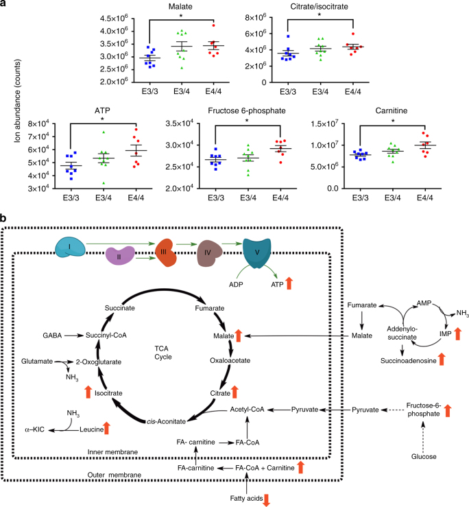
Critical to this study, we also observed an APOE4-specific upregulation of a number of mitochondrial metabolites that are involved in cellular energy metabolism (Fig. 3). These include several TCA-cycle metabolites (malate, citrate, and isocitrate), fructose-6-phosphate, which is an important glycolysis intermediate, and carnitine, which is required for the transport of fatty acids into the mitochondrial matrix for energy utilization via beta-oxidation. Importantly, we also observed an APOE4-specific upregulation of ATP. In addition, we observed an APOE4-specific upregulation of several metabolites that are involved in mitochondria-related ammonia regulation, including leucine, which is used for ammonia shuttling between astrocytes and neurons, and inosine monophosphate and succinoadenosine, which are involved in the ammonia-generating purine nucleotide cycle (see Fig. 3b for an overall schematic). With the exception of carnitine, which was upregulated in both the EC and PVC and may play a role in the decreased levels of various fatty acids observed in these regions, the other energy-related metabolites were only found to be upregulated in the EC. This APOE4-specific increase in energy-related metabolites in the EC represents a molecular correlate to the observed hypermetabolism/hyperexcitability revealed by the previously described fMRI and in vivo electrophysiological experiments.
Fig. 3.
APOE4 is associated with increased expression of small molecules related to energy metabolism. Targeted metabolite profiling was performed on small-molecule metabolites extracted from the EC and PVC of aged APOE mice (14–15 months old; 8 APOE3/3, 9 APOE3/4, and 7 APOE4/4 males). a Scatter dot plots depicting metabolite abundance in the EC for five energy-related metabolites found to be upregulated (Mann–Whitney; malate, p = 0.037; citrate/isocitrate, p = 0.028; ATP, p = 0.049; fructose-6-phosphate, p = 0.011; carnitine, p = 0.021) in APOE4 EC (black lines represent mean values and SEM). b Schematic of the energy metabolism-related metabolites found to be dysregulation in APOE4 EC. (*p ≤ 0.05)
Hyperexcitability and reduced inhibitory tone in APOE4 mice
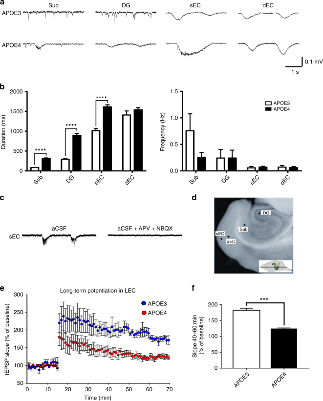
In order to further study the basis of APOE4-associated hyperactivity, we performed in vitro electrophysiology on 400 μM thick horizontal slices consisting of ventral hippocampus, subicular complex, entorhinal, perirhinal, and temporal cortices, prepared as previously described52, from aged APOE mice (mean age = 20 months; 5 APOE3 and 4 APOE4 males). First, we investigated the overall excitability in these slices by placing electrodes in the superficial (II/III) and deep (V/VI) layers of the lateral EC, as well as in the subiculum and DG, from which we recorded spontaneous extracellular field potentials (sEFPs) at 34 °C (Fig. 4). sEFPs occurring at different frequencies were detected (Fig. 4a, b). Compared to the APOE3 mice, superficial layers of the EC had significantly longer sEFPs in APOE4 mice (p < 0.0001), with no differences in frequency (p = 0.58), while no significant differences were observed in the duration of sEFPs in deep layers of the EC between APOE3 and APOE4 mice (p = 0.11). Additionally, we observed significantly longer sEFPs in both the subiculum (p < 0.0001) and the DG (p < 0.0001) of APOE4 mice, with no significant differences in the frequency in the subiculum (p = 0.29) or the DG (p = 0.85). In every case, we blocked transmission by adding (2R)-amino-5-phosphonovaleric acid (APV, 30 μM) and 2,3-dihydroxy-6-nitro-7-sulfamoyl-benzo[f]quinoxaline-2,3-dione (NBQX, 10 μM) to the bath, demonstrating that these are mainly synchronized glutamatergic synaptic events (Fig. 4c). Given the afferent projections from the superficial layers of the EC to the Sub and DG53, 54, it is possible that the increased activity observed in the subiculum and DG are a downstream result of the observed EC activation, as has been shown previously in the case of EC to DG activation53.
Fig. 4.
APOE4 is associated with abnormal excitability and synaptic plasticity in the hippocampal formation. In vitro electrophysiology was performed on horizontal hippocampal brain slices from aged APOE mice (mean age = 20 months; 5 APOE3 and 4 APOE4 males). a Representative traces of spontaneous extracellular field potentials (sEFPs) recorded in the subiculum (Sub), the dentate gyrus (DG), superficial layers (II/III) of the EC (sEC), and deep layers (V/VI) of the EC (dEC) in APOE3 and APOE4 mice. b Event durations and frequencies comparing APOE3 and APOE4 mice in the different regions recorded (mean values and SEM). Event durations in the Sub, DG, and sEC were found to be increased in aged APOE4 mice (Mann–Whitney; p < 0.001). c Representative sEFPs in APOE4 sEC before and after bath application (25 min) of AP-V (30 μM) and NBQX (10 μM). d A representative horizontal hippocampal brain slice used in this study, with asterisks denoting the location where each electrode was placed and the insert depicting the location of the horizontal slice in the mouse brain. e Cortico-cortical LTP in superficial EC induced by high-frequency stimulation (HFS, 3 trains of 100 pulses at 100 Hz, 10 s interval). Stimulation electrode was placed in lateral EC layer II, and the recording electrode was placed in lateral EC layers III/II. Stimulus intensity was selected to be 50–60% of the maximum amplitude of the fEPSP observed in the input–output curve, and a baseline for 15 mins was obtained before HFS. The slope of the initial component of the fEPSP (10–90%) was normalized to baseline and compared between APOE3 and APOE4 mice (repeated measures ANOVA; F (1,6) = 15.001, p = 0.008). f Mean value of potentiation between 40 and 60 min after LTP induction, comparing APOE3 and APOE4 (t (6) = 3.878, p = 0.008). (**p < 0.01)
We also studied evoked EC synaptic activity by stimulating EC layer II and recording at EC layer II/III (Fig. 4e, f). After obtaining an input–output relationship and an appropriate baseline, we elicited synaptic long-term potentiation (LTP) by high-frequency stimulation (HFS) in EC layer II, as reported previously55. The mean potentiation (late phase) elicited by HFS in APOE4 mice between 40 and 60 min after HFS was 140 ± 13%, which was significantly lower than in APOE3 mice (190 ± 15%), suggesting that the electrophysiological differences observed in the EC result in impaired long-term synaptic plasticity in this region.
In order to understand the mechanism responsible for the observed hyperactivity, we performed a series of whole-cell patch-clamp experiments in layer II EC of aged APOE mice (mean age = 20 months). Excitatory neurons in this region fall into two distinct electrophysiological and morphological categories: the pyramidal neurons and non-pyramidal neurons such as stellate and fan cells56, 57. We concentrated on pyramidal neurons here (n = 6 cells per group). Current clamp recordings demonstrated that APOE3 mice had larger amplitude and wider evoked action potentials (with constant current injection, see Methods section) as compared to APOE4 mice (Supplementary Fig. 3) (amplitude F = 88.0, p < 0.0001 and width F = 19.9, p < 0.001). Additionally, we induced focal seizure-like discharges (fSLDs) by direct pressure application of NMDA (1 mM) for 200 ms in the vicinity of the recorded neurons (Supplementary Fig. 4a, b). Simultaneously, calcium-induced fluorescence changes were also monitored upon NMDA application. The results revealed that NMDA-evoked fSLDs were not significantly different between the two groups in the multiple measurements obtained (Supplementary Fig. 4c–e). NMDA-evoked calcium transients, evaluated by changes in fluorescence (ΔF) in the soma of EC layer II pyramidal neurons, were also similar between the two groups (Supplementary Fig. 4f). These data suggest that the observed increases in sEFPs in the EC of aged APOE4 mice are not due to increased excitability of pyramidal cells of this region and may point to a defect in inhibitory signaling as the primary source of this hyperexcitability. Moreover, the reduced amplitude and narrower evoked action potentials observed in the APOE4 mice may represent an adaptive response to the decreased inhibitory tone of the EC in these mice.
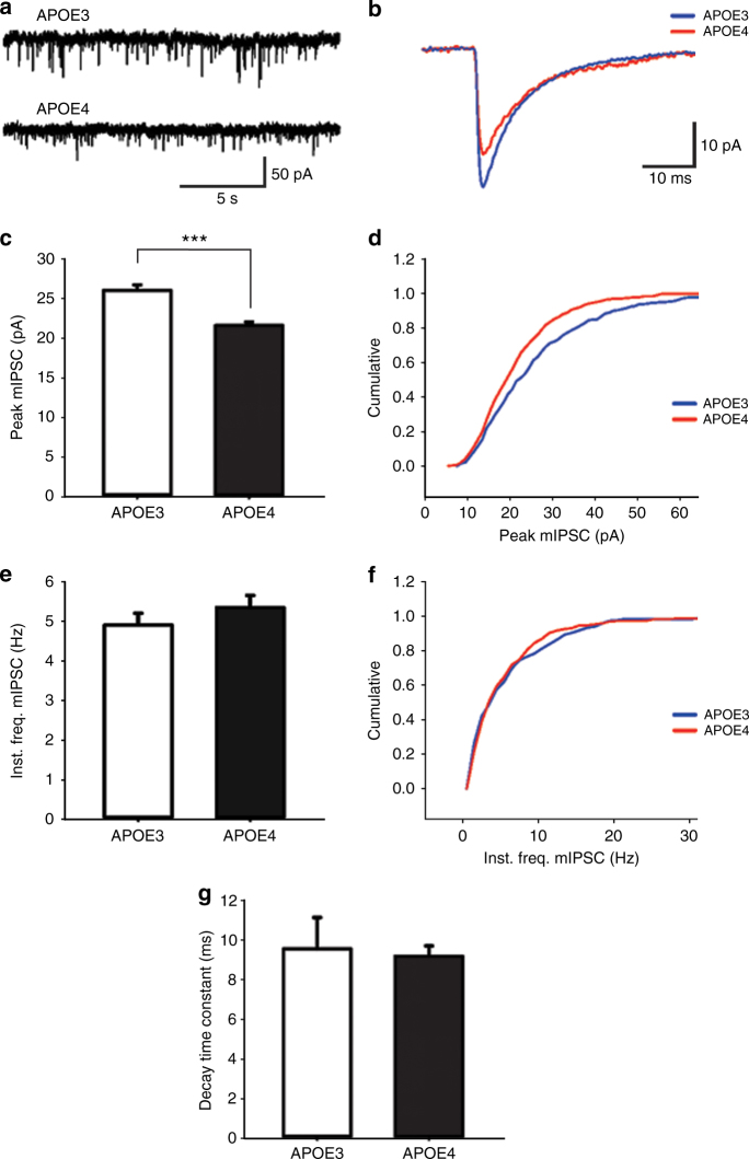
In order to investigate this possibility, we recorded miniature inhibitory post-synaptic currents (mIPSCs) in layer II EC pyramidal neurons (n = 7 cells from APOE3 and n = 8 cells from APOE4) of aged APOE mice (mean age = 20 months) (Fig. 5a, b). The peak amplitude of mIPSCs were significantly smaller in the APOE4 pyramidal neurons as compared to APOE3 neurons (Fig. 5c). Cumulative probability distributions showed that the entire distribution of mIPSC amplitudes were shifted toward smaller values for APOE4 mice as compared to APOE3 (Fig. 5d). Conversely, a comparison of instantaneous frequencies revealed no significant differences between groups (Fig. 5e, f). Individual measurements of decay time constant also showed no significant differences between conditions (Fig. 5g). The lack of effect on frequency and kinetics suggests that there is no significant difference between APOE4 and APOE3 in the spatial segregation of inhibitory inputs onto the pyramidal neurons, and in consequence, no difference in electrotonic filtering. The present data suggest that the observed difference in APOE4 mIPSC amplitude is through a post-synaptic change, either in receptor (GABA-A) number or function, or in overall synaptic number or morphology. We also cannot rule out the possibility that there may be additional presynaptic changes, such as the GABA content in synaptic vesicles. However, this possibility of decreased vesicular GABA content may be difficult to evaluate due to the fact that miniature release is highly variable from synapse to synapse58. In summary, mIPSC data indicate that the action potential-independent background synaptic inhibition in layer II of EC is decreased in APOE4 as compared to APOE3, and differences in the properties of such inhibition can explain the hyperexcitability observed in the EC of APOE4 mice.
Fig. 5.
APOE4 is associated with decreased background synaptic inhibition in layer II entorhinal cortex pyramidal neurons. Miniature inhibitory post-synaptic currents (mIPSCs) were recorded using the patch-clamp whole-cell modality on pyramidal cells (n = 7 cells from APOE3 and n = 8 cells from APOE4) in the layer II EC of aged APOE mice (mean age = 20 months). a Representative traces of mIPSCs of layer II EC pyramidal neurons of APOE3 and APOE4 mice from recordings in voltage-clamp modality (holding at −65 mV) in the presence of APV (10 µM), NBQX (16 µM), and TTX (1 µM). b Representative average mIPSCs from a 3 min recording of an APOE3 and APOE4 mouse. c Plotted are mean + SEM values of peak amplitude of mIPSCs in the different genotypes as indicated (APOE3 26.09 ± 0.73 pA; APOE4 21.6 ± 0.48 pA; p < 0.001). d Cumulative probability distribution of peak amplitude in APOE3 and APOE4 mice, showing lower values for APOE4 in the entire distribution. e Shown are mean + SEM values of instantaneous frequencies in the different genotypes (APOE3 4.92 ± 0.3 Hz; APOE4 5.36 ± 0.31 Hz; t(748) = −1.035, p = 0.301). f Cumulative probability distribution of instantaneous frequencies in APOE3 and APOE4 mice, showing no significant differences. g Plotted are mean + SEM values of decay time constant in both groups (APOE3 9.56 ± 1.61 ms; APOE4 9.19 ± 0.53; p = 0.819). (***p < 0.001)
Lack of overt amyloid or tau pathology in APOE4 mice
The brains of APOE4 mice have been reported to have slightly elevated levels of Aβ and phosphorylated tau59, and older mice (over 18 months) develop some vascular amyloid deposits in the frontal cortex and hippocampus60. To examine whether this was apparent in the EC of the aged APOE4 mice used for the imaging studies, we performed postmortem studies in which brain sections were immunolabeled with an antibody recognizing murine APP/Aβ (4G8) and an antibody against murine phospho-tau (CP13). Results showed no evidence of AD-related histopathology anywhere in the brain (Supplementary Fig. 1a, b).
Discussion
Neuronal hyperactivity has been demonstrated in AD transgenic mouse models14–17, as well as in humans with MCI18–22 and in presynaptic carriers of FAD-mutations23, 24. In addition, numerous neuroimaging studies have sought to elucidate the effect of APOE4 on overall brain metabolism, but they have generated contradictory results33–35. We hypothesize that some of this contradiction may be due to varying degrees of incipient AD pathology in the brains of the study participants. Therefore, in order to investigate the effects of differential APOE gene variants on brain metabolism in a pathology-free environment, we performed CBV-fMRI in young and old transgenic mice expressing human APOE3 or APOE4. This analysis revealed hypermetabolism in the hippocampal formation of aged APOE4 mice, most notably in the EC. (Fig. 1). Subsequent in vivo electrophysiological analysis revealed an increase in activity in the EC of awake, freely moving aged APOE4 mice, with LFP recordings showing an APOE4-specific increase in theta, beta, and gamma oscillations, and single unit recordings showing an increase in the firing rates of excitatory neurons (Fig. 2). In addition, using an MS-based metabolite profiling platform, we observed an upregulation of numerous small molecules related to energy metabolism in the EC of aged APOE4 mice, including ATP (Fig. 3). Finally, in vitro electrophysiological analysis revealed increased durations of spontaneous synchronized events in superficial layers of the EC of aged APOE4 mice, as well as in the subiculum and DG, with a corresponding deficit in LTP in the superficial layers of the EC of these mice (Fig. 4). Additional in vitro electrophysiology revealed a decreased background inhibitory tone on EC pyramidal neurons, likely caused by reduced responsiveness to GABAergic inhibitory inputs.
Each of these four methods—fMRI, in vitro and in vivo electrophysiology, and metabolomics—have strengths and limitations, with each compensating for the limitations of the others. While fMRI is a powerful tool for detecting overall brain metabolism, the direct cause of the observed differences in brain metabolism is not always known. Electrophysiology, on the other hand, is a rigorous method for directly measuring neuronal activity; however, the molecular mechanisms of any detected activity differences can be difficult to discern. And while metabolite profiling can detect specific differences in the levels of hundreds of molecular species, the resulting effects on brain activity and morphology cannot be determined using this method. Cumulatively, however, when the results from these diverse techniques are viewed as a whole, they provide a cohesive and consistent picture of a hypermetabolic/hyperactive state that is present in the EC of aged APOE4 mice. In addition, the increased firing rate of excitatory neurons revealed by single unit in vivo electrophysiology, combined with the observation of decreased mIPSC amplitudes (but not frequency) recorded via in vitro electrophysiology, reveal a distinct mechanism of hyperactive excitatory neurons with decreased inhibitory tone, likely caused by reduced responsiveness to inhibitory inputs from GABAergic interneurons in the aged APOE4 EC.
These results corroborate and expand upon previous data that have been reported on these mice. For example, it has been reported that aged APOE4 mice demonstrate increased excitatory synaptic activity in the amygdala61, a region directly connected with the EC. Furthermore, APOE4 mice have been reported to have an age-associated seizure phenotype62, which may be a downstream consequence of the observed hyperactivity in the EC. In addition, a recent report has shown that APOE4 mice possess an age-dependent reduction in slow gamma oscillations during sharp wave ripples (SWRs)63, which are unique high-frequency bursts that occur primarily in the CA1 region of the hippocampus during periods of slow-wave sleep or awake immobility. Interestingly, the timing of SWRs have previously been shown to be affected by activity changes in the EC, either directly or by way of the DG64, suggesting that EC hyperactivity may play a role in the observed abnormalities in slow gamma oscillations during SWRs.
Increased brain activity has been shown to accelerate AD pathology. In addition to causing increased production and secretion of Aβ25–27, increased neuronal activity may also affect tau pathogenesis, as tau release from cells is increased in vivo and in vitro when agents such as picrotoxin are administered28, 29, and hyperstimulating manipulations can exacerbate tau pathology30. Given that the EC is one of the first regions to be affected by overt tauopathy in the earliest stages of human AD44, our data showing that APOE4 is associated with neuronal hyperactivity in this region could indicate that APOE4 may accelerate the accumulation and propagation of Aβ or tau (or both), thereby accelerating the onset of AD in APOE4 carriers. Intriguingly, EC hyperactivity has also been reported in non-demented adults with Down syndrome, another group at significant risk of developing AD65, suggesting that this may be a common mechanism among populations that are vulnerable to AD.
Functional imaging studies using fMRI40, 66, 67 or fluorodeoxyglucose positron emission tomography68, 69 have documented EC hypometabolism at various stages of AD, including a recent study from our lab70. Interestingly, APP metabolites such as Aβ have been shown to be synaptotoxic71–73, and tauopathy is known to correlate with cell loss74 and synaptic dysfunction75. As a decrease in CBV correlates with synaptic loss76, it is possible that the neurotoxic effects exerted by the overt accumulation of these pathological proteins could lead to the observed hypometabolism in AD. Given these observations, we hypothesize that APOE4-associated hyperactivity, particularly in the EC, may drive the accumulation of Aβ and tau, which would then lead to neurodegeneration and ultimately result in a hypoactive state once the disease process has reached a critical stage. In this way, the APOE4-associated hyperactivity reported here may act as a central mediator of AD pathogenesis among APOE4 carriers and may therefore represent a prime target for therapeutic intervention in AD.
Methods
Mice
Human APOE-targeted replacement mice were first developed by Sullivan et al.37, 38. All mice used in this study were treated in accordance with the National Institutes of Health Guide for the Care and Use of Laboratory Animals and approved by the Columbia University Medical Center and SUNY Medical Center Institutional Animal Care and Use Committee (IACUC). For the fMRI analysis, two groups of mice were utilized, one with a mean age of 8 months, and one with a mean age of 20 months. For the in vivo electrophysiology experiment, mice were aged to a mean age of 18 months. For metabolite profiling, mice possessed a mean age of 14.5 months. And for in vitro electrophysiology experiments, mice with a mean age of 20 months were utilized. All experiments were performed on male APOE mice.
Mouse fMRI
A steady-state gadolinium enhancement approach was used to generate CBV maps, as previously described40. Briefly, images were acquired with a Bruker AVANCE 400WB spectrometer outfitted with an 89-mm bore, 9.4-T vertical Bruker magnet (Oxford Instruments), a 30-mm inner-diameter birdcage radio frequency coil, and a shielded gradient system (100 G/cm) before and 37.5 min after intraperitoneal injections of the contrast agent gadolinium (10 mmol/kg). Mice were anesthetized using a gaseous mixture of 30% O2, 70% N2, and isoflurane (3% volume for induction, 1.1–1.5% for maintenance at 1 liter per min air flow, via a nose cone). T2-weighted images were acquired perpendicular to the hippocampal long axis with a fast-spin echo acquisition (time to repeat, 2000 ms; time to echo, 70 ms; rapid acquisition and relaxation enhancement factor, 16; in plane resolution, 86 m; slice thickness, 500 m). Relative CBV maps were generated according to the formula CBV ∞ ∆R2 = ln (Spre/Spost) / TE; where TE = effective echo time; Spre = signal before contrast; Spost = signal contrast agent reaches steady state. Relative CBV was then derived from this image by normalizing its signal to the mean CBV value present in internal jugular vein.
For voxel-based analysis, image processing was conducted using custom bash and MATLAB scripts. Volumes were co-registered into a group-wise template space using an inverse-inconsistent diffeomorphic co-registration algorithm, as described previously77. Briefly, the co-registration incorporates a stationary vector field that smoothly warps images in their native coordinate space while conserving topology. Furthermore, each iteration of the co-registration was optimized by a Jacobian weighting term that encodes local distortions generated by the warp. This method yields robust and accurate alignment of images within their native coordinate space. The co-registration consisted of three steps. Anatomical images were first aligned to a randomly selected image in their data set using a linear transformation with 12 degrees of freedom. Next, the linearly aligned images were used to determine the optimal diffeomorphic transformation necessary to warp each image in the data set to an unbiased template image. Each iteration of the pairwise diffeomorphic registration between each image and the template generated a normalized warp with an average deformation of zero, ensuring that the template image generated was not biased by relative local volume loss or expansion between groups. Finally, the linear transformation and nonlinear warps were applied to corresponding CBV maps for each subject.
CBV maps were analyzed using a general linear model implemented in SPM8 (Wellcome Department of Imaging Neuroscience). Data were modeled in a factorial framework with genotype as the between subject factor. Individual genotypes were contrasted using Student’s t test. Results were height thresholded and corrected for multiple comparisons at the cluster level using a Monte-Carlo simulation run for 10,000 iterations to yield a cluster volume corrected to p < 0.05. Thresholded t maps from individual group comparisons were then overlaid onto their respective groupwise templates in cross-section using 3DSlicer (www.slicer.org) and displayed as maximum intensity projections over volume ray cast mesh models of the groupwise template and hippocampal formation, also generated in 3DSlicer. Mesh models of the whole-brain template were generated from manual segmentations of individual mice, which were transformed into template space by applying the non-linear warp. Individual segmentations of the hippocampal formation were transformed similarly. The hippocampal formation ROI was segmented using operational criteria described previously45. Specifically, this ROI included high-resolution segmentation of the DG, CA3, CA2, CA1, subiculum, pre-subiculum, para-subiculum, medial EC, and lateral EC.
In vivo electrophysiology recordings
Custom-made, reusable 16-channel microdrives (Axona, UK) were constructed as described previously46. Tetrodes were created by twisting four 25 mm thick platinum-iridium wires (California wires) and heat bonding them. Four tetrodes were inserted into the inner cannula of the microdrive and connected to the wires of the microdrive. 1 day prior to surgery, the tetrodes were cut to an appropriate length and plated with a platinum solution until the impedance dropped to about 150 ohm. On the day of surgery, mice were anesthetized with isoflurane (1–2%) and monitored for depth of anesthesia before proceeding. Mice were then fixed within the stereotaxic frame with the use of zygomatic process cuff holders, after which an incision was made to expose the skull and about 3–4 jeweler’s screws were inserted into the skull to support the microdrive implant. An additional screw connected with wire was also inserted into the skull, which served as a ground/reference for LFP recordings. A 2 mm hole was made on the right hemisphere of the skull at 4.3 mm lateral and 3.4 mm posterior to bregma. Tetrodes were then lowered to about 2.5 mm from the surface of the brain (below dura). Dental cement was spread across the exposed skull and secured with the microdrive. Mice usually recovered within 24 h, after which the tetrodes were lowered and recording began. All recording depths reported are from the surface of the brain.
The mice explored a white box (45 × 45 cm) and underwent two recording sessions per day, with ≥4 h between sessions. Tetrode positions were moved not more than 100 µm at a time, and only after the last recording session of the day, allowing >12 h of stable electrode positioning prior to the next recording session. Neuronal signals from experimental mice were recorded using the Axona DacqUSB system. The signals were amplified 15,000–30,000 times and band pass-filtered between 0.8 and 6.7 kHz. LFPs were recorded from four channels of the electrodes. The LFP was amplified 15,000 times, low pass-filtered at 125 Hz, and sampled at 250 Hz. Notch filter was used to eliminate 60 Hz noise. The recording system tracked the position of the infrared LED on the head stage (sampling rate 50 Hz) by means of an overhead infrared video camera. Position data were speed-filtered, with only speeds of 5–20 cm/s used for LFP measurements. Tracking artifacts were removed by deleting samples greater than 100 cm/s and missing positions were interpolated with total durations <1 s, and then smoothing the path with a 21-sample boxcar window filter (400 ms; 10 samples on each side). For single unit recordings, a total of 155 excitatory neurons and 26 interneurons were detected at a depth of 3.1–3.3 mm in the nine mice used in this study. Putative excitatory cells were distinguished from putative interneurons based on waveform width (>300 µs), as described previously48. Investigators were blinded to genotype assignments during surgery, recordings, and single unit validation.
Metabolite profiling
Mice were killed by cervical dislocation to maintain the brain environment, and individual brain regions were immediately removed and snap-frozen on dry ice. Tissues were stored at −80°C prior to extraction. Metabolite extraction was performed using a methyl tert-butyl ether (MTBE)/methanol extraction protocol modified from previous reports78, 79. Briefly, individual EC or PVC tissues were homogenized in 400 μl of ice-cold methanol using a bead mill homogenizer (TissueLyser II, Qiagen) at 25 beats per s, 2× for 45 s each. Following homogenization, samples were incubated in 1200 μl of MTBE for 1 h at room temperature to separate organic-soluble lipids from aqueous-soluble lipids and other small molecules. Finally, 360 μl of ultrapure water was added (for a final ratio of 3:1:0.9 MTBE:methanol:water) to resolve the two liquid phases, and the samples were centrifuged at 10,000×g for 10 min. The lower aqueous phase and the upper organic phase was collected from each sample and stored in separate tubes, and the remaining protein pellets were resuspended in 25 mM ammonium bicarbonate, pH 8, with 2.5% SDS. A BCA protein assay was performed on each protein fraction, and both the aqueous phase and organic phase were normalized to their protein concentration equivalent with 50% and 100% methanol, respectively. All samples were then stored at −80°C prior to analysis.
Metabolite profiling was performed using an Agilent Model 1200 liquid chromatography system coupled to an Agilent Model 6230 time-of-flight mass analyzer as described previously49. Metabolite separation was accomplished using aqueous normal phase gradient chromatography on a Diamond Hydride column (Microsolv), with mobile phase buffer A consisting of 50% isopropanol and 0.025% acetic acid, and mobile phase buffer B consisting of 90% acetonitrile and 5 mM ammonium acetate. Each aqueous phase sample was analyzed in both positive and negative ion detection mode for comprehensive coverage of metabolites with a mass of 50–1000 Da. Prior to analysis, it was observed that one APOE3/4 PVC sample possessed a chromatographic error and was removed from all future analysis.
Following MS analysis, the raw data was analyzed using Agilent Profinder (version B.06.00) and Mass Profiler Professional (version 12.0) software. Briefly, Profinder performs batch recursive feature extraction, which consists of a first-pass identification of features (ions) that possess a common elution profile (e.g., identical mass-to-charge (m/z) ratios and retention times), followed by a recursive analysis to re-mine each sample for the presence of features that were identified in at least one sample. Feature extraction was limited to the top 2000 metabolites per ion detection mode, with each metabolite possessing a minimum abundance of 600 ion counts. Each feature was also manually validated following extraction (with investigators blinded to genotype assignments during validation), and re-integration was performed when necessary. In addition to this untargeted feature extraction, we also used ProFinder to perform a targeted feature extraction, matching features against a proprietary database of 626 biologically relevant metabolites whose mass and retention times were previously determined using pure chemical standards. For this analysis, we allowed for a maximum mass deviation of 10 mDa and a retention time deviation of 1 min, and identified features were manually validated following extraction. For both untargeted and targeted feature extraction, Profinder analysis was followed up by multivariate and differential expression analysis using Mass Profiler Professional. Principle component analysis and hierarchical clustering analysis were used to examine data sets for common expression patterns, expected and unexpected clusters, and the presence of outlying samples. Following differential expression analysis, metabolites identified as differentially expressed were further validated for chromatographic and biological accuracy, and non-validated metabolites were removed.
In vitro electrophysiological recordings
Mice were anesthetized with Ketamine/Xylazine (100/10 mg/kg). Mice were decapitated with an animal guillotine, and the brains were rapidly dissected out of the skull cavity. The cutting solution contained the following in mM: 130 potassium gluconate, 5 KCl, 20 HEPES acid, 25 glucose, 0.05 kynurenic acid, 0.05 EGTA-K, and pH equilibrated at 7.4 with KOH. Recordings were performed at an approximate position A/P −3.8 mm, M/L 3.0 mm, and D/V −5.6 mm (based on the C57Bl/6J mouse brain atlas http://www.mbl.org/atlas/atlas.php). The horizontal brain slices contained all the regions of the hippocampal formation: DG, CA3, CA1, subiculum, pre/para subiculum, lateral and medial EC and perirhinal cortices (this slice has an orientation that is similar to the middle hippocampus MRI slice used). After slicing, the tissue was placed in artificial CSF (aCSF) that contained the following in mM: 157 Na+, 136 Cl−, 2.5 K+, 1.6 Mg2+, 2 Ca2+, 26 HCO3−, and 11 d-glucose. Perfusion rate for aCSF during experiments was set at 1 ml/min with a peristaltic pump (Rabbit MiniPuls 2, Rainin Intruments Co.). Experiments were performed in an interface chamber (Fine Scientific Tools, Vancouver Canada). For recordings of sEFPs in the hippocampal formation, we used an extracellular aCSF containing 5 mM K+, which is known to reduce apparent synaptic failures and increase neuronal excitability, without promoting convulsant activity54. Slices were perfused with aCSF continuously bubbled with 95% O2/5% CO2, to maintain pH near 7.4 and the temperature was set at 34 °C. In all experiments, the slices were allowed to recover from the isolation procedure for at least 1.5 h before the beginning of the experiments. Borosilicate electrodes (1.5 mm outer diameter with filament, World Precision Instruments, Inc.) of 2–3 MΩ filled with 150 mM NaCl were used to measure LFPs. The stimulating electrode was connected to a stimulus isolation unit (Grass S88). An input–output relationship curve was obtained (basal synaptic transmission evaluation), and for the rest of the experiment, a stimulus evoking ~50–60% of the maximum field excitatory post-synaptic potential (fEPSP) response was selected. Extracellular recordings were performed using an Axoclamp 2B amplifier (Molecular Devices, Palo Alto, CA), filter (0.1 Hz–10 KHz using −6 dB/octave). Voltage signals were digitized and stored in a PC using a digidata 1200 A (Molecular Devices) for off line analysis. Data was collected for 20 min per regions per slice and analyzed using Neuromatic routines run in an Igor platform (Wavemetrics), with the threshold for events being a defection in amplitude 3 SD from noise and >20 ms in duration.
For the LTP induction, a stimulus as described above was used and a baseline was obtained (15 min with an inter-stimulus interval of 30 s). HFSs (3 trains of 100 pulses at 100 Hz, 10 seconds interval) were then applied, and responses were recorded for 50 min after tetanization and measured as fEPSP slope (10–90%), expressed as percentage of baseline. Basal fEPSP slopes, as well as the input–output relationships, were similar before stimulation, and they were normalized separately. To ensure that elicited fEPSP were indeed glutamatergic, at the end of each recording, NMDA- and AMPA-mediated currents were blocked with APV (30 μM) and NBQX (10 μM).
For the current clamp and NMDA-induced burst experiments, horizontal brain slices as described above were obtained. Pyramidal neurons (n = 6 cells per group) were selected for analysis. Glass pipettes were pulled from borosilicate glass (1.5 mm outer diameter with filament, World Precision Instruments, Inc). Pipette resistance was 4–5 MΩ. Intracellular solution was based on potassium gluconate (in mM): 166, MgCl2 4.6, HEPES acid 10, ATP-Na 4, GTP-Na 0.4, and pH was balanced at 7.3–7.4 with KOH. Oregon Green 488 BAPTA-1 (OG1; Thermo Fischer Scientific) was used as a non-ratiometric calcium indicator, and added to the intracellular solution (final concentration of 50 µM). Series resistance was compensated, and access resistance (10–20 MΩ) was monitored regularly. Liquid junction potential was calculated and compensated for the holding potential. Signals were acquired using an amplifier Axopatch 200B (Molecular Devices), digitized with a Digidata 1550B (Molecular Devices), and processed with pClamp 10 (Molecular Devices). Signals were filtered at 5 kHz and digitized at 10 kHz. Neurons were discarded if the leak current was larger than 100 pA at voltage holding of −70 mV, or if the access resistance was larger than 20 MΩ. In the whole-cell configuration, pyramidal neurons were recorded in current clamp mode, and responses to current injection hyperpolarizing and depolarizing were used as indicated in each experiment. Neuronal firing was compared between the groups by injecting 250 pA for 4 s. For calcium imaging, an OptoLED system (Cairn, UK) was used as the light source, and excitation light was provided by a 470-nm LED. Light signals were captured by an iXon DV887 EM-CCD camera (Andor, USA). Software developed by Andor (Solis) was used with an image acquisition of 20 ms-frame (effective pixel size after ×60 objective = 266.7 nm; 1 × 1 binning). An in-house routine developed in an IGOR Pro programming environment (WaveMetrics, Lake Oswego, OR) was used for the analysis of calcium signals. After focal application of NMDA (1 mM, 200 ms) calcium signals were measured by calculating the average fluorescence in ROIs as a function of time. The size of the ROI was chosen as to just enclose the apical dendrite. Values are expressed as the percentage of change in fluorescence with respect to control, ΔF/F 0 = 100 (F−F 0)/(F 0−B), where: F corresponds to the fluorescence signal from the ROI in the apical dendrite after the induction of the calcium transient at any given time; F 0 corresponds to the average basal fluorescence signal previous to the induction of the calcium transients; and B is the average value at each time point of the background fluorescence from four regions of the imaged field that do not contain any part of the dye-filled cell. A glass pipette filled with NMDA and a pressure system (Picospritzer II, General Valve Corporation) positioned near the recorded neurons was used to induce fSLDs in cells kept in current clamp, with no current injection. fSLDs were blocked completed by APV 10 uM.
For mIPSC recordings, a new group of mice of the same age were anesthetized with Ketamine/Xylazine, and horizontal brain slices were obtained, as described above. All recordings were performed from layer II EC. In whole-cell configuration, pyramidal neurons (n = 7 cells from APOE3 and n = 8 cells from APOE4) were recorded in voltage-clamp mode (Vh = −65 mV) in the presence of TTX (1 µM). A different internal solution (in mM: KCl 120, MgCl2 4.6, EGTA 10, CaCl2 1, HEPES acid 10, ATP-Na 4, GTP-Na 0.4) was used for these recordings. Additionally, AMPA receptors and NMDA receptors were blocked with bath applied NBQX (16 µM) and 2 APV (10 µM), respectively. At the end of the experiment, a GABA-A antagonist (picrotoxin, 200 µM) was used to confirm the recording of GABAergic synapses, as has been reported previously80. In every case, the synaptic currents were fully blocked by picrotoxin. The recorded mIPSCs were analyzed with Clampfit (Molecular Devices). Individual mIPSC events were detected with a template (threshold of 3.5) which was three times the SD from baseline noise and visually inspected to exclude artifacts. When data were pooled for this analysis, randomly selected 50 events were obtained from a continuous recording period of 3 min for each neuron. We have also included mean values in our analysis, as they more closely reflect the population distributions comprising our cumulative probability data. For all in vitro electrophysiology experiments, investigators were blinded to genotype assignment during recordings and analyses.
Histological analysis
Mouse brains were removed after transcardial perfusion, and drop-fixed with 4% paraformaldehyde overnight, followed by cryoptrotection in 30% sucrose in PBS for 16 h. Free-floating brain sections from brains sectioned in the horizontal plane were used for immunohistochemistry using SuperPicTure™ polymer detection kit (Zymed, San Francisco, CA). Sections were washed with PBS for 10 min, and then treated with 3% H2O2 in PBS for 10 min. The sections were then transferred to a microfuge tube that contained primary antibody diluted in PBS containing 0.3% Triton and 5% normal serum, and incubated at 4 °C overnight on a rotator. After three washes with PBS-T (0.1% Tween 20), the sections were incubated for 10 min with HRP polymer conjugate. Following three washes with PBS-T, the immunoreactive material was visualized using DAB as a chromogen. The stained sections were mounted on slides and inspected by light microscopy. For murine tau detection, antibody CP13 (pS202) was utilized (a gift from Dr. P. Davies). Monoclonal antibody 4G8 was used to detect murine APP/Aβ.
Statistical analysis
For each experiment, mouse numbers were selected to be similar to those used in previous experiments utilizing mouse fMRI52, 70, in vivo electrophysiology30, metabolite profiling81, 82, and in vitro electrophysiology52. For fMRI analysis, statistical analysis was performed using SPM8 software, as described above. For in vivo electrophysiological recordings, statistical analysis for LFP and single unit measurements was performed with Prism 6 (Graphpad) using an unpaired Student’s t test between groups with Welch’s correction. For metabolite profiling, differential expression analysis was performed using Mann–Whitney tests, with a Benjamini Hochberg correction applied to adjust for FDRs considering multiple group comparisons. For untargeted comparisons, reporting was restricted to those metabolites with an FDR-corrected p value of <0.05, while for targeted metabolite analyses, the window was widened to include metabolites with an uncorrected p value of <0.05. For in vitro electrophysiological recordings, statistical analysis for sEFP measurements was performed using non-parametric Mann–Whitney tests to generate p values. For LTP measurements, a repeated measures ANOVA with unequal variance was used to generate F and p values, and a Student’s t test with unequal variance was used to generate t and p values for the mean potentiation comparison. For current clamp and NMDA-induced burst experiments, we used one-way ANOVA for the statistical analysis. For mIPSC recordings, mIPSC amplitude, cumulative amplitudes, cumulative frequencies, instantaneous frequencies, and decay time constants were compared and analyzed (according to their distribution) using t test or Kolmogorov–Smirnov test (K–S) with the significance level set at p < 0.05.
Data availability
The metabolomics data generated during the study are available through MetaboLights: http://www.ebi.ac.uk/metabolights/MTBLS530. All other data are available from the authors upon request.
Electronic supplementary material
Acknowledgements
We acknowledge Dr. W.-H. Yu and Dr. A. Marty for advice on this project, as well as Dr. H. Fu for assistance with tissue collection, L. Piroli for assistance with genotyping, and Dr. G. Rodriguez for assistance with optimization of in vivo electrophysiological recordings. This work was supported by grants from NIA and NINDS to K.E.D. (AG048408 and NS071836), a grant from NIA to S.A.H. and K.E.D. (AG050425), grants from NICHD and NHLBI to S.S.G. (HD67244 and HL87062), fellowships from NIA (AG047797) and the Brightfocus Foundation (A2014200F) to T.N., grants from NIA to H.M. (AG051556) and S.A.S. (AG08702), and funding from Cure Alzheimer’s Fund to K.E.D. and T.N.
Author contributions
This study was designed and managed by T.N., S.L.A., U.K., S.A.H., H.M., S.A.S., and K.E.D. Animal care and breeding was performed by H.F. Mouse fMRI was performed by U.K., in the laboratory of S.A.S., and by C.F., in the laboratory of K.E.D. Histological analysis was performed by L.L., S.E., and A.A. In vitro electrophysiology was performed by S.L.A., L.B., and E.C.-P., in the laboratory of H.M. In vivo electrophysiology and analysis was performed by S.A.H., M.H., G.B., and V.S. MS analysis was performed by Q.C. and T.N., in the laboratory of S.S.G. MS bioinformatic analysis was performed by T.N., A.A., and E.G. Manuscript preparation was performed by T.N., with input from S.S.G., S.A.H., H.M., U.K., S.A.S., and K.E.D.
Competing interests
The authors declare no competing financial interests.
Footnotes
Electronic supplementary material
Supplementary Information accompanies this paper at doi:10.1038/s41467-017-01444-0.
Publisher's note: Springer Nature remains neutral with regard to jurisdictional claims in published maps and institutional affiliations.
Contributor Information
Herman Moreno, Email: Herman.Moreno@downstate.edu.
Scott A. Small, Email: sas68@columbia.edu
Karen E. Duff, Email: ked2115@columbia.edu
References
- 1.Farrer LA, et al. Effects of age, sex, and ethnicity on the association between apolipoprotein E genotype and Alzheimer disease. A meta-analysis. APOE and Alzheimer Disease Meta Analysis Consortium. JAMA. 1997;278:1349–1356. doi: 10.1001/jama.1997.03550160069041. [DOI] [PubMed] [Google Scholar]
- 2.Mahley RW, Rall SC., Jr. Apolipoprotein E: far more than a lipid transport protein. Annu. Rev. Genomics Hum. Genet. 2000;1:507–537. doi: 10.1146/annurev.genom.1.1.507. [DOI] [PubMed] [Google Scholar]
- 3.Han X. The role of apolipoprotein E in lipid metabolism in the central nervous system. Cell Mol. Life Sci. 2004;61:1896–1906. doi: 10.1007/s00018-004-4009-z. [DOI] [PMC free article] [PubMed] [Google Scholar]
- 4.Holtzman DM, Herz J, Bu G. Apolipoprotein E and apolipoprotein E receptors: normal biology and roles in Alzheimer disease. Cold Spring Harb. Perspect. Med. 2012;2:a006312. doi: 10.1101/cshperspect.a006312. [DOI] [PMC free article] [PubMed] [Google Scholar]
- 5.Bales KR, et al. Lack of apolipoprotein E dramatically reduces amyloid beta-peptide deposition. Nat. Genet. 1997;17:263–264. doi: 10.1038/ng1197-263. [DOI] [PubMed] [Google Scholar]
- 6.Castano EM, et al. Fibrillogenesis in Alzheimer’s disease of amyloid beta peptides and apolipoprotein E. Biochem. J. 1995;306(Pt 2):599–604. doi: 10.1042/bj3060599. [DOI] [PMC free article] [PubMed] [Google Scholar]
- 7.Rebeck GW, Reiter JS, Strickland DK, Hyman BT. Apolipoprotein E in sporadic Alzheimer’s disease: allelic variation and receptor interactions. Neuron. 1993;11:575–580. doi: 10.1016/0896-6273(93)90070-8. [DOI] [PubMed] [Google Scholar]
- 8.Schmechel DE, et al. Increased amyloid beta-peptide deposition in cerebral cortex as a consequence of apolipoprotein E genotype in late-onset Alzheimer disease. Proc. Natl Acad. Sci. USA. 1993;90:9649–9653. doi: 10.1073/pnas.90.20.9649. [DOI] [PMC free article] [PubMed] [Google Scholar]
- 9.Ma J, et al. Amyloid-associated proteins alpha 1-antichymotrypsin and apolipoprotein E promote assembly of Alzheimer beta-protein into filaments. Nature. 1994;372:92–94. doi: 10.1038/372092a0. [DOI] [PubMed] [Google Scholar]
- 10.Castellano JM, et al. Human apoE isoforms differentially regulate brain amyloid-beta peptide clearance. Sci. Transl. Med. 2011;3:89ra57. doi: 10.1126/scitranslmed.3002156. [DOI] [PMC free article] [PubMed] [Google Scholar]
- 11.Holtzman DM, et al. Apolipoprotein E isoform-dependent amyloid deposition and neuritic degeneration in a mouse model of Alzheimer’s disease. Proc. Natl Acad. Sci. USA. 2000;97:2892–2897. doi: 10.1073/pnas.050004797. [DOI] [PMC free article] [PubMed] [Google Scholar]
- 12.Huang Y. Abeta-independent roles of apolipoprotein E4 in the pathogenesis of Alzheimer’s disease. Trends Mol. Med. 2010;16:287–294. doi: 10.1016/j.molmed.2010.04.004. [DOI] [PubMed] [Google Scholar]
- 13.Wolf AB, et al. Apolipoprotein E as a beta-amyloid-independent factor in alzheimer’s disease. Alzheimers Res. Ther. 2013;5:38. doi: 10.1186/alzrt204. [DOI] [PMC free article] [PubMed] [Google Scholar]
- 14.Busche MA, et al. Decreased amyloid-beta and increased neuronal hyperactivity by immunotherapy in Alzheimer’s models. Nat. Neurosci. 2015;18:1725–7. doi: 10.1038/nn.4163. [DOI] [PubMed] [Google Scholar]
- 15.Busche MA, et al. Clusters of hyperactive neurons near amyloid plaques in a mouse model of Alzheimer’s disease. Science. 2008;321:1686–1689. doi: 10.1126/science.1162844. [DOI] [PubMed] [Google Scholar]
- 16.Davis KE, Fox S, Gigg J. Increased hippocampal excitability in the 3xTgAD mouse model for Alzheimer’s disease in vivo. PLoS ONE. 2014;9:e91203. doi: 10.1371/journal.pone.0091203. [DOI] [PMC free article] [PubMed] [Google Scholar]
- 17.Palop JJ, et al. Aberrant excitatory neuronal activity and compensatory remodeling of inhibitory hippocampal circuits in mouse models of Alzheimer’s disease. Neuron. 2007;55:697–711. doi: 10.1016/j.neuron.2007.07.025. [DOI] [PMC free article] [PubMed] [Google Scholar]
- 18.Hamalainen A, et al. Increased fMRI responses during encoding in mild cognitive impairment. Neurobiol. Aging. 2007;28:1889–1903. doi: 10.1016/j.neurobiolaging.2006.08.008. [DOI] [PubMed] [Google Scholar]
- 19.Kircher TT, et al. Hippocampal activation in patients with mild cognitive impairment is necessary for successful memory encoding. J. Neurol. Neurosurg. Psychiatry. 2007;78:812–818. doi: 10.1136/jnnp.2006.104877. [DOI] [PMC free article] [PubMed] [Google Scholar]
- 20.Bakker A, et al. Reduction of hippocampal hyperactivity improves cognition in amnestic mild cognitive impairment. Neuron. 2012;74:467–474. doi: 10.1016/j.neuron.2012.03.023. [DOI] [PMC free article] [PubMed] [Google Scholar]
- 21.Dickerson BC, et al. Increased hippocampal activation in mild cognitive impairment compared to normal aging and AD. Neurology. 2005;65:404–411. doi: 10.1212/01.wnl.0000171450.97464.49. [DOI] [PMC free article] [PubMed] [Google Scholar]
- 22.Miller SL, et al. Hippocampal activation in adults with mild cognitive impairment predicts subsequent cognitive decline. J. Neurol. Neurosurg. Psychiatry. 2008;79:630–635. doi: 10.1136/jnnp.2007.124149. [DOI] [PMC free article] [PubMed] [Google Scholar]
- 23.Quiroz YT, et al. Hippocampal hyperactivation in presymptomatic familial Alzheimer’s disease. Ann. Neurol. 2010;68:865–875. doi: 10.1002/ana.22105. [DOI] [PMC free article] [PubMed] [Google Scholar]
- 24.Sepulveda-Falla D, Glatzel M, Lopera F. Phenotypic profile of early-onset familial Alzheimer’s disease caused by presenilin-1 E280A mutation. J. Alzheimers Dis. 2012;32:1–12. doi: 10.3233/JAD-2012-120907. [DOI] [PubMed] [Google Scholar]
- 25.Das U, et al. Activity-induced convergence of APP and BACE-1 in acidic microdomains via an endocytosis-dependent pathway. Neuron. 2013;79:447–460. doi: 10.1016/j.neuron.2013.05.035. [DOI] [PMC free article] [PubMed] [Google Scholar]
- 26.Cirrito JR, et al. Synaptic activity regulates interstitial fluid amyloid-beta levels in vivo. Neuron. 2005;48:913–922. doi: 10.1016/j.neuron.2005.10.028. [DOI] [PubMed] [Google Scholar]
- 27.Yamamoto K, et al. Chronic optogenetic activation augments abeta pathology in a mouse model of Alzheimer disease. Cell Rep. 2015;11:859–865. doi: 10.1016/j.celrep.2015.04.017. [DOI] [PubMed] [Google Scholar]
- 28.Pooler AM, Phillips EC, Lau DH, Noble W, Hanger DP. Physiological release of endogenous tau is stimulated by neuronal activity. EMBO Rep. 2013;14:389–394. doi: 10.1038/embor.2013.15. [DOI] [PMC free article] [PubMed] [Google Scholar]
- 29.Yamada K, et al. Neuronal activity regulates extracellular tau in vivo. J. Exp. Med. 2014;211:387–393. doi: 10.1084/jem.20131685. [DOI] [PMC free article] [PubMed] [Google Scholar]
- 30.Wu JW, et al. Neuronal activity enhances tau propagation and tau pathology in vivo. Nat. Neurosci. 2016;19:1085–92. doi: 10.1038/nn.4328. [DOI] [PMC free article] [PubMed] [Google Scholar]
- 31.Filippini N, et al. Distinct patterns of brain activity in young carriers of the APOE-epsilon4 allele. Proc. Natl Acad. Sci. USA. 2009;106:7209–7214. doi: 10.1073/pnas.0811879106. [DOI] [PMC free article] [PubMed] [Google Scholar]
- 32.Bookheimer SY, et al. Patterns of brain activation in people at risk for Alzheimer’s disease. N. Engl. J. Med. 2000;343:450–456. doi: 10.1056/NEJM200008173430701. [DOI] [PMC free article] [PubMed] [Google Scholar]
- 33.Trachtenberg AJ, Filippini N, Mackay CE. The effects of APOE-epsilon4 on the BOLD response. Neurobiol. Aging. 2012;33:323–334. doi: 10.1016/j.neurobiolaging.2010.03.009. [DOI] [PubMed] [Google Scholar]
- 34.Fleisher AS, et al. Cerebral perfusion and oxygenation differences in Alzheimer’s disease risk. Neurobiol. Aging. 2009;30:1737–1748. doi: 10.1016/j.neurobiolaging.2008.01.012. [DOI] [PMC free article] [PubMed] [Google Scholar]
- 35.Wierenga CE, et al. Effect of mild cognitive impairment and APOE genotype on resting cerebral blood flow and its association with cognition. J. Cereb. Blood Flow Metab. 2012;32:1589–1599. doi: 10.1038/jcbfm.2012.58. [DOI] [PMC free article] [PubMed] [Google Scholar]
- 36.Jack CR, Jr, et al. Tracking pathophysiological processes in Alzheimer’s disease: an updated hypothetical model of dynamic biomarkers. Lancet Neurol. 2013;12:207–216. doi: 10.1016/S1474-4422(12)70291-0. [DOI] [PMC free article] [PubMed] [Google Scholar]
- 37.Sullivan PM, et al. Targeted replacement of the mouse apolipoprotein E gene with the common human APOE3 allele enhances diet-induced hypercholesterolemia and atherosclerosis. J. Biol. Chem. 1997;272:17972–17980. doi: 10.1074/jbc.272.29.17972. [DOI] [PubMed] [Google Scholar]
- 38.Sullivan PM, Mace BE, Maeda N, Schmechel DE. Marked regional differences of brain human apolipoprotein E expression in targeted replacement mice. Neuroscience. 2004;124:725–733. doi: 10.1016/j.neuroscience.2003.10.011. [DOI] [PubMed] [Google Scholar]
- 39.Moreno H, Hua F, Brown T, Small SA. Longitudinal mapping of mouse cerebral blood volume with MRI. NMR in Biomedicine. 2006;19:535–543. doi: 10.1002/nbm.1022. [DOI] [PubMed] [Google Scholar]
- 40.Moreno H, et al. Imaging the abeta-related neurotoxicity of Alzheimer disease. Arch. Neurol. 2007;64:1467–1477. doi: 10.1001/archneur.64.10.1467. [DOI] [PubMed] [Google Scholar]
- 41.Raichle ME. Positron emission tomography. Annu. Rev. Neurosci. 1983;6:249–267. doi: 10.1146/annurev.ne.06.030183.001341. [DOI] [PubMed] [Google Scholar]
- 42.Leenders KL, et al. Cerebral blood flow, blood volume and oxygen utilization. Normal values and effect of age. Brain. 1990;113:27–47. doi: 10.1093/brain/113.1.27. [DOI] [PubMed] [Google Scholar]
- 43.Gonzalez RG, et al. Functional MR in the evaluation of dementia: correlation of abnormal dynamic cerebral blood volume measurements with changes in cerebral metabolism on positron emission tomography with fludeoxyglucose F 18. Am. J. Neuroradiol. 1995;16:1763–1770. [PMC free article] [PubMed] [Google Scholar]
- 44.Braak H, Braak E. Neuropathological stageing of Alzheimer-related changes. Acta. Neuropathol. 1991;82:239–259. doi: 10.1007/BF00308809. [DOI] [PubMed] [Google Scholar]
- 45.Richards K, et al. Segmentation of the mouse hippocampal formation in magnetic resonance images. Neuroimage. 2011;58:732–740. doi: 10.1016/j.neuroimage.2011.06.025. [DOI] [PubMed] [Google Scholar]
- 46.Hussaini SA, Kempadoo KA, Thuault SJ, Siegelbaum SA, Kandel ER. Increased size and stability of CA1 and CA3 place fields in HCN1 knockout mice. Neuron. 2011;72:643–653. doi: 10.1016/j.neuron.2011.09.007. [DOI] [PMC free article] [PubMed] [Google Scholar]
- 47.Kohara K, et al. Cell type-specific genetic and optogenetic tools reveal hippocampal CA2 circuits. Nat. Neurosci. 2014;17:269–279. doi: 10.1038/nn.3614. [DOI] [PMC free article] [PubMed] [Google Scholar]
- 48.Quirk MC, Sosulski DL, Feierstein CE, Uchida N, Mainen ZF. A defined network of fast-spiking interneurons in orbitofrontal cortex: responses to behavioral contingencies and ketamine administration. Front. Syst. Neurosci. 2009;3:13. doi: 10.3389/neuro.06.013.2009. [DOI] [PMC free article] [PubMed] [Google Scholar]
- 49.Chen Q, et al. Untargeted plasma metabolite profiling reveals the broad systemic consequences of xanthine oxidoreductase inactivation in mice. PLoS ONE. 2012;7:e37149. doi: 10.1371/journal.pone.0037149. [DOI] [PMC free article] [PubMed] [Google Scholar]
- 50.Wang X, Michaelis ML, Michaelis EK. Functional genomics of brain aging and Alzheimer’s disease: focus on selective neuronal vulnerability. Curr. Genomics. 2010;11:618–633. doi: 10.2174/138920210793360943. [DOI] [PMC free article] [PubMed] [Google Scholar]
- 51.Minoshima S, et al. Metabolic reduction in the posterior cingulate cortex in very early Alzheimer’s disease. Ann. Neurol. 1997;42:85–94. doi: 10.1002/ana.410420114. [DOI] [PubMed] [Google Scholar]
- 52.Cepeda-Prado E, et al. R6/2 Huntington’s disease mice develop early and progressive abnormal brain metabolism and seizures. J. Neurosci. 2012;32:6456–6467. doi: 10.1523/JNEUROSCI.0388-12.2012. [DOI] [PMC free article] [PubMed] [Google Scholar]
- 53.Walther H, Lambert JD, Jones RS, Heinemann U, Hamon B. Epileptiform activity in combined slices of the hippocampus, subiculum and entorhinal cortex during perfusion with low magnesium medium. Neurosci. Lett. 1986;69:156–161. doi: 10.1016/0304-3940(86)90595-1. [DOI] [PubMed] [Google Scholar]
- 54.Kunitake A, Kunitake T, Stewart M. Differential modulation by carbachol of four separate excitatory afferent systems to the rat subiculum in vitro. Hippocampus. 2004;14:986–999. doi: 10.1002/hipo.20016. [DOI] [PubMed] [Google Scholar]
- 55.Criscuolo C, et al. BDNF prevents amyloid-dependent impairment of LTP in the entorhinal cortex by attenuating p38 MAPK phosphorylation. Neurobiol. Aging. 2015;36:1303–1309. doi: 10.1016/j.neurobiolaging.2014.11.016. [DOI] [PubMed] [Google Scholar]
- 56.Canto CB, Witter MP. Cellular properties of principal neurons in the rat entorhinal cortex. I. The lateral entorhinal cortex. Hippocampus. 2012;22:1256–1276. doi: 10.1002/hipo.20997. [DOI] [PubMed] [Google Scholar]
- 57.Alonso A, Klink R. Differential electroresponsiveness of stellate and pyramidal-like cells of medial entorhinal cortex layer II. J. Neurophysiol. 1993;70:128–143. doi: 10.1152/jn.1993.70.1.128. [DOI] [PubMed] [Google Scholar]
- 58.Prange O, Murphy TH. Correlation of miniature synaptic activity and evoked release probability in cultures of cortical neurons. J. Neurosci. 1999;19:6427–6438. doi: 10.1523/JNEUROSCI.19-15-06427.1999. [DOI] [PMC free article] [PubMed] [Google Scholar]
- 59.Liraz O, Boehm-Cagan A, Michaelson DM. ApoE4 induces Abeta42, tau, and neuronal pathology in the hippocampus of young targeted replacement apoE4 mice. Mol. Neurodegener. 2013;8:16. doi: 10.1186/1750-1326-8-16. [DOI] [PMC free article] [PubMed] [Google Scholar]
- 60.Sullivan PM, Mace BE, Estrada JC, Schmechel DE, Alberts MJ. Human apolipoprotein E4 targeted replacement mice show increased prevalence of intracerebral hemorrhage associated with vascular amyloid deposition. J. Stroke Cerebrovasc. Dis. 2008;17:303–311. doi: 10.1016/j.jstrokecerebrovasdis.2008.03.011. [DOI] [PubMed] [Google Scholar]
- 61.Klein RC, Acheson SK, Mace BE, Sullivan PM, Moore SD. Altered neurotransmission in the lateral amygdala in aged human apoE4 targeted replacement mice. Neurobiol. Aging. 2014;35:2046–2052. doi: 10.1016/j.neurobiolaging.2014.02.019. [DOI] [PMC free article] [PubMed] [Google Scholar]
- 62.Hunter JM, et al. Emergence of a seizure phenotype in aged apolipoprotein epsilon 4 targeted replacement mice. Brain Res. 2012;1467:120–132. doi: 10.1016/j.brainres.2012.05.048. [DOI] [PubMed] [Google Scholar]
- 63.Gillespie AK, et al. Apolipoprotein E4 causes age-dependent disruption of slow gamma Oscillations during Hippocampal sharp-wave ripples. Neuron. 2016;90:740–751. doi: 10.1016/j.neuron.2016.04.009. [DOI] [PMC free article] [PubMed] [Google Scholar]
- 64.Sullivan D, et al. Relationships between hippocampal sharp waves, ripples, and fast gamma oscillation: influence of dentate and entorhinal cortical activity. J. Neurosci. 2011;31:8605–8616. doi: 10.1523/JNEUROSCI.0294-11.2011. [DOI] [PMC free article] [PubMed] [Google Scholar]
- 65.Haier RJ, et al. Temporal cortex hypermetabolism in down syndrome prior to the onset of dementia. Neurology. 2003;61:1673–1679. doi: 10.1212/01.WNL.0000098935.36984.25. [DOI] [PubMed] [Google Scholar]
- 66.Small SA, Perera GM, DeLaPaz R, Mayeux R, Stern Y. Differential regional dysfunction of the hippocampal formation among elderly with memory decline and Alzheimer’s disease. Ann. Neurol. 1999;45:466–472. doi: 10.1002/1531-8249(199904)45:4<466::AID-ANA8>3.0.CO;2-Q. [DOI] [PubMed] [Google Scholar]
- 67.Small SA, Tsai WY, DeLaPaz R, Mayeux R, Stern Y. Imaging hippocampal function across the human life span: is memory decline normal or not? Ann. Neurol. 2002;51:290–295. doi: 10.1002/ana.10105. [DOI] [PubMed] [Google Scholar]
- 68.de Leon MJ, et al. Prediction of cognitive decline in normal elderly subjects with 2-[(18)F]fluoro-2-deoxy-D-glucose/poitron-emission tomography (FDG/PET) Proc. Natl Acad. Sci. USA. 2001;98:10966–10971. doi: 10.1073/pnas.191044198. [DOI] [PMC free article] [PubMed] [Google Scholar]
- 69.Karow DS, et al. Relative capability of MR imaging and FDG PET to depict changes associated with prodromal and early Alzheimer disease. Radiology. 2010;256:932–942. doi: 10.1148/radiol.10091402. [DOI] [PMC free article] [PubMed] [Google Scholar]
- 70.Khan UA, et al. Molecular drivers and cortical spread of lateral entorhinal cortex dysfunction in preclinical Alzheimer’s disease. Nat. Neurosci. 2014;17:304–311. doi: 10.1038/nn.3606. [DOI] [PMC free article] [PubMed] [Google Scholar]
- 71.Walsh DM, et al. Naturally secreted oligomers of amyloid beta protein potently inhibit hippocampal long-term potentiation in vivo. Nature. 2002;416:535–539. doi: 10.1038/416535a. [DOI] [PubMed] [Google Scholar]
- 72.Shankar GM, et al. Amyloid-beta protein dimers isolated directly from Alzheimer’s brains impair synaptic plasticity and memory. Nat. Med. 2008;14:837–842. doi: 10.1038/nm1782. [DOI] [PMC free article] [PubMed] [Google Scholar]
- 73.Kamenetz F, et al. APP processing and synaptic function. Neuron. 2003;37:925–937. doi: 10.1016/S0896-6273(03)00124-7. [DOI] [PubMed] [Google Scholar]
- 74.Gomez-Isla T, et al. Profound loss of layer II entorhinal cortex neurons occurs in very mild Alzheimer’s disease. J. Neurosci. 1996;16:4491–4500. doi: 10.1523/JNEUROSCI.16-14-04491.1996. [DOI] [PMC free article] [PubMed] [Google Scholar]
- 75.Hoover BR, et al. Tau mislocalization to dendritic spines mediates synaptic dysfunction independently of neurodegeneration. Neuron. 2010;68:1067–1081. doi: 10.1016/j.neuron.2010.11.030. [DOI] [PMC free article] [PubMed] [Google Scholar]
- 76.Brickman AM, Small SA, Fleisher A. Pinpointing synaptic loss caused by Alzheimer’s disease with fMRI. Behav. Neurol. 2009;21:93–100. doi: 10.1155/2009/246892. [DOI] [PMC free article] [PubMed] [Google Scholar]
- 77.Sabuncu MR, Yeo BT, Van Leemput K, Vercauteren T, Golland P. Asymmetric image-template registration. Med. Image Comput. Comput. Assist. Interv. 2009;12:565–573. doi: 10.1007/978-3-642-04268-3_70. [DOI] [PMC free article] [PubMed] [Google Scholar]
- 78.Matyash V, Liebisch G, Kurzchalia TV, Shevchenko A, Schwudke D. Lipid extraction by methyl-tert-butyl ether for high-throughput lipidomics. J. Lipid Res. 2008;49:1137–1146. doi: 10.1194/jlr.D700041-JLR200. [DOI] [PMC free article] [PubMed] [Google Scholar]
- 79.Giavalisco P, et al. Elemental formula annotation of polar and lipophilic metabolites using (13) C, (15) N and (34) S isotope labelling, in combination with high-resolution mass spectrometry. Plant J. 2011;68:364–376. doi: 10.1111/j.1365-313X.2011.04682.x. [DOI] [PubMed] [Google Scholar]
- 80.Woodhall GL, Bailey SJ, Thompson SE, Evans DI, Jones RS. Fundamental differences in spontaneous synaptic inhibition between deep and superficial layers of the rat entorhinal cortex. Hippocampus. 2005;15:232–245. doi: 10.1002/hipo.20047. [DOI] [PubMed] [Google Scholar]
- 81.von Kienlin M, et al. Altered metabolic profile in the frontal cortex of PS2APP transgenic mice, monitored throughout their life span. Neurobiol. Dis. 2005;18:32–39. doi: 10.1016/j.nbd.2004.09.005. [DOI] [PubMed] [Google Scholar]
- 82.Salek RM, et al. A metabolomic study of the CRND8 transgenic mouse model of Alzheimer’s disease. Neurochem. Int. 2010;56:937–947. doi: 10.1016/j.neuint.2010.04.001. [DOI] [PubMed] [Google Scholar]
Associated Data
This section collects any data citations, data availability statements, or supplementary materials included in this article.
Supplementary Materials
Data Availability Statement
The metabolomics data generated during the study are available through MetaboLights: http://www.ebi.ac.uk/metabolights/MTBLS530. All other data are available from the authors upon request.


