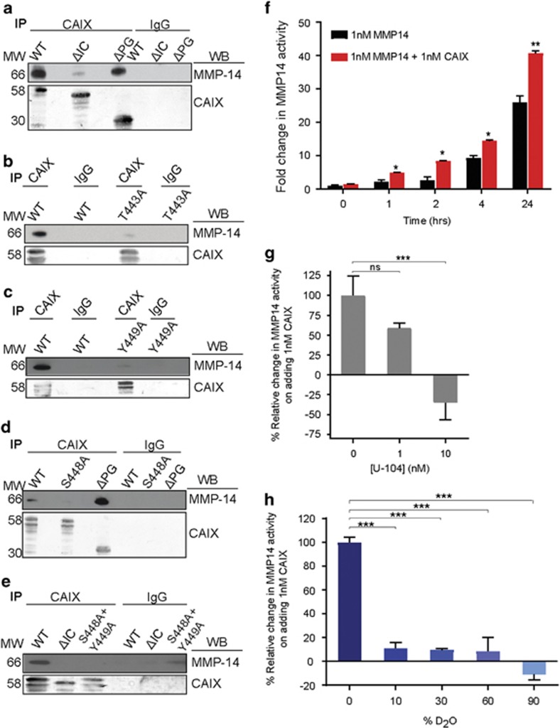
Figure 6.
CAIX associates with MMP14 through its intracellular domain and potentiates MMP14 activity. (a) Co-IP of huCAIX and MMP14 from 4T1shCAIX cells cultured in hypoxia and expressing WT, ΔIC or ΔPG variants of huCAIX. (b) Co-IP of huCAIX and MMP14 from 4T1shCAIX cells cultured in hypoxia and expressing WT huCAIX or the T443A point mutant. (c) Co-IP of huCAIX and MMP14 from 4T1shCAIX cells cultured in hypoxia and expressing WT or the Y449A point mutant. (d) Co-IP of huCAIX and MMP14 from 4T1shCAIX cells grown in hypoxia and expressing WT huCAIX, the S448A point mutant or the ΔPG mutant. (e) Co-IP of huCAIX and MMP14 from 4T1shCAIX cells in hypoxia expressing WT huCAIX, the ΔIC truncation or the double point mutant S448A+Y449A. Isotype-specific IgG was used as a control for co-IPs. (f) Time-dependent stimulation of MMP14 activity in the presence (red bars) or absence (black bars) of equimolar concentrations of CAIX. Data show the mean±s.e.m. of technical replicates (n=5) and are representative of two independent experiments. *P<0.05, **P<0.01. (g) Dose-dependent inhibition of CAIX-mediated stimulation of MMP14 activity in the presence of increasing concentrations of U-104. Mean±s.e.m. of technical replicates (n=5). Data are representative of two independent experiments. ***P<0.001. (h) Inhibition of CAIX-mediated stimulation of MMP14 activity in the presence of increasing amounts of D2O (heavy water). Mean±s.e.m. of technical replicates (n=5). Data are representative of three independent experiments. ***P<0.001. Statistical analysis was performed using Student’s t-test (f) or ANOVA (g,h).

