Abstract
The structural analysis of class B G protein-coupled receptors (GPCR), cell surface proteins responding to peptide hormones, has until recently been restricted to the extracellular domain (ECD). Cor-ticotropin-releasing factor receptor type 1 (CRF1R) is a class B receptor mediating stress response and also considered a drug target for depression and anxiety. Here we report the crystal structure of the trans-membrane domain of human CRF1R in complex with the small-molecule antagonist CP-376395 in a hex-agonal setting with translational non-crystallographic symmetry. Molecular dynamics and metadynamics simulations on this novel structure and the existing TMD structure for CRF1R provides insight as to how the small molecule ligand gains access to the induced-fit allosteric binding site with implications for the observed selectivity against CRF2R. Furthermore, molecular dynamics simulations performed using a full-length receptor model point to key interactions between the ECD and extracellular loop 3 of the TMD providing insight into the full inactive state of multidomain class B GPCRs.
Keywords: CRF1R, translational non-crystallographic symmetry, molecular dynamics, GPCR, metadynamics, CP-376395
1. Introduction
The secretin subfamily of Class B G protein-coupled receptors (GPCRs) includes many important and clinically validated drug targets. These include the glucagon like peptide receptor GLP1 for diabetes, calcitonin gene related peptide receptor for migraine and the parathyroid hormone receptor PTH1 for osteoporosis [1]. Corticotropin releasing factor receptor (CRF1R) itself is an important drug target across a range of different disease areas outlined elsewhere in accompanying chapters. In particular a focus of interest for many years by pharmaceutical companies is its role in stress related disorders such as depression [2]. Despite extensive efforts, Class B GPCRs have proved very intractable as drug targets and to date no small molecule modulators have reached the market. The CRF1 receptor is one of the few Class B receptors where small molecule antagonists, such as CP-376395 have been identified by high throughput screening [3]. In order to fully understand the precise mechanism of action of CP-376395 we set out to solve the X-ray structure of the CRF1R bound to CP-376395.
Major technological advances in the area of protein expression, purification and crystallization together with techniques to address the lack of stability and conformational flexibility of GPCRs have enabled the structures of over 20 Class A receptors to be solved. However, structures of Class B GPCRs have lagged behind. This is due in part to the lack of small molecule ligands for co-crystallization and the multi-domain architecture of class B GPCRs consisting of the membrane spanning 7-transmembrane domain and the extracellular peptide ligand binding domain. GPCR crystal structures are highly enabling for structure based drug design approaches [4] and also permitting homology models to be built for related receptors. However X-ray structures represent a ‘snap-shot’ of one particular conformation of the protein/ligand complex providing limited information on the flexibility of the protein and no information relating to the dynamics of ligand binding. Molecular dynamic (MD) simulations are increasingly being applied to study GPCRs and are being actively used in drug design to provide information on the dynamics of conformational changes by the receptor as well as the complex interplay between the protein, water networks and ligand receptor interactions [5]. Here we have also used MD and metadynamics simulations to study ligand receptor interactions in CRF1R and the conformational changes which may occur in the full length receptor.
2. Materials and Methods
2.1. Protein Expression and Purification
The full-length human CRF1R with an intact ICL2 (no T4L insertion) was used as background for generation of the StaR using a mutagenesis approach described previously and mutants analyzed for thermostability in the presence of the selective radioligand [3H]CP-376395. While wild type CRF1R displayed an apparent thermostability (Tm) of 18.4 °C (±2.0 °C) in n-dodecyl-β-D-maltopyranoside (DDM), the CRF1R StaR (full-length receptor with 12 thermostabilizing mutations) displayed a Tm of 44.7 °C (±2.2 °C) in an identical assay, dropping to a Tm of 37.5 °C (±0.7 °C) upon fusion of the T4L. The CRF1R-TMD carrying a T4 lysozyme fusion in intracellular loop 2 and a C-terminal deca-histidine tag was expressed in Trichoplusia ni (High Five) cells in EX-CELL 405 medium (Sigma-Aldrich) supplemented with 10% (v/v) fetal bovine serum (Sigma-Aldrich), 1% (v/v) CD lipid concentrate (GIBCO) and 1% (v/v) Penicillin/Streptomycin (PAA Laboratories). Cells were infected at a density of 2 x 10⁶ cells/ml with 10 ml of baculovirus per liter of culture, corresponding to an approximate multiplicity of infection (MOI) of 1. Cultures were grown at 27 °C with constant shaking and harvested 72 hours post infection. Cells were pelleted and washed with 250 ml PBS and stored at -80 °C. All subsequent purification steps were carried out at 4 °C unless indicated differently. To prepare membranes, cells were thawed at room temperature and resuspended in 400 ml ice-cold 50 mM Tris-HCl pH 8.0, 500 mM NaCl supplemented with EDTA-free protease inhibitors (Roche). The cell suspension was incubated with 0.3 µM CP376395 (Tocris) for 1 hour to allow the ligand to bind. Cells were disrupted by ultra-sonication and cell debris was removed by centrifugation at 10.000 x g. Membranes were collected by ultracentrifugation at 140.000 x g, resuspended and stored at -80 °C until further use. Membranes were thawed at room temperature and solubilized with 2% (w/v) DM for 1.5 hours. Insoluble material was removed by ultra-centrifugation and the receptors were immobilized by batch binding to TALON metal-affinity resin (Clontech) for 2 hours. The resin was packed into a XK-16 column (GE Healthcare) and washed with steps of 8 and 30 mM imidazole in 50 mM Tris-HCl pH 8.0, 500 mM NaCl, 0.15% (w/v) DM, and 0.3 µM CP376395 for a total of 15-20 column volumes before bound material was eluted with 200 mM imidazole. The protein was then concentrated using an Amicon Ultra-15 centrifugal filter unit (Millipore) and subjected to preparative gel filtration in 20 mM Tris-HCl pH 8.0, 150 mM NaCl, 0.15% (w/v) DM, and 0.3 µM CP376395 on a Superdex 200 10/300 GL gel filtration column (GE Healthcare) to remove remaining contaminating proteins and aggregates. Receptor purity was analyzed using SDS-PAGE and mass spectrometry and receptor mono-dispersity was assayed by FSEC monitoring tryptophan fluorescence. Protein concentration was determined with a NanoDrop spectrophotometer using the receptor’s calculated extinction coefficient at 280 nm (e280, calc = 1.6 (mg/ml x cm)-1).
2.2. Crystallization
The CRF1R-TMD was crystallized in lipidic cubic phase (LCP) at 22.5 °C. The protein was concentrated to 20-30 mg/ml by ultrafiltration and mixed with monoolein (Nu-Check) supplemented with 10% (w/w) cholesterol (Sigma) and 5 µM CP376395 using the twin-syringe method [6] with a final protein:lipid ratio of 1:1.5 (w/w). A Mosquito LCP (TTP Labtech) was used to dispense 40-60 nl boli on 96-well Laminex Glass Bases (Molecular Dimensions), overlaid with 0.75 µl precipitant solution and sealed with Laminex Film Covers (Molecular Dimensions). 20-30 µm crystals of construct CRF1R-#76 were obtained in 100 mM Na-citrate pH 5.5, 200 mM Li2SO4, 30% (v/v) polyethylene glycol 400, 0.6 µM CP376395. Crystals were flash cooled in liquid nitrogen without additional cryoprotectant.
2.3. Diffraction Data Collection and Processing
X-ray diffraction data were measured on a Pilatus 6M hybrid-pixel detector at Diamond Light Source beamline I24 using a 5 μm diameter microbeam. Crystals displayed isotropic diffraction to beyond 3.0 Å following exposure to an unattenuated beam for 8 seconds per degree of oscillation. Consequently radiation damage set in quickly and less than 5 degrees of oscillation data per crystal could be used in subsequent data merging. Data from individual crystals were integrated using XDS [7]. The final dataset included data from 21 crystals (with reindexing as required) and was scaled to 3.18 Å using the microdiffraction assembly method as described previously [8, 9] with a final overall completeness of 93.7%. Crystals belonged to hexagonal space group P6 with unit cell dimension of a = b = 189.4 Å, c = 88.6 Å, α = β = 90 ˚ γ = 120 ˚. The resulting multi-record reflection file was scaled using AIMLESS from the CCP4 suite [10, 11]. Data collection statistics are presented in Table 1.
Table 1.
Crystallographic table of statistics.
| Data Collection | |
|---|---|
| Space Group | P6 |
| Cell Dimensions a, b, c, (Å) | 189.4, 189.4, 88.6 |
| Cell Angles α, β, γ (°) | 90, 90, 120 |
| Resolution (Å) | 3.18 |
| Rmerge | 0.158 (0.627) |
| I / σ I * | 6.4 (1.8) |
| Completeness (%) | 93.7 (82.0) |
| Redundancy | 3.8 (2.5) |
| REFINEMENT | |
| Resolution (Å) | 19.91 – 3.18 |
| No. Reflections | 28,393 |
| Rwork / Rfree | 24.4 / 28.9 |
| No. atoms Protein Ligand |
9,980 264 |
|
B-factors Protein Ligand |
90.7 85.5 |
| R.m.s deviations Bond lengths (Å) Bond Angles (°) |
0.006 0.882 |
| Ramachandran Plot: Preferred (%) Allowed (%) Outlier (%) |
96.4 3.4 0.2 |
*Statistics in parentheses refer to outer resolution shell.
2.4. Structure Solution and Refinement
The CRF1R-#76 crystals belong to hexagonal space group P6 exhibiting a 30% off-origin peak in a native Patterson map, indicating translational non-crystallographic symmetry (tNCS). Previously, it was possible to modulate the construct in terms of the TMD and T4 Lysozyme (T4L) linker resulting in construct CRF1R-#105 which crystallized in the same conditions as CRF1R-#76 yet belonged to an orthorhombic spacegroup displaying no tNCS and which was subsequently solved and refined (PDB ID: 4K5Y) [9]. The structure of CRF1R-#76 was solved by molecular replacement (MR) with the program Phaser [12] utilising corrections for the statistical effects of tNCS function [13] with two independent search models, T4L from CRF1R and the TMD of CRF1R (PDB ID 4K5Y). Solutions were found for all three copies of the T4L and TMD in the asymmetric unit.
The nature of the tNCS was unusual. The peak in the native Patterson map indicated a tNCS translation of approximately 1/3,2/3,0, from which one might expect three copies in the asymmetric unit to be generated by successive applications of the same translation vector, corresponding to an approximate tripling of a smaller unit cell. However, the tNCS likelihood target [13] was about 1600 units higher when assuming two tNCS-related copies instead of three. A molecular replacement search for two copies each of the TMD and T4L models gave an unambiguous solution, in which a crystallographic 3-fold axis generated hexamers from the two copies. The crystal packing left a hole around the crystallographic 6-fold axis, sufficient to place an additional copy generating a hexamer, but surprisingly the molecular replacement search for an additional copy each of the TMD and T4L placed them in an inverted orientation, so the third copy was not in fact related by translation to the first two.
Manual model building was performed in COOT [14] using sigma-A weighted 2m|Fo|-|DFc|, m|Fo|-D|Fc| maps calculated using Phenix [15]. Initial refinement was carried out with REFMAC5 [11, 16] using maximum-likelihood restrained refinement in combination with the jelly-body protocol. Late stages of the refinement were performed with Phenix.refine [17] with positional and individual isotropic B-factor refinement and TLS. The structure was validated using MolProbity [18]. The final refinement statistics are presented in Table 1. Figures were prepared using PyMOL [19].
2.5. Structural Analysis
Cα RMSD calculation between different copies of the CRF1R-TMD structures was performed using Superpose [11]. The following amino-acid ranges were used 125-140(TM1), 154-164(TM2), 190-209(TM3), 241-249(TM4), 272-291(TM5), 314-322(TM6) and 350-365(TM7).
2.6. Molecular Dynamics Simulation
The three dimensional coordinates of CRF1R (space group P22121, CRF1RP22121) in complex with the small molecule antagonist CP-376395 [9] were downloaded from the Protein Data Bank [20]. The receptor was prepared with the Protein Preparation Wizard in Maestro [21]: only the protein and the ligand in chain C have been included, hydrogen atoms have been added and the H-bond network has been optimized through an exhaustive sampling of hydroxyl and thiol moieties, tautomeric and ionic state of His and 180° rotations of the terminal dihedral angle of amide groups of Asp and Gln. His1552.50 has been considered to be protonated. Hydrogen atoms have been energy minimized using the OPLS2.1 force field. The same protocol has been applied to chain A of CRF1R in the hexagonal space group P6 (CRF1RP6), while for the MD simulation of the apo state of CRF1RP22121 the ligand CP-376395 has been deleted. The homology model of the receptor including the extracellular domain (ECD-CRF1RP22121) has been created using CRF1RP22121 chain A for the TMD and the crystal structure of the N-terminal extracellular domain available in the Protein Data Bank (PDB ID: 3EHS) [22]. The 6 residues missing between the two crystal structures (from E109 to V114) have been assumed to be helical creating one and half helical turn connecting the top of TM1 helix to the helix at the end of the ECD. The continuous helical nature of the link between the two domains determined their final relative orientation. The T4L has been removed, the ICL2 conformation has been predicted using Prime [23] and the final system has been prepared with the same protocol described above with the Protein Preparation Wizard in Maestro.
The 4 prepared systems (CRF1RP22121, CRF1RP6, apo CRF1RP22121 and ECD-CRF1RP22121) have been analysed using standard MD simulations with the following protocol. The AMBER99SB force field (ff) [24] parameters were used for the protein and the GAFF ff [25] for the ligands using AM1-BCC partial charges [26]. The system has been embedded in a triclinic box including an equilibrated membrane consisting of 256 DMPC (1,2-dimyristoyl-sn-glycero-3-phosphocholine) lipids [27] and 24513 waters using g_membed [28] in GROMACS (v4.6.5). The SPC water model was used and ions were added to neutralize the system (final concentration 0.01 M). CRF1RP22121 included a total of 103,468 atoms (24,233 water molecules, 7 Na+, 16 Cl- and 226 lipids), while CRF1RP6 was composed by 103,590 atoms (24,190 water molecules, 7 Na+, 18 Cl- and 226 lipids). An energy minimization protocol based on 200 steps steepest-descent algorithm followed by 500 steps conjugate gradient algorithm has been applied to the system. The membrane has been equilibrated using 2 ns MD simulation with a time step of 2.5 fs, using LINCS on all bonds and keeping the protein and ligand restrained applying a force of 100 kJ mol-1 nm-1. Lennard-Jones and Coulomb interactions were treated with a cut-off of 1.069 nm with particle-mesh Ewald electrostatics (PME) [29]. The MD has been executed in the NPT ensemble using v-rescale [30] (tau_t = 0.5 ps) for the temperature coupling to maintain the temperature of 303 K and using Parrinello-Rahman [31] (tau_p = 10.0 ps) for the semi-isotropic pressure coupling to maintain the pressure of 1.013 bar. Finally a 50 ns MD simulation has been performed using the same settings described above, but without applying any positional restraints. The CRF1R double mutant has been generated introducing the His199Val3.40b and Met276Ile5.44b mutations in the equilibrated system. ECD-CRF1RP22121 has been generated and equilibrated with a comparable protocol and resulted in a system composed by a total of 124,752 atoms (30,717 water molecules, 8 Na+, 17 Cl- and 228 lipids).
2.7. Metadynamics Simulation
Well-tempered Metadynamics (WTMetaD) [32] simulations have been used to evaluate: 1) the lowest energy binding path of CP-376395 to CRF1RP22121, 2) low energy conformations of the ECD relative to the receptor TMD. All simulations have been carried out using GROMACS with the same MD protocol described above and PLUMED (v2.0.2) [33] with a time step of 2 fs.
Possible ligand binding and dissociation paths were initially generated using a steered MD [34] protocol. The method was based on 24 steps of 250 ps MD each for a total of 6 ns: 3 ns for the ligand binding and 3 ns for the dissociation event simulation, 12 steps each. Using a python script the protocol started with a target RMSD from the ligand bound conformation value of 10 Å and a force constant of 1 kJ mol-1 nm-1. To ensure the final ligand bound state was reached, the target RMSD value was consecutively decreased and kappa increased by 10-fold in the first 12 steps. For the dissociation 12 steered MD steps with the same settings were applied using as target position the initial unbound conformation of the ligand. Two possible binding-dissociation paths for the small molecule have been considered: one accessing the ligand binding site from the extracellular side and one from the membrane. These have been achieved positioning the ligand in different starting locations at about 20 Å from the bound state, respectively in the extracellular side close to the orthosteric site and into the membrane close to TM5. The obtained binding routes have been used to define a path collective variable (CV) for the WTMetaD with the following settings: simulated temperature 300 K, bias factor 90, initial energy bias Gaussian height of 0.3 kJ/mol with a deposition frequency of 500 MD steps. A geometric based hills width scheme [35] has been applied starting with a sigma value of 0.1. Two path collective variables have been defined [36] one defining the position on the path and the other the distance from the path. A Lambda value of 3.0 has been applied. A total of 58 ns WTMetaD starting from the ligand bound state were required to reach the lowest energy barrier allowing the ligand to dissociate from the receptor. The same procedure has been used for the study of the CRF1R double mutant (His199Val3.40b and Met276Ile5.44b).
A similar protocol has been used to identify stable “open” and “closed” conformations of the ECD relative to the receptor helical bundle. In this case the path CV has been defined using the starting and final protein conformations from the 50 ns MD of the ECD-CRF1RP22121. Two restraining potential walls have been applied to the CV describing the distance from the path at CV values of 0.3 and -0.3 (k=500 kJ mol-1 nm-1, exp=3). Two simulations for a total of 114 ns WTMetaD starting from the “open” and “closed” ECD states have been performed. This simulation length was not sufficient to obtain a converged energy landscape of the full conformational change for the full length receptor moving from the “open” to the “closed” ECD states. The protocol described identifies representative low energy ECD conformations of the two states.
3. Results
3.1. CRF1R Crystal Structures
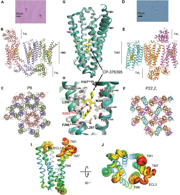
In 2013 the crystal structure of the TMD of CRF1R was reported at 3.0 Å resolution [9]. CRF1R was crystallised in Lipidic Cubic Phase (LCP) (Fig. 1-D) using a conformational thermostabilisation approach to generate the stabilised receptor (StaR) and fusion of T4-Lysozyme (T4L) within intracellular loop 2 (ICL2) of the receptor to aid crystallisation (this construct is referred to as CRF1R-#105 henceforth). The resulting structure of the human CRF1R receptor TMD in complex with the small molecule antagonist CP-376395 (2-aryloxy-4-alkylaminopyridine) in an orthorhombic setting (referred to as CRF1RP22121 henceforth) provided an exciting inaugural view of the architecture of the CRF1R receptor and that of class B GPCRs in general.
Fig. (1).
Overview of the CRF1R crystal structures solved in complex with CP-376395. A - Left) Crystals grown in lipidic cubic phase of CRF1R-#76 – hexagonal setting. B) The overall structure of CRF1R-#76 solved with three copies in the asymmetric unit. CRF1R-T4L fusion is shown in ribbon representation and coloured by chain, pink, blue and green. The CP-376395 small-molecule is depicted in space fill representation with carbon, nitrogen and oxygen atoms coloured yellow, blue and red respectively. CRF1R-TMD and T4L fusions are denoted. C) Crystal packing of CRF1R in the hexagonal setting – view down unique c axis. Receptor copies coloured as in (B). D - Right) Crystals grown in lipidic cubic phase of CRF1R-#105 – orthorhombic setting. E) The overall structure of CRF1R-#105 solved with three copies in the asymmetric unit. CRF1R-T4L fusion is shown in ribbon representation and coloured by chain, pink, blue and green. The CP-376395 small-molecule is depicted in space fill representation coloured as in (B), CRF1R-TMD and T4L fusions are denoted. F) Crystal packing of CRF1R in the orthorhombic setting – view down a axis. Receptor copies coloured as in (B). G - Centre) Superposition of all 6 CRF1R-TMD copies in ribbon representation coloured in varying shades of cyan as viewed from a plane parallel to the membrane. TM helices are labeled. CP-376395 is labeled and shown in stick representation coloured as in B. H) Close-up view of the induced-fit small-molecule allosteric site in CRF1R – receptor copies coloured as in (G). Important receptor residues are labeled and shown in stick representation with carbon, nitrogen and oxygen atoms coloured white, blue and red respectively. CP-376395 is labeled and shown in stick representation coloured as in B. Hydrogen bonds depicted as dashed red lines. I) Superposition of all 6 CRF1R-TMD copies as in (G) shown in sausage representation coloured using a relative B-factor spectrum, blue=low; red=high. J) Representation as in (I) rotated to view from extracellular space. (The color version of the figure is available in the electronic copy of the article).
However, these were not the first crystals of CRF1R to be obtained. In addition to CRF1R crystals grown using bicelles, and classical vapour diffusion (data not shown) the first generation of CRF1R TMD crystals grown (Fig. 1-A) using the in meso method utilised a near identical construct to CRF1R-#105 but which incorporated two extra residues from intracellular loop 2 – one of which had been previously identified as a thermostabilising mutation (S222L) in the fusion to T4-Lysozyme (this construct is referred to as CRF1R-#76 henceforth). The CRF1R-#76 crystals belong to hexagonal space group P6 exhibiting a 30% off-origin peak in a native Patterson map, indicating translational non-crystallographic symmetry (tNCS). Although a complete dataset to 3.18 Å resolution could be generated through merging data collected from multiple crystals, extensive trials to solve the structure by molecular replacement failed, due in part to the presence of tNCS and / or the low structural similarity between search model and target. With the CRF1RP22121 structure in hand it was finally possible to solve the hexagonal tNCS data (see methods) with three copies in the asymmetric unit, thereby generating a second structure of the CRF1R receptor in complex with CP-376395 referred to as CRF1RP6 henceforth (Table 1) and which doubles the structural information available for this receptor (Fig. 1-B,C).
Despite fundamental differences in crystal contacts / packing between the orthorhombic and hexagonal lattices, superposition of the 6 CRF1R-TMD structures (3 copies in the asymmetric unit from both CRF1RP22121 and CRF1RP6) demonstrate the structures are all in close agreement (root-mean-square deviation RMSD less than 0.4 Å across core TM residue Cα atoms – see methods) (Fig. 1-G,H). In the hexagonal CRF1RP6 crystal system interactions between receptors occur exclusively via parallel packing with TM1 and TM7 from one receptor copy interacting with TM4 and the N-terminus of TM3 for both non-crystallographic and symmetry related copies (Fig. 1-C). In the orthorhombic CRF1RP22121 crystal form interactions between receptors occur in both parallel and antiparallel fashion. Parallel interactions are observed between ECL3/TM6 to ECL3/TM6, TM1 to TM1, and TM1 to TM4/N-terminus of TM3, with a single antiparallel interaction from TM4 to TM4 (Fig. 1-F).
The open extracellular conformation of the peptidic agonist orthosteric pocket initially revealed in the CRF1R P22121 structure is maintained across the 3 CRF1RP6 copies. One side of this “chalice-like” conformation is provided by TM2-TM5, and the other by TM1, TM6, and TM7. As previously reported, in CRF1R the slightly bent extracellular portion of TM1 packs against and stabilizes a kink in TM7 contributing to the open nature of the receptor extracellular vestibule. The highly conserved S1301.50b on TM1 hydrogen bonds to the backbone Nitrogen of F3577.51b and main-chain carbonyl of S3537.47b, which flank G3567.50b on TM7. This results in the extracellular halves of TM6, TM7 and extracellular loop 3 (ECL3) tilting away from the central helical bundle [9]. Furthermore, the extracellular portions of TM6, TM7 and ECL3 (along with the N-terminus of TM1) demonstrate the highest B-factor values and structural variation across all 6 CRF1R-TMD structures, pointing to the inherent structural flexibility of this region in CRF1R (Fig. 1-I,J). Indeed in only 2 of the 6 copies of the CRF1R receptor from both the CRF1RP22121 and CRF1RP6 is ECL3 ordered and visible in the electron density.
Finally in all 3 copies of CRF1RP6 the CP-376395 small molecule is again visible in the extraordinary position towards the intracellular side of the receptor with a single hydrogen-bond supplied by N2835.50b, while TM3, TM5 and TM6 provide the residues that constitute the rest of the hydrophobic pocket towards the intracellular side of the receptor, as previously observed and in close agreement with the CRF1RP22121 structure (Fig. 1-G,H).
3.2. Structural Insights into CP-376395 Binding and CRF1R Selectivity
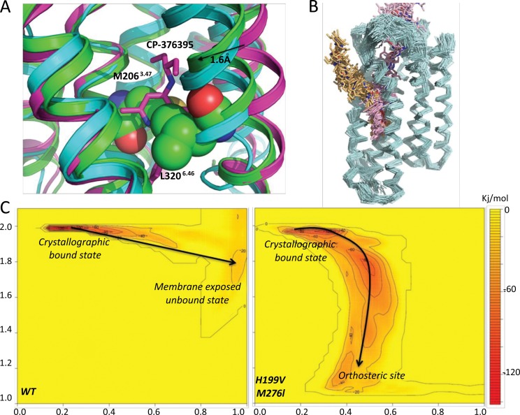
The striking position of CP-376395 and its interactions with the receptor resulted in a very stable configuration across a 50 ns Molecular dynamics (MD) simulation within an explicit water-membrane environment. For both crystal forms (CRF1RP22121 and CRF1RP6) the average ligand RMSD during simulation was ~1 Å. To evaluate whether the allosteric small-molecule binding site in CRF1R was induced by CP-376395 we also analyzed an apo CRF1R model using an identical MD simulation protocol. In this case the allosteric pocket appeared very unstable. TM6 quickly moved closer to TM3 in a position similar to that adopted in the glucagon receptor crystal structure (Fig. 2-A). In particular residues M2063.47b and L3206.46b occupied and collapsed the binding site. These two residues have recently proposed to be part of the hydrophobic core of the receptor [1, 37] playing a crucial role in stabilizing the inactive receptor state by controlling the movement of the N-terminus of TM6 during activation, an essential structural prerequisite that is required for G protein docking on the intracellular surface of the receptor.
Fig. (2).
Analysis of CP-376395 binding to CRF1R. A) Comparison of CP-376395-CRF1RP22121 complex (in magenta), apo CRF1RP22121 (in green) and GCGR crystal structure (in cyan). Residues M2063.47 and L3206.46 are shown in space fill representation with carbon, nitrogen and oxygen atoms coloured green, blue and red respectively. CP-376395 is shown in stick representation with carbon, nitrogen and oxygen atoms coloured magenta, blue and red respectively B) The two predicted ligand binding paths are compared, in pink starting from the orthosteric site and in yellow from within the membrane. Binding paths are shown using snapshots of the ligand position during the simulation of binding and dissociation. The protein backbone is shown in cyan as ribbon. C) Free energy landscape predicted by the WTMetaD simulation for the dissociation event of CP-376395 in the wild type receptor (left) and in the double mutant H199V, M276I (right). Y axis represents the path CV defining the position on the path, while the X axis the distance from the path CV. The free energy surface is colour-coded from yellow to red (0 to -137 kJ/mol) and the positions of the bound and dissociated states are indicated. (The color version of the figure is available in the electronic copy of the article).
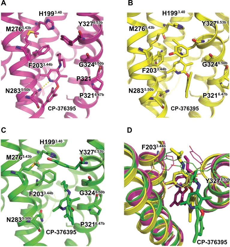
To further investigate the nature of the induced-fit CP-376395 pocket within CRF1R, ligand binding and dissociation paths were generated using a Steered MD protocol. Two different starting positions of the small molecule were evaluated: one accessing the binding site from extracellular space; and one from within the membrane. Starting positions for CP-376395 were located ~20 Å from the crystallographic ligand bound position: in the first case this was on the extracellular side of the receptor close to the orthosteric site, while in the second it was within the membrane - in close proximity to TM5 (Fig. 2-B). The Steered MD trajectories obtained were subsequently used for a well-tempered metadynamics (WTMetaD) simulation protocol starting from the bound crystallographic state. WTMetaD permitted the evaluation of the free energy surface of ligand dissociation (Fig. 2-C). The most favourable and lowest energy escape route for the ligand from the induced-fit pocket was between TM5 and TM6 and towards the membrane environment. Analysis of the simulation trajectory reveals crucial movements of F2033.44b and Y3276.53b changing rotameric states during ligand dissociation (Fig. 3-A,B). These key conformational changes permit the initial movement of the ligand from the bound crystallographic state up and towards the extracellular side of the receptor creating a high-energy transition state conformation where the H-bond between CP-376395 and N2835.50 is broken. From this position the ligand can access the membrane between TM5 and TM6 in a location close to G3246.50b and P3216.47b (Fig. 3-C). Both G3246.50b and P3216.47b contribute to a bent / flexible local conformation of TM6 to create a sterically viable exit for CP-376395 from the receptor TMD. Finally, the movement of F2033.44b and Y3276.53b during ligand dissociation (Fig. 3-D) is influenced by M2765.44b and H1993.40b. M2765.44b and H1993.40b have previously been demonstrated to be important for the observed selectivity of CP-376395 for CRF1R over CRF2R, where they are instead found to be Ile2725.44b and Val1953.40b respectively [38, 39]. To evaluate the importance of these two residues we applied the same WTMetaD protocol to the CRF1R double mutant His199Val3.40 Met276Ile5.44. These mutations prevent ligand unbinding toward the membrane, creating a favourable route for unbinding in the direction of the orthosteric site (Fig. 2-C). This is possibly the result of Val1993.40 and Ile2765.44 effecting the rotameric states of F2033.44b and Y3276.53b that are compatible (decrease the energy barrier) for ligand unbinding toward the orthosteric site. Overall the crystallographic bound state conformation is predicted 1.4 kcal/mol less stable in the double mutant compared to wild type.
Fig. (3).
Key conformations identified during the WTMetaD ligand dissociation path. The ligand and relevant receptor residues are shown in stick representation. N2835.50 provides the only H-bond with the CP-376395 ligand while G3246.50 and P3216.47 modulate the bent trajectory of TM6. F2033.44 and Y3276.53 change rotameric states during ligand dissociation and are controlled by M2765.44 and H1993.40. In CRF2R these two residues are an I2725.44 and V1953.40 respectively. A) CP-376395 in the bound crystallographic conformation. B) Predicted first step in CP-376395 dissociation associated with breaking the H-bond with N2835.50 and changes in the conformation of F2033.44 and Y3276.53 to permit initial movement of the ligand toward the extracellular side of the receptor. C) The CP-376395 exit route between TM5 and TM6 close to G3246.50 and P3216.47. D) Comparison of the three ligand dissociation states (starting in magenta, first step in yellow and final step in green). The movement of F2033.44 and Y3276.53 are depicted in line representation. (The color version of the figure is available in the electronic copy of the article).
3.3. Structural Insights into CRF1R Inter-Domain Interactions
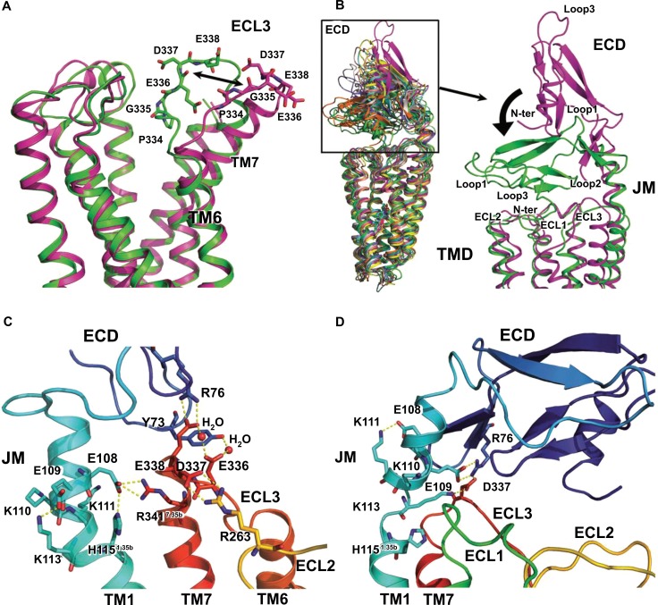
As expected, analysis of CRF1RP22121 and CRF1RP6 dynamic behaviour using MD simulation confirmed the conformations of both structures to be stable and in agreement. Across a 50 ns MD simulation in an explicit water-membrane environment the average RMSD for all protein Cα atoms was measured at ~2 Å. Root mean square fluctuation analysis of the trajectories highlighted a high degree of flexibility for extracellular loop 3 (ECL3) from residue N333 to E338 (Fig. 4-A). This is in agreement with the high resultant B-factors and structural variation for residues in this region across both crystal forms and six receptor copies obtained for the CRF1R-TMD (Fig. 1-I,J). To investigate the observed flexibility of ECL3 and any potential structural role in the context of the full-length receptor, a full-length model using the CRF1RP22121 TMD and the crystal structure of the extracellular domain (ECD) (PDB ID: 3EHS) [22] was built. The 6 residues (E109 to V114) connecting the ECD to the TMD of the receptor which are not resolved in any of the available crystal structures have been assumed to be alpha helical and therefore modelled as 1.5 helical turns connecting the C-terminus of the ECD with the N-terminus / top of TM1. The assumption of continuous alpha helix in ab initio modelling of these 6 residues, connecting experimentally resolved regions of alpha helix which flank either side, determined the relative orientation of the two protein domains in the final full-length CRF1R model.
Fig. (4).
Analysis of the extracellular surface of CRF1R. A) Side view of the extracellular region of the CRF1R receptor TMD highlighting the flexibility of ECL3 during MD simulation. Residues predicted to be crucial in determining the loop flexibility are shown in stick representation. B) On the left, superimposition of different snapshots from MD simulation of ECD-CRF1RP22121. On the right, comparison between the starting (in magenta) and final (in green) conformations of the receptor ECD. Representative lowest energy conformations identified by the WTMetaD protocol for the full length receptor with the ECD in the “open” (C) and “closed” (D) conformations are shown. Relevant residues are represented as sticks and the backbone as cartoon. The protein is colour coded as rainbow, from blue (N terminus) to red (C terminus). Key interactions are shown as yellow dotted lines. (The color version of the figure is available in the electronic copy of the article).
The conformational stability between the CRF1R –TMD and ECD was initially analyzed using a standard MD simulation within an explicit water-membrane environment. In the starting conformation the main interactions between the two domains are between ECL3 of the receptor TMD and Loop 2 of the ECD. During the 50 ns MD simulation the ECD changed its relative position to adopt a final “closed” conformation on top of the receptor TMD. In the final state model Loop 1, Loop 2 and the C-terminus of the ECD adopt a position closer to ECL1 and ECL2 of the TMD. After structural superimposition of the starting and final state full-length models using the TMD regions only, the Cα RMSD for the ECDs was ~18 Å, with a maximum change for Loop 3 in the ECD greater than 45 Å. In order to identify the most stable “open” and “closed” conformations of the ECD in the full length receptor the system was analyzed using a WTMetaD protocol.
The analysis highlighted the potential integral role of charged residues and electrostatic interactions in mediating interactions in the juxtamembranous (JM) region connecting TM1 to the ECD, and between ECL3 of the TMD to the ECD in controlling the relative position of the two CRF1R domains. In the “open” conformation D337 and E338 within ECL3 form salt bridges with ECL2 (R263) and the ECD (R76) respectively. In addition R3417.35b interacts with E108 in the JM region (Fig. 4-C) and together with the adjacent E109, K110, K111 and K113 appears to play a key role in stabilizing the full-length receptor “open” state. In the “closed” conformation the JM region partially unwinds allowing D337 in ECL3 to interact with R76 and K113. These interactions result in a stable position of the ECD over the TM helical bundle occluding the entrance to the orthosteric site further locking full-length CRF1R in the “closed” conformation (Fig. 4-D).
4. Discussion
The crystal structure of the CRF1R TMD in a hexagonal setting represents a doubling of the structural information that exists for this receptor in the public domain. That the CRF1RP6 and CRF1RP22121 structures are in close agreement in terms of structural superposition of the TMD regions, with molecular features maintained across crystal systems / fundamentally different lattices (and exhibit a comparable and extremely stable dynamics behaviour) further increases confidence that the molecular features of the ground state of the receptor TMD are captured across these structures. Additionally the structure of the human class B glucagon receptor TMD was reported at 3.4 Å resolution in 2013 in complex with the antagonist NNC0640 [40]. Though the position of NNC0640 was not determined in the glucagon crystal structure, superposition of the TMD of CRF1R and glucagon receptors demonstrates considerable structural conservation of the canonical 7TM helix arrangement (particularly over TMs 1-5) [1]. Taken together this represents a reliable structural framework from which to investigate the complexity of the inactive CRF1R conformation, and potentially multidomain class B receptors in general. Additionally insight can be gained into the nature of the CRF1R small-molecule allosteric binding site, the protein conformational changes required for the ligand binding event and potential implications for selectivity. Finally, a molecular model of full-length CRF1R in the inactive state has been assembled and optimized using unbiased and biased MD.
MD simulation using an apo CRF1R model strongly suggests that the allosteric pocket (found deep towards the intracellular side of the receptor) is induced by ligand binding (Fig. 2-A). Steered MD supports two possible access routes for the ligand to the induced-fit site in CRF1R: one from the putative orthosteric site in the extracellular side of the receptor and a second from within the membrane, in a location close to TM5 and TM6 (Fig. 2-B). WTMetaD analysis of the free energy surface of the ligand dissociation suggests that the lowest energy escape route for the ligand from the induced-fit pocket is between TM5 and TM6 toward the membrane (Fig. 2-C). During ligand dissociation F2033.44b and Y3276.53b change rotameric states (Fig. 3-D) permitting exit of the small-molecule from the CRF1R-TMD in a region close to G3246.50b and P3216.47b (Fig. 3-C). These predicted conformational changes required for ligand binding and dissociation also provide a potential rationale for the selectivity of CP-376395 to the human CRF1R subtype. The functional antagonist activity of CP-376395 for CRF1R is 12 nM, while >10000 nM for CRF2R [3]. Sequence identity within the helical bundle between the receptor subtypes is high (78%) and residues participating in direct interactions with CP-376395 in CRF1R are completely conserved in CRF2R. Across the second shell of residues in proximity of CP-376395 (less than 5 Å) only two residues differ between CRF1R and CRF2R, these being H1993.40b and M2765.43b corresponding to V1953.40b and I2725.43b respectively. Mutation of these residues in CRF1R to the corresponding amino acids in CRF2R has been demonstrated to reduce the binding affinity of standard non-peptide antagonists in CRF1R while concomitantly having little effect on peptide ligand binding [38, 39]. From the WTMetaD simulation M2765.44b and H1993.40b appear to play a crucial role in modulating the conformational changes of F2033.44b and Y3276.53b required for CP-376395 binding / dissociation. In CRF2R the corresponding V1953.40b and I2725.43b may potentially lock F3.44b and Y6.53b in a conformation incompatible with CP-376395 gaining access to the induced-fit allosteric site.
The ECD of CRF1R has been shown previously to act as an intrinsic negative regulator of receptor activity, with ECL3 of the TMD playing an important role in mediating crosstalk with the ECD [41, 42]. Using a full-length CRF1R model and WTMetaD protocol, representative low energy conformations of the ECD in “open” and “closed” states relative to the TMD have been identified. Mutagenesis, photoaffinity labelling and cysteine trapping studies all provide experimental evidence that in addition to roles played by the extracellular loops, the JM region (connecting TM1 to the ECD) plays an important role in the structure, function and context of full-length multidomain class B receptors [43]. The results presented here implicate electrostatic interactions between ECL3, the ECD, and the JM region as the major determinants in the relative positioning of the two receptor domains. In particular ECL3 appears to play a critical role in mediating interactions between the TMD and the ECD, maintaining the CRF1R ECD in an inactive “closed” conformation with the receptor TMD, as recently proposed for the glucagon receptor [41]. Studies of the multidomain calcitonin gene related peptide receptor have also previously identified point mutations in ECL3 that result in a significant increase of both basal and ligand-induced activity for this receptor [44]. The result of the WTMetaD highlights the role of E338 from ECL3 in forming a salt bridge with R76 from the ECD, indeed mutagenesis of R76 in CRF1R to the corresponding residue in either human vasoactive intestinal peptide receptor type 2 (hVIP-R2) or CRF1R from Xenopus laevis abolishes CRF1R peptide ligand binding [45]. Finally E336 in ECL3 is predicted to interact with Y73 from loop 2 on the CRF1R ECD in the “closed” state and mutation of the corresponding residue to alanine in the ECD of the glucagon receptor (Y65A) increases basal activity almost fivefold [41]. Taken together this provides experimental evidence corroborating the modelled interactions in CRF1R that are important for positioning the ECD relative to the TMD in a functional context. In terms of activation it is possible that following initial binding of the 41 amino acid corticotropin-releasing hormone to the ECD [46, 47], the peptide agonist then relieves receptor inactivation through affecting ECD interactions with ECL3 before subsequently binding to the TMD to induce receptor activation.
Conclusion
The structure of CRF1R in complex with the small molecule CP-376395 at 3.0 Å [9] and in a hexagonal setting at 3.2 Å as presented here have greatly increased our understanding of the architecture of class B receptors including the configuration of the TMD, the open chalice-like nature of the extracellular vestibule and peptidic orthosteric peptide binding pocket and finally the location of a small-molecule induced-fit allosteric pocket deep within the TMD towards the intracellular side of the receptor. The molecular model of full length CRF1R corroborates the role of the ECD as an intrinsic negative regulator of the receptor activity. To further our understanding of the mode of action of CRF1R, and Class B GPCRs in general, structures of the full-length receptor are now required and will represent a significant advance for the field, either in the antagonist close conformation, or in the full agonist state with the corticotropin-releasing hormone (CRH) peptide or a peptide mimetic bound.
Co-ordinates and structure factors have been deposited in the Protein Data Bank under the accession code 4Z9G
Author Contributions
A.S.D. designed crystallization constructs, established the platform/protocols for, and carried out LCP crystallization and designed crystal optimization, harvested crystals, collected and processed X-ray diffraction data and refined the structure. R.K.Y.C. performed expression / purification and grew crystals in bicelles, vapour diffusion and collected X-ray diffraction data. K.H. designed and characterized truncation / fusion and crystallisation constructs, established procedures for, and carried out expression and purification, established the platform/protocols for and carried out lipidic cubic phase (LCP) crystallization, collected and processed X-ray diffraction data. R.J.R. solved the novel CRF1R structure. Computational analysis of the structure and modeling was carried out by A.B. The manuscript was prepared by A.S.D., A.B. and F.H.M.
ACKNOWLEDGEMENTS
We thank G. Evans, R. Owen and D. Axford at I24, Diamond Light Source, Oxford, UK for technical support. We thank A. W. Leslie and R. Henderson at the MRC - Laboratory of Molecular Biology, Cambridge, UK. We also thank R. Nonoo and other colleagues at Heptares Therapeutics Ltd. for suggestions and comments. R.J.R is supported by a Principal Research Fellowship from the Wellcome Trust (grant No. 082961/Z/07/Z).
CONFLICT OF INTEREST
The authors from Heptares Therapeutics declare competing financial interests.
References
- 1.Bortolato A., Doré A.S., Hollenstein K., Tehan B.G., Mason J.S., Marshall F.H. Structure of Class B GPCRs: new horizons for drug discovery. Br. J. Pharmacol. 2014;171:3132–3145. doi: 10.1111/bph.12689. [DOI] [PMC free article] [PubMed] [Google Scholar]
- 2.Bale T.L., Vale W.W. CRF and CRF receptors: role in stress responsivity and other behaviors. Annu. Rev. Pharmacol. Toxicol. 2004;44:525–557. doi: 10.1146/annurev.pharmtox.44.101802.121410. [DOI] [PubMed] [Google Scholar]
- 3.Chen Y.L., Obach R.S., Braselton J., Corman M.L., Forman J., Freeman J., Gallaschun R.J., Mansbach R., Schmidt A.W., Sprouse J.S., Tingley Iii F.D., Winston E., Schulz D.W. 2-Aryloxy-4-alkylaminopyridines: discovery of novel corticotropin-releasing factor 1 antagonists. J. Med. Chem. 2008;51:1385–1392. doi: 10.1021/jm070579c. [DOI] [PubMed] [Google Scholar]
- 4.Jacobson K.A., Costanzi S. New insights for drug design from the X-ray crystallographic structures of G-protein-coupled receptors. Mol. Pharmacol. 2012;82:361–371. doi: 10.1124/mol.112.079335. [DOI] [PMC free article] [PubMed] [Google Scholar]
- 5.Tautermann C.S., Seeliger D., Kriegl J.M. What can we learn from molecular dynamics simulations for GPCR drug design? Comput. Struct. Biotechnol. 2015;13:111–121. doi: 10.1016/j.csbj.2014.12.002. [DOI] [PMC free article] [PubMed] [Google Scholar]
- 6.Caffrey M., Cherezov V. Crystallizing membrane proteins using lipidic mesophases. Nat. Protoc. 2009;4:706–731. doi: 10.1038/nprot.2009.31. [DOI] [PMC free article] [PubMed] [Google Scholar]
- 7.Kabsch W. Integration, scaling, space-group assignment and post-refinement. Acta Crystallogr. D Biol. Crystallogr. 2010;66:133–144. doi: 10.1107/S0907444909047374. [DOI] [PMC free article] [PubMed] [Google Scholar]
- 8.Hanson M.A., Roth C.B., Jo E., Griffith M.T., Scott F.L., Reinhart G., Desale H., Clemons B., Cahalan S.M., Schuerer S.C., Sanna M.G., Han G.W., Kuhn P., Rosen H., Stevens R.C. Crystal Structure of a lipid G protein-coupled receptor. Science. 2012;335:851–855. doi: 10.1126/science.1215904. [DOI] [PMC free article] [PubMed] [Google Scholar]
- 9.Hollenstein K., Kean J., Bortolato A., Cheng R.K., Doré A.S., Jazayeri A., Cooke R.M., Weir M., Marshall F.H. Structure of class B GPCR corticotropin-releasing factor receptor 1. Nature. 2013;499:438–443. doi: 10.1038/nature12357. [DOI] [PubMed] [Google Scholar]
- 10.Evans P.R., Murshudov G.N. How good are my data and what is the resolution? Acta Crystallogr. D Biol. Crystallogr. 2013;69:1204–1214. doi: 10.1107/S0907444913000061. [DOI] [PMC free article] [PubMed] [Google Scholar]
- 11.Collaborative Computational Project, Number 4 The CCP4 Suite: Programs for Protein Crystallography. Acta Crystallogr. D Biol. Crystallogr. 1994;50:760–763. doi: 10.1107/S0907444994003112. [DOI] [PubMed] [Google Scholar]
- 12.McCoy A.J., Grosse-Kunstleve R.W., Adams P.D., Winn M.D., Storoni L.C., Read R.J. Phaser crystallographic software. J. Appl. Cryst. 2007;40:658–674. doi: 10.1107/S0021889807021206. [DOI] [PMC free article] [PubMed] [Google Scholar]
- 13.Read R.J., Adams P.D., McCoy A.J. Intensity statistics in the presence of translational non-crystallographic symmetry. Acta Crystallogr. D Biol. Crystallogr. 2013;69:176–183. doi: 10.1107/S0907444912045374. [DOI] [PMC free article] [PubMed] [Google Scholar]
- 14.Emsley P., Lohkamp B., Scott W.G., Cowtan K. Features and development of Coot. Acta Crystallogr. 2010;D66:486–501. doi: 10.1107/S0907444910007493. [DOI] [PMC free article] [PubMed] [Google Scholar]
- 15.Adams P.D., Afonine P.V., Bunkóczi G., Chen V.B., Davis I.W., Echols N., Headd J.J., Hung L-W., Kapral G.J., Grosse-Kunstleve R.W., McCoy A.J., Moriarty N.W., Oeffner R., Read R.J., Richardson D.C., Richardson J.S., Terwilliger T.C., Zwart P.H. PHENIX: a comprehensive Python-based system for macromolecular structure solution. Acta Crystallogr. 2010;D66:213–221. doi: 10.1107/S0907444909052925. [DOI] [PMC free article] [PubMed] [Google Scholar]
- 16.Murshudov G.N., Skubák P., Lebedev A.A., Pannu N.S., Steiner R.A., Nicholls R.A., Winn M.D., Long F., Vagin A.A. REFMAC5 for the refinement of macromolecular crystal structures. Acta Crystallogr. 2011;D67:355–367. doi: 10.1107/S0907444911001314. [DOI] [PMC free article] [PubMed] [Google Scholar]
- 17.Afonine P.V., Grosse-Kunstleve R.W., Echols N., Moriarty N.W., Mustyakimov M., Terwilliger T.C., Urzhumtsev A., Zwart P.H., Adams P.D. Towards automated crystallographic structure refinement with phenix.refine. Acta Crystallogr. Bio Crystallogr. 2012;68:352–367. doi: 10.1107/S0907444912001308. [DOI] [PMC free article] [PubMed] [Google Scholar]
- 18.Chen V.B., Arendall W.B., III, Headd J.J., Keedy D.A., Immormino R.M., Kapral G.J., Murray L.W., Richardson J.S., Richardson D.C. MolProbity: all-atom structure validation for macromolecular crystallography. Acta Crystallogr. D Biol. Crystallogr. 2010;66:12–21. doi: 10.1107/S0907444909042073. [DOI] [PMC free article] [PubMed] [Google Scholar]
- 19.Badawi M.M., Fadl Alla A.A., Alam S.S., Mohamed W.A., Nasr-Eldin Osman D.A., Abd Alrazig Ali S.A., Ahmed E.M., Adam A.A., Abdullah R.O., Salih M.A. Immunoinformatics Predication and in silico Modeling of Epitope-Based Peptide Vaccine Against virulent Newcastle Disease Viruses. Am. J. Infectious Dis. Microbiol. The PyMOL Molecular Graphics System, Version 1.7.4 Schrödinger, LLC. Available: http://pubs.sciepub.com/ajidm/4/3/3/index.html. 2016;4(3):61–71. [Google Scholar]
- 20.Berman H.M., Westbrook J., Feng Z., Gilliland G., Bhat T.N., Weissig H., Shindyalov I.N., Bourne P.E. The Protein Data Bank. Nucleic Acids Res. 2000;28:235–242. doi: 10.1093/nar/28.1.235. [DOI] [PMC free article] [PubMed] [Google Scholar]
- 21.Maestro, version 9.8. New York, NY: Schrödinger, LLC; 2014. [Google Scholar]
- 22.Pioszak A.A., Parker N.R., Suino-Powell K., Xu H.E. Molecular recognition of corticotropin-releasing factor by its G-protein-coupled receptor CRFR1. J. Biol. Chem. 2008;283:32900–32912. doi: 10.1074/jbc.M805749200. [DOI] [PMC free article] [PubMed] [Google Scholar]
- 23.Prime, version 3.6. New York, NY: Schrödinger, LLC; 2014. [Google Scholar]
- 24.Lindorff-Larsen K., Piana S., Palmo K., Maragakis P., Klepeis J.L., Dror R.O., Shaw D.E. Improved side-chain torsion potentials for the Amber ff99SB protein force field. Proteins. 2010;78:1950–1958. doi: 10.1002/prot.22711. [DOI] [PMC free article] [PubMed] [Google Scholar]
- 25.Wang J., Wolf R.M., Caldwell J.W., Kollman P.A., Case D.A. Development and testing of a general amber force field. J. Comput. Chem. 2004;25:1157–1174. doi: 10.1002/jcc.20035. [DOI] [PubMed] [Google Scholar]
- 26.Jakalian A., Jack D.B., Bayly C.I. Fast, efficient generation of high-quality atomic charges. AM1-BCC model: II. Parameterization and validation. J. Comput. Chem. 2002;23:1623–1641. doi: 10.1002/jcc.10128. [DOI] [PubMed] [Google Scholar]
- 27.Jämbeck J.P., Lyubartsev A.P. Derivation and systematic validation of a refined all-atom force field for phosphatidylcholine lipids. J. Phys. Chem. B. 2012;116:3164–3179. doi: 10.1021/jp212503e. [DOI] [PMC free article] [PubMed] [Google Scholar]
- 28.Wolf M.G., Hoefling M., Aponte-Santamaría C., Grubmüller H., Groenhof G. g_membed: Efficient insertion of a membrane protein into an equilibrated lipid bilayer with minimal perturbation. J. Comput. Chem. 2010;31:2169–2174. doi: 10.1002/jcc.21507. [DOI] [PubMed] [Google Scholar]
- 29.Darden T., York D., Pedersen L. Particle mesh Ewald: An N⋅log(N) method for Ewald sums in large systems. J. Chem. Phys. 1993;98:10089–10092. [Google Scholar]
- 30.Bussi G., Donadio D., Parrinello M. Canonical sampling through velocity rescaling. J. Chem. Phys. 2007;126:014101. doi: 10.1063/1.2408420. [DOI] [PubMed] [Google Scholar]
- 31.Parrinello M., Rahman A. Polymorphic transitions in single crystals: A new molecular dynamics method. J. Appl. Phys. 1981;52:7182–7190. [Google Scholar]
- 32.Barducci A., Bussi G., Parrinello M. Well-tempered metadynamics: a smoothly converging and tunable free-energy method. Phys. Rev. Lett. 2008;100:020603. doi: 10.1103/PhysRevLett.100.020603. [DOI] [PubMed] [Google Scholar]
- 33.Tribello G.A., Bonomi M., Branduardi D., Camilloni C., Bussi G. PLUMED 2: New feathers for an old bird. Comput. Phys. Commun. 2014;185:604–613. [Google Scholar]
- 34.Grubmüller H., Heymann B., Tavan P. Ligand binding: molecular mechanics calculation of the streptavidin-biotin rupture force. Science. 1996;271:997–999. doi: 10.1126/science.271.5251.997. [DOI] [PubMed] [Google Scholar]
- 35.Branduardi D., Bussi G., Parrinello M. Metadynamics with adaptive Gaussians. J. Chem. Theory Comput. 2012;8:2247–2254. doi: 10.1021/ct3002464. [DOI] [PubMed] [Google Scholar]
- 36.Branduardi D., Gervasio F.L., Parrinello M. From A to B in free energy space. J. Chem. Phys. 2007;126:054103. doi: 10.1063/1.2432340. [DOI] [PubMed] [Google Scholar]
- 37.Tehan B.G., Bortolato A., Blaney F.E., Weir M.P., Mason J.S. Unifying family A GPCR theories of activation. Pharmacol. Ther. 2014;143:51–60. doi: 10.1016/j.pharmthera.2014.02.004. [DOI] [PubMed] [Google Scholar]
- 38.Liaw C.W., Grigoriadis D.E., Lorang M.T., De Souza E.B., Maki R.A. Localization of agonist- and antagonist-binding domains of human corticotropin-releasing factor receptors. Mol. Endocrinol. 1997;11:2048–2053. doi: 10.1210/mend.11.13.0034. [DOI] [PubMed] [Google Scholar]
- 39.Hoare S.R., Brown B.T., Santos M.A., Malany S., Betz S.F., Grigoriadis D.E. Single amino acid residue determinants of non-peptide antagonist binding to the corticotropin-releasing factor1 (CRF1) receptor. Biochem. Pharmacol. 2006;72:244–255. doi: 10.1016/j.bcp.2006.04.007. [DOI] [PubMed] [Google Scholar]
- 40.Siu F.Y., He M., de Graaf C., Han G.W., Yang D., Zhang Z., Zhou C., Xu Q., Wacker D., Joseph J.S., Liu W., Lau J., Cherezov V., Katritch V., Wang M., Stevens R.C. Structure of the human glucagon class B G-protein-coupled receptor. Nature. 2013;499:444–449. doi: 10.1038/nature12393. [DOI] [PMC free article] [PubMed] [Google Scholar]
- 41.Koth C.M., Murray J.M., Mukund S., Madjidi A., Minn A., Clarke H.J., Wong T., Chiang V., Luis E., Estevez A., Rondon J., Zhang Y., Hötzel I., Allan B.B. Molecular basis for negative regulation of the glucagon receptor. Proc. Natl. Acad. Sci. USA. 2012;109:14393–14398. doi: 10.1073/pnas.1206734109. [DOI] [PMC free article] [PubMed] [Google Scholar]
- 42.Sydow S., Flaccus A., Fischer A., Spiess J. The role of the fourth extracellular domain of the rat corticotropin-releasing factor receptor type 1 in ligand binding. Eur. J. Biochem. 1999;259:55–62. doi: 10.1046/j.1432-1327.1999.00007.x. [DOI] [PubMed] [Google Scholar]
- 43.Dong M., Koole C., Wootten D., Sexton P.M., Miller L.J. Structural and functional insights into the juxtamembranous amino-terminal tail and extracellular loop regions of class B GPCRs. Br. J. Pharmacol. 2014;171:1085–1101. doi: 10.1111/bph.12293. [DOI] [PMC free article] [PubMed] [Google Scholar]
- 44.Barwell J., Conner A., Poyner D. R. Extracellular loops 1 and 3 and their associated transmembrane regions of the calcitonin receptor-like receptor are needed for CGRP receptor function. 2011. [DOI] [PMC free article] [PubMed]
- 45.Wille S., Sydow S., Palchaudhuri M.R., Spiess J., Dautzenberg F.M. Identification of amino acids in the N-terminal domain of corticotropin-releasing factor receptor 1 that are important determinants of high-affinity ligand binding. J. Neurochem. 1999;72:388–395. doi: 10.1046/j.1471-4159.1999.0720388.x. [DOI] [PubMed] [Google Scholar]
- 46.Dong M., Pinon D.I., Cox R.F., Miller L.J. Importance of the amino terminus in secretin family G protein-coupled receptors. Intrinsic photoaffinity labeling establishes initial docking constraints for the calcitonin receptor. J. Biol. Chem. 2004;279:1167–1175. doi: 10.1074/jbc.M305719200. [DOI] [PubMed] [Google Scholar]
- 47.Parthier C., Reedtz-Runge S., Rudolph R., Stubbs M.T. Passing the baton in class B GPCRs: peptide hormone activation via helix induction? Trends Biochem. Sci. 2009;34:303–310. doi: 10.1016/j.tibs.2009.02.004. [DOI] [PubMed] [Google Scholar]


