Abstract
After endovascular treatment became the standard of care procedure for acute ischaemic stroke with large artery occlusion in 2015 the number of performed interventions has increased dramatically. Especially because age is no exclusion criterion for endovascular treatment, a relevant number of patients with difficult to access carotid arteries has to be treated. In these patients a direct puncture of the carotid is a valuable tool but is associated with severe complications and an initial learning curve. We therefore developed the so called retriever first embolectomy (ReFirE) technique in which a stentretriever is deployed over a 5F diagnostic catheter and a microcatheter to establish a stable anchor prior to accessing the internal carotid artery/intracranial vasculature with an 8F guide catheter and a 5F/6F intermediate catheter. We hereby report the first case in which we performed a thrombectomy applying our novel technique.
Keywords: Endovascular, stentretriever, stroke
Introduction
Multiple randomized trials have demonstrated the superiority of endovascular therapy (EVT) in acute ischemic stroke with underlying large artery occlusion (LAO).1 As a consequence the number of interventions has already and will further increase in the next years.2 According to current guidelines, patients should be treated by endovascular means within the first six hours of symptom onset if a LAO is present and an intracranial haemorrhage is ruled out.3 Several studies have shown EVT to lead to a favourable result in older patients.1,4 As a consequence of this more patients with challenging cervical anatomy are treated. The percentage of patients with challenging anatomy of the aortic arch has been reported to be as high as 5% of the treated population. Moreover, when access to the carotid is difficult, recanalization rates are significantly lower and a favourable clinical outcome is less likely.5 In these cases a direct puncture of the carotid artery is an option to ensure a fast access to the intracranial vasculature. However, this technique requires a broad experience and it is associated to severe complications like neck haematoma. In addition, until now there has been no vascular closure system effectively addressing the issue of direct carotid artery punctures.6 Hence a direct carotid access in centres without a vascular surgery may be dangerous and difficult to handle.
Therefore we developed a novel strategy to get access to the extra- and intracranial vasculature by deploying a stentretriever first which is delivered through a 5F diagnostic catheter and a microcatheter (e.g. SIM2 (MERIT MEDICAL, South Jordan, UT, USA), 5F Envoy (Codman, Raynham, MA, USA) or another 5F catheter with an inner lumen large enough for microcatheter delivery) prior to accessing the extracranial internal carotid artery with a large 8F guide and an 6F aspiration catheter (guided only by the wire of the stentretriever).
Case report
A 76 year old male patient was referred to us from a regional stroke centre after he developed a hemiparesis and aphasia (NIHSS 18) at 04:00 h. The initial computed tomography (CT) scan showed a dense artery sign of the left middle cerebral artery (MCA) and an intracranial haemorrhage was excluded (ASPECTS 4). The patient underwent colo-rectal surgery two days before he developed stroke symptoms and was therefore not appropriate for intravenous tissue plasminogen activator. After arrival at 07:10 h at our angio suite a flat detector CT scan (FDCT) and a flat detector multiphase CT angiography (FDCTA) were performed (complete infarct of the MCA territory was ruled out by FDCT). FDCTA depicted a carotid-T occlusion of the left side and the femoral artery was punctured at 07:27 h.
Treatment
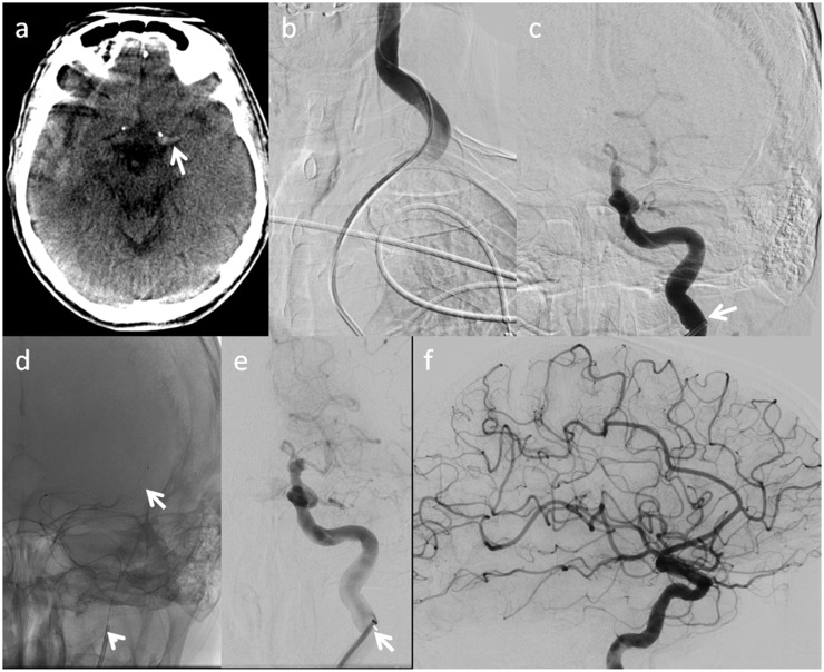
The treatment was performed under conscious sedation. A short femoral sheath was introduced into the right femoral artery (07:27 h). The left common carotid artery (CCA) was elongated (aortic arch type I), but could be accessed with a 8F Vista (Cordis, Milpitas, CA, USA) catheter for the first time at 07:37 h. Several attempts were made to access the internal carotid artery (ICA) with the 8F Vista or with the aspiration catheter (with a normal wire, stiff wire, exchange wire, with and without a 5F SIM2 as a guide for the 8F Vista as well as with a combination of an ACE68 aspiration catheter, Trevo pro 18 microcatheter and Transend EX Platinum microwire), but all attempts failed due to the tortuous anatomy of the CCA and the ostium of the ICA. Afterwards, the 5F SIM2 alone was advanced to the subpetrous level (07:58 h) of the left ICA over a 0.035-inch guide wire and subsequently a 0.021-inch microcatheter (Trevo pro 18, Stryker, Kalamazoo, MI, USA) was advanced to the superior M2 division of the left MCA. A control injection was performed to verify the correct position and a stentretriever (Trevo XP pro vue, 4 mm × 30 mm, Stryker) was deployed in the left MCA M1/M2. Notably the position of the 5F SIM2 alone was stable enough to deliver the microcatheter and the stentretriever. The stentretriever was deployed at 08:01 h (3 min after the SIM2 was placed in the distal internal carotid artery). An ACE 68 (Penumbra, Alameda, CA, USA) and a 8F Vista (Cordis) were then advanced in a bi-axial manner over the wire of the Trevo into the ICA/intracranial vasculature (at 08:03 h). Finally, thrombectomy was carried out using the so called Stentretriever Assisted Vacuum locked Embolectomy as described previously,13 resulting in a TICI3 result after three attempts (08.16 h) (Figure 1).
Figure 1.
(a) Initial flat detector computed tomography showing a dense artery sign of the distal internal carotid artery (ICA) and middle cerebral artery (arrow). (b) Injection into the left common carotid artery (CCA) through an 8F Vista (which could not be placed distally in the CCA or in the left ICA) depicting the elongation of the CCA. (c) Injection through a 5F SIM2 (arrow) placed in the distal ICA depicting an occlusion of the left carotid-T. (d) Unsubtracted image showing the deployed stentretriever (arrow) and the 5F SIM2 in the ICA (arrow head). (e) Angiogram showing the 8F Vista (arrow) placed in the ICA after the first (unsuccessful) thrombectomy manoeuvre. (f) Lateral angiogram showing the final results (TICI3) after three Stentretriever Assisted Vacuum locked Embolectomy manoeuvres.
Clinical outcome
Despite a complete recanalization within 4.5 h of symptom onset the patient suffered from a large MCA infarct of the left side and died two days after admission.
Discussion
After the publication of several prospective randomized trials in 2015, EVT is the standard of care in acute ischaemic stroke with underlying LAO.1,3 In particular, the results of the MR CLEAN trial have shown that comparatively loose inclusion criteria (including no upper age limit) are sufficient to identify patients that will profit from EVT.7 As a consequence we and others have adopted the MR CLEAN inclusion criteria and the number of treatments has risen significantly over recent years (from 40 in 2014 to more than 140 in 2016 at our centre). In addition we currently treat all patients within 6 h of onset regardless of their initial ASPECTS after the publication of the HERMES meta-analysis, which concluded that these patients might also profit from EVT.8 In summary, more patients are treated and, especially due to the fact that there is no upper age limit for EVT, more patients with tortuous cervical vessel anatomy are treated. A rate of 5% patients with difficult to access carotids in the setting of acute stroke treatment has been described.5 In these cases, the time loss to establish carotid access or the failure to access the carotid over a femoral route leads to worse clinical outcomes.5 A direct carotid artery puncture or a transradial access have been described as alternative access options in these patients.6,9 However, operators without a broad experience in interventional surgery have little or no experience with these options. As opposed to a direct puncture of the carotid or a transradial access, our approach to deliver a stentretriever first over a 5F diagnostic catheter and a microcatheter offers a technique most interventional neuroradiologists are familiar with and which, in our experience, should be possible in almost all cases despite a difficult anatomy of the aortic arch or a tortuous anatomy of the carotid artery. Once the stentretriever is deployed and the microcatheter as well as the 5F diagnostic catheter are withdrawn the implanted stentretriever can be easily used as a ‘grappling hook’ to deliver an intermediate and guide catheter to the cervical ICA or the intracranial vasculature, respectively.10 A similar approach has also been described before as the so called ‘anchor technique’, in which a stentretriever is used to deliver a large bore aspiration catheter to the intracranial vasculature in cases with tortuous cervical ICA anatomy.11 The wire of the stentretriever has been shown to be stable enough for advancement of a carotid stent/balloon in cases of tandem occlusions or carotid dissections.12 In our case, the total manoeuvre to place the stent and advance the guide catheter over the stentwire took 5 min after we had lost over 30 min in accessing the ICA with conventional methods. As a result, the stent-deployment as a first step may reduce procedural time in cases with difficult to access carotids. Notably, the deployed stent may allow for antegrade flow through the occluded vessel until a stable carotid access is achieved and thrombectomy can be performed, which might be another substantial advantage in the setting of a difficult to access carotid artery.
This novel strategy has to be evaluated carefully in terms of safety, success and procedural time in the near future; however, it offers another troubleshooting tool when access to the ICA with a large guide catheter seems impossible or is time consuming.
Additionally the treatment of patients with low initial ASPECTS should always be considered carefully because the current data from the prospective trials do not prove EVT to be superior in patients with low ASPECTS. Therefore imaging should always be repeated at the treating centre to avoid futile treatments.
Conclusion
The retriever first embolectomy (ReFirE) technique is an easy to handle alternative to direct carotid artery puncture and may be considered in cases with difficult to access carotid arteries.
Declaration of conflicting interests
The authors declared the following potential conflicts of interest with respect to the research, authorship, and/or publication of this article: DB: travel grants from Stryker (DGNR, ESMINT), MNP: travel grants and consultancy for Siemens and Stryker, both none regarding the content of the manuscript. MK: consultancy for Siemens.
Funding
The authors received no financial support for the research, authorship, and/or publication of this article.
References
- 1.Goyal M, Menon BK, van Zwam WH, et al. Endovascular thrombectomy after large-vessel ischaemic stroke: A meta-analysis of individual patient data from five randomised trials. Lancet 2016; 387: 1723–1731. [DOI] [PubMed] [Google Scholar]
- 2.Kuntze Soderqvist A, Andersson T, Ahmed N, et al. Thrombectomy in acute ischemic stroke: Estimations of increasing demands. J Neurointerv Surgery Epub ahead of print 26 August 2016. doi: 10.1136/neurintsurg-2016-012575. [DOI] [PubMed] [Google Scholar]
- 3.Wahlgren N, Moreira T, Michel P, et al. Mechanical thrombectomy in acute ischemic stroke: Consensus statement by ESO-Karolinska Stroke Update 2014/2015, supported by ESO, ESMINT, ESNR and EAN. Int J Stroke 2016; 11: 134–147. [DOI] [PubMed] [Google Scholar]
- 4.Mohlenbruch M, Pfaff J, Schonenberger S, et al. Endovascular stroke treatment of nonagenarians. AJNR Am J Neuroradiol 2017; 38: 299–303. [DOI] [PMC free article] [PubMed] [Google Scholar]
- 5.Ribo M, Flores A, Rubiera M, et al. Difficult catheter access to the occluded vessel during endovascular treatment of acute ischemic stroke is associated with worse clinical outcome. J Neurointerv Surg 2013; 5(Suppl. 1): i70–i73. [DOI] [PubMed] [Google Scholar]
- 6.Mokin M, Snyder KV, Levy EI, et al. Direct carotid artery puncture access for endovascular treatment of acute ischemic stroke: Technical aspects, advantages, and limitations. J Neurointerv Surg 2015; 7: 108–113. [DOI] [PubMed] [Google Scholar]
- 7.Berkhemer OA, Fransen PS, Beumer D, et al. A randomized trial of intraarterial treatment for acute ischemic stroke. N Engl J Med 2015; 372: 11–20. [DOI] [PubMed] [Google Scholar]
- 8.Goyal M, Menon BK, van Zwam WH, et al. Endovascular thrombectomy after large-vessel ischaemic stroke: A meta-analysis of individual patient data from five randomised trials. Lancet 2016; 387: 1723–1731. [DOI] [PubMed] [Google Scholar]
- 9.Dahm JB, van Buuren F, Hansen C, et al. The concept of an anatomy related individual arterial access: Lowering technical and clinical complications with transradial access in bovine- and type-III aortic arch carotid artery stenting. Vasa 2011; 40: 468–473. [DOI] [PubMed] [Google Scholar]
- 10.Hui FK, Hussain MS, Spiotta A, et al. Merci retrievers as access adjuncts for reperfusion catheters: The grappling hook technique. Neurosurgery 2012; 70: 456–460. [DOI] [PubMed] [Google Scholar]
- 11.Singh J, Wolfe SQ, Janjua RM, et al. Anchor technique: Use of stent retrievers as an anchor to advance thrombectomy catheters in internal carotid artery occlusions. Interventional neuroradiology: Journal of peritherapeutic neuroradiology, surgical procedures and related neurosciences 2015; 21: 707–709. [DOI] [PMC free article] [PubMed] [Google Scholar]
- 12.Behme D, Knauth M and Psychogios MN. Retriever wire supported carotid artery revascularization (ReWiSed CARe) in acute ischemic stroke with underlying tandem occlusion caused by an internal carotid artery dissection: Technical note. Interv Neuroradiol 2017: 1591019917690916. [DOI] [PMC free article] [PubMed]
- 13.Maus V, Behme D, Kabbasch C, et al. Maximising first-pass complete reperfusion with SAVE. Clin Neuroradiol. Epub ahead of print 13 February 2017. doi: 10.1007/s00062-017-0566-z. [DOI] [PubMed]


