Abstract
Objective:
To investigate the plan quality of tri-Co-60 intensity-modulated radiation therapy (IMRT) plans for spine stereotactic ablative radiotherapy (SABR).
Methods:
A total of 20 patients with spine metastasis were retrospectively selected. For each patient, a tri-Co-60 IMRT plan and a volumetric-modulated arc therapy (VMAT) plan were generated. The spinal cords were defined based on MR images for the tri-Co-60 IMRT, while isotropic 1-mm margins were added to the spinal cords for the VMAT plans. The VMAT plans were generated with 10-MV flattening filter-free photon beams of TrueBeam STx™ (Varian Medical Systems, Palo Alto, CA), while the tri-Co-60 IMRT plans were generated with the ViewRay™ system (ViewRay inc., Cleveland, OH). The initial prescription dose was 18 Gy (1 fraction). If the tolerance dose of the spinal cord was not met, the prescription dose was reduced until the spinal cord tolerance dose was satisfied.
Results:
The mean dose to the target volumes, conformity index and homogeneity index of the VMAT and tri-Co-60 IMRT were 17.8 ± 0.8 vs 13.7 ± 3.9 Gy, 0.85 ± 0.20 vs 1.58 ± 1.29 and 0.09 ± 0.04 vs 0.24 ± 0.19, respectively. The integral doses and beam-on times were 16,570 ± 1768 vs 22,087 ± 2.986 Gy cm3 and 3.95 ± 1.13 vs 48.82 ± 10.44 min, respectively.
Conclusion:
The tri-Co-60 IMRT seems inappropriate for spine SABR compared with VMAT.
Advances in knowledge:
For spine SABR, the tri-Co-60 IMRT is inappropriate owing to the large penumbra, large leaf width and low dose rate of the ViewRay system.
INTRODUCTION
Stereotactic ablative radiotherapy (SABR) is an effective tool for treating spine metastasis owing to its strong local disease control ability in conjunction with minimal complications and rapid recovery time for the patient.1–3 The most challenging part of spine SABR is ensuring that the dose delivered to the spinal cord is lower than its tolerance dose while still delivering a prescription dose to the target volume, as the target volume of spine SABR is close to the spinal cord.4,5 In certain cases, the target volume of spine SABR wraps around the spinal cord; therefore, a steep dose gradient between the target volume and the spinal cord, i.e. highly conformal dose distribution, is necessary in terms of treatment planning. From the perspective of plan delivery to a patient, highly accurate target localization is necessary for the delivery of the highly conformal dose distribution to guarantee local control as well as to prevent side effects due to radiotherapy. Therefore, image-guided radiation therapy (IGRT) has an important role in guiding spine SABR.1,6 The IGRT with MRI seems particularly beneficial for spine SABR, since the spinal cord can be distinguished from the cerebrospinal fluid in the MRI, which allows the spinal cord to be accurately defined.
Recently, a commercial IGRT system based on MR images acquired with a 0.35-T magnetic field, the ViewRay™ system (ViewRay inc., Cleveland, OH), has become clinically available. The ViewRay system uses a total of three Co-60 sources located at intervals of 120° in a ring-type bore as treatment radiation sources. The ViewRay system is equipped with double-focused multileaf collimators (MLCs) for each Co-60 source, which makes it possible to deliver static intensity-modulated radiation therapy (IMRT) plans. The width of each leaf of the MLC is 1.05 cm at the isoplane located 105 cm from the source. For treatment planning and patient localization before treatment, three-dimensional volumetric MRI can be acquired with the ViewRay system. During treatment, sagittal planar MR images can be acquired in almost real time for the monitoring of internal organ motion of the patient as well as for respiratory gating with anatomical information. Park et al7 showed the commissioning results of the ViewRay system, which was compatible with international guidelines such as the American Association of Physicists in Medicine Task Group (TG)-142 and TG-40. The IMRT capability of the ViewRay system was tested by Wooten et al based on the American Association of Physicists in Medicine TG-119 guidelines.8,9 Several treatment planning studies with the ViewRay system have been performed to identify the capability and benefits of the system. Wooten et al10 demonstrated the quality of the IMRT plans using the ViewRay system with a total of 33 patients with various diseases in the abdominal, pelvic, thorax and head and neck regions. They showed the clinically acceptable plan quality of the ViewRay system in comparison with the IMRT plans generated with conventional linear accelerator systems. Kishan et al11 showed that liver SABR plans generated with the ViewRay system could pass the mandatory Radiation Therapy Oncology Group (RTOG) 1112 organ at risk (OAR) constraints. Kim et al12 showed the 0.35-T magnetic field effect on the treatment plans for accelerated partial breast irradiation. Merna et al13 and Park et al14 showed that the ViewRay system was a feasible method for treating lung SABR. Various studies have investigated the treatment plan quality of the ViewRay system; however, no treatment planning study has been performed for spine SABR. Since the ViewRay imaging system is based on MRI, the ViewRay system seems beneficial for spine SABR owing to its ability to allow accurate definition of the spinal cord, which could reduce the volume of the spinal cord when compared with CT-based imaging systems. Accurate definition of the spinal cord using MRI could provide increased separation between the target volume and the spinal cord, which might reduce dose to the spinal cord. Moreover, the ViewRay system can monitor the spinal cord as well as cerebrospinal fluid flow during treatment in real time, allowing a more accurate beam delivery than conventional radiotherapy systems. Therefore, the MRI system of the ViewRay system seems to provide some benefits for spine SABR. However, the ViewRay system has some disadvantages in comparison with conventional linear accelerator systems in terms of beam delivery system. The penumbra of the ViewRay system is larger than that of most linear accelerators as it uses Co-60 sources. To minimize this disadvantage, the ViewRay system adopted double-focused MLCs. In terms of beam energy, the penetrating power of the Co-60 gamma ray is generally lower than that of conventional linear accelerators. Moreover, the MLC leaf width is relatively large compared with those of conventional linear accelerators, which is 1 cm at the source-to-surface distance (SSD) of 100 cm, while those of the Millennium 120™ MLC and the high definition (HD) 120™ (Varian Medical Systems, Palo Alto, CA) MLC are 0.5 and 0.25 cm, respectively. Therefore, it is unclear whether the tri-Co-60 IMRT plans for spine SABR with the ViewRay system would be better than those of IMRT or volumetric-modulated arc therapy (VMAT) with a linear accelerator. In this study, we investigated the tri-Co-60 IMRT plan quality of the ViewRay system in comparison with VMAT plans with a conventional linear accelerator. The spinal cord for the tri-Co-60 IMRT planning was determined based on MRI and a slightly larger spinal cord was defined for the VMAT planning. Clinically relevant dose–volumetric parameters were examined by comparing the tri-Co-60 IMRT plans with the VMAT plans.
METHODS AND MATERIALS
Patient selection and simulation
After institutional review board approval, we retrospectively selected a total of 20 patients with spine metastasis (T9–T12) in our institution. All patients received SABR using VMAT technique. Every patient underwent CT scans using the Brilliance CT Big Bore™ (Philips, Amsterdam, Netherlands) with a slice thickness of 1.5 mm.
Volumetric-modulated arc therapy planning for spine stereotactic ablative radiotherapy
The initial prescription dose was 18 Gy in a single fraction. If the delivered dose to the spinal cord was higher than the spinal cord tolerance dose, the prescription dose was reduced until it satisfied the tolerance level of the spinal cord. The clinical target volume was defined as the target volume for both VMAT planning and tri-Co-60 IMRT planning in this study. The VMAT plans were generated with the Eclipse™ system (Varian Medical Systems, Palo Alto, CA) with two full arcs and 10 MV in flattening filter-free (FFF) mode of TrueBeam STx™ (Varian Medical Systems, Palo Alto, CA). The VMAT plans were generated using the HD 120 MLCs, whose inner leaf width is 2.5 mm (outer leaf width = 5 mm). During optimization, dose constraints of the RTOG 0631 study were followed for sparing OARs to avoid complications.15 The progressive resolution optimizer 3 v. 10 (Varian Medical Systems, Palo Alto, CA) was used for optimization. For dose calculation, the anisotropic analytic algorithm v. 10 (Varian Medical Systems, Palo Alto, CA) was used with a dose calculation grid of 2 mm. All VMAT plans were normalized to cover 85% of the target volume with 100% of the prescription dose. The spinal cord for the VMAT planning was defined based on MR image registration to the CT images, and 1-mm isotropic margins were added.
Tri-Co-60 intensity-modulated radiation therapy planning for spine stereotactic ablative radiotherapy
For each patient, CT images and structures used for VMAT planning were also used for the planning of tri-Co-60 IMRT in order to eliminate disturbance factors induced by the deformable registration of the CT images to MR images. The CT image set and the structure set were exported from the Eclipse system in digital imaging and communications in medicine format. After that, the digital imaging and communications in medicine-formatted CT images and the structure set of each patient were imported to the ViewRay treatment planning system (TPS), which is the MRIdian™ system (ViewRay inc., Cleveland, OH). To determine the optimal number of treatment fields for spine SABR with the ViewRay system, we investigated the ViewRay plan quality for spine SABR, increasing the number of treatment fields. For the target volumes, the quality of the ViewRay plans with 12 fields (12-field plan) was similar to that of the plans with 27 fields (27-field plan). The differences in the minimum, maximum and mean doses between the 12-field plans and the 27-field plans were 0.3 ± 1.8, 0.6 ± 0.8 and 0.4 ± 0.5%, respectively (all with p > 0.05). The other dose–volumetric parameters of the target volume showed differences <1% between the 12-field plans and the 27-field plans (all with p > 0.05). Similarly, no noticeable differences were observed for OARs (all with p > 0.05). However, the calculated beam-on time of the 27-field plans increased by 17% on an average in comparison with that of the 12-field plans (p < 0.001). Therefore, a total of 4 beam groups, i.e. a total of 12 fields, were used for tri-Co-60 IMRT plans in this study.1,16 The gantry angles of each field were 100, 220 and 340° (Group 1); 30, 150 and 270° (Group 2); 70, 190 and 310° (Group 3); and 50, 170 and 290° (Group 4). The value of IMRT efficiency was 0.1 to allow high modulation of the fluence, which determines the level of smoothing of the fluence map. The value of level, which determines the number of beam segments for each field, was set to 20. The dose calculation algorithm used was the Monte Carlo calculation algorithm developed by the manufacturer ViewRay inc. (Cleveland, OH), with a dose calculation resolution of 3 mm. To be realistic, dose distributions were calculated with the presence of the magnetic field and imaging surface coils. Interdigitation of the MLCs was allowed. Target coverage for the tri-Co-60 IMRT was planned just as the VMAT plans, i.e. 85% of the target volume was covered by 100% of the prescription dose. During optimization, we followed RTOG 0631 dose–volumetric constraints for OARs.15 We defined a spinal cord based on the MR images and added no margins, which was a different method than that used for the VMAT plans. If the dose–volumetric constraints for OARs were not met, the prescription dose was reduced until it satisfied the tolerance doses for OARs. After finishing planning, the calculated dose distributions from the MRIdian system were exported and imported to the Eclipse system. Dose–volumetric analyses for the tri-Co-60 IMRT plans were performed with the Eclipse system.
Evaluation of the treatment plans
For both VMAT and tri-Co-60 IMRT plans, clinically relevant dose–volumetric parameters were evaluated according to the guidelines of the RTOG 0631 study.15 For the target volume, the minimum dose to 95% of the target volume (D95%), D90%, D80%, D5%, D1%, maximum dose, minimum dose and mean dose were calculated. The conformity index (CI) and homogeneity index (HI) were calculated as follows:17,18
| (1) |
| (2) |
where V100% is the volume receiving 100% of the prescription dose.
For the spinal cord, the volume irradiated by least 14 Gy of dose (V14 Gy), V10 Gy and D10% were calculated for both VMAT and tri-Co-60 IMRT plans. The values of V10.6 Gy and V8.4 Gy were calculated for each kidney. For lungs, the value of of both lungs was calculated. The V14.3 Gy of the colon, V11.2 Gy of the stomach, V14 Gy of the brachial plexus, V11.9 Gy of the oesophagus and V14 Gy of the cauda equina were calculated for each VMAT and tri-Co-60 IMRT plan. For the entire body, integral dose was calculated by multiplication of the body mean dose and the body volume. To examine doses higher than 50% of the prescription dose, the gradient measure (GM) and the gradient index (GI) by Paddick and Lippitz were calculated as follows:19,20
| (3) |
| (4) |
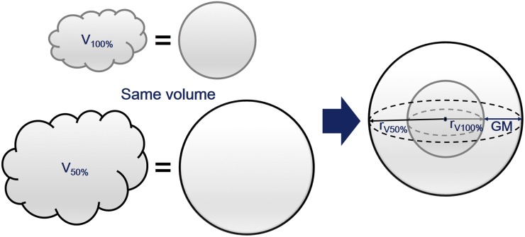
where is the equivalent sphere radius of n% of the prescription isodose, where the equivalent sphere is a sphere with the same volume as the one inside a given isodose surface and is the volume of n% of the prescription isodose. An illustration explaining the GM is shown in Figure 1.
Figure 1.
An example of an equivalent sphere of a structure and gradient measure (GM) is shown. The value of GM is calculated by subtracting the radius (r) of the equivalent sphere of volume receiving 100% of the prescription dose (V100%) from the radius of the equivalent sphere of volume receiving 50% of the prescription dose (V50%).
The statistical significance of the differences in the dose–volumetric parameters between VMAT and tri-Co-60 IMRT plans was analyzed with paired t-test.
To investigate the capability of VMAT and tri-Co-60 IMRT to generate steep dose gradients, we calculated the dose gradient (percent per millimetre). At the CT slice of the target volume centre in the craniocaudal direction, we drew a line from the centroid of the target volume to that of the spinal cord. After that we acquired the percent dose difference relative to the prescription dose between the boundary of the target volume and the boundary of the spinal cord. After that we defined the dose gradient by dividing the percent dose difference by the distance between boundaries of the target volume and the spinal cord. In addition, to identify the differences in the penumbra between photon beams generated with a conventional linear accelerator, and gamma rays generated with the Co-60 source of the ViewRay system, we calculated off-axis beam profiles with field sizes of 10 c × 10 cm and 4 × 4 cm at an SSD of 100 cm at depths of 3, 5, 10 and 15 cm in a water phantom with dimensions of 30 c × 30 × 30 cm.
RESULTS
Prescription doses of volumetric-modulated arc therapy and tri-Co-60 intensity-modulated radiation therapy plans
The average monitor unit of the VMAT plans was 6037 ± 799 MU. The mean prescription dose to the clinical target volume of the VMAT plans was 17.5 ± 0.9 Gy, while that of the tri-Co-60 IMRT plans was 12.6 ± 3.3 Gy. The mean spinal cord volumes of the VMAT plans and the tri-Co-60 IMRT plans were 4.4 ± 2.7 and 1.8 ± 1.0 cm3, respectively (p = 0.001). In the case of VMAT, 4 patients out of a total of 20 patients could not receive the initial prescription dose of 18 Gy to the target volume because irradiation of the spinal cord exceeded the tolerance dose due to the proximity of the spinal cord and target volume. Those four patients received a prescription dose of 16 Gy to ensure the delivered dose to the spinal cord was lower than the tolerance level. In the case of tri-Co-60 IMRT, only 3 patients out of a total of 20 patients could receive the initial prescription dose of 18 Gy to the target volume owing to high irradiation of the spinal cord. Eight patients could receive prescription doses equal to or less than 10 Gy to the target volume, which is the tolerance dose of the spinal cord according to the RTOG 0631 study (the volume ≤0.35 cm3 of the spinal cord should not be irradiated by a dose larger than 10 Gy).15 Therefore, those eight patients could not derive benefit from the IMRT technique as provided by the ViewRay system.
Dose–volumetric parameters of the target volume
The clinically relevant dose–volumetric parameters of the VMAT and tri-Co-60 IMRT plans are shown in Table 1. For the target volume, the differences in all dose–volumetric parameters examined in this study between the VMAT plans and the tri-Co-60 IMRT plans were statistically significant (with p < 0.05), except for the minimum dose (p = 0.155). Since the prescription doses of the VMAT plans were higher than those of the tri-Co-60 IMRT plans, the values of D95%, D90%, D80%, D5%, D1%, maximum dose, minimum dose and mean dose were higher in the VMAT plans than in the tri-Co-60 IMRT plans. The target conformity as well as dose homogeneity in the target volumes of the VMAT plans was better than that of the tri-Co-60 IMRT plans with statistical significance (0.85 vs 1.58 with p = 0.020 for CI and 0.09 vs 0.24 with p = 0.003 for HI). The dose–volumetric quality of the VMAT plans was superior to the quality of the tri-Co-60 IMRT plans for the target volume.
Table 1.
Average values of dose–volumetric parameters of volumetric-modulated arc therapy (VMAT) and tri-Co-60 intensity-modulated radiation therapy (IMRT) plans for spine stereotactic ablative radiotherapy
| Parameter | VMAT | Tri-Co-60 IMRT | p-value |
|---|---|---|---|
| Target volume | |||
| Volume (cm3) | 57.6 ± 46.4 | 57.6 ± 46.4 | – |
| D95% (Gy) | 16.8 ± 1.2 | 11.4 ± 2.7 | <0.0001 |
| D90% (Gy) | 17.5 ± 0.8 | 12.1 ± 3.0 | <0.0001 |
| D80% (Gy) | 17.7 ± 0.8 | 12.9 ± 3.4 | <0.0001 |
| D5% (Gy) | 18.4 ± 0.8 | 15.2 ± 4.9 | 0.010 |
| D1% (Gy) | 18.6 ± 0.8 | 15.5 ± 5.0 | 0.012 |
| Maximum dose (Gy) | 19.3 ± 0.9 | 15.9 ± 5.3 | 0.010 |
| Minimum dose (Gy) | 10.3 ± 3.2 | 9.1 ± 2.2 | 0.155 |
| Mean dose (Gy) | 17.8 ± 0.8 | 13.7 ± 3.9 | <0.001 |
| CI | 0.85 ± 0.20 | 1.58 ± 1.29 | 0.020 |
| HI | 0.09 ± 0.04 | 0.24 ± 0.19 | 0.003 |
| OAR | |||
| Spinal cord V14 Gy (cm3) | 0.0 ± 0.0 | 0.0 ± 0.0 | 0.332 |
| Spinal cord V10 Gy (cm3) | 0.3 ± 0.6 | 0.1 ± 0.1 | 0.325 |
| Spinal cord D10% (Gy) | 8.2 ± 2.5 | 10.1 ± 0.9 | 0.002 |
| Right kidney V10.6 Gy (%) | 0.3 ± 0.7 | 0.3 ± 0.7 | 0.879 |
| Right kidney V8.4 Gy (cm3) | 1.3 ± 3.1 | 1.1 ± 3.2 | 0.810 |
| Left kidney V10.6 Gy (%) | 0.0 ± 0.1 | 0.8 ± 2.0 | 0.123 |
| Left kidney V8.4 Gy (cm3) | 0.8 ± 1.3 | 3.9 ± 11.2 | 0.227 |
| Both lung (Gy) | 0.0 ± 0.0 | 0.0 ± 0.0 | – |
| Colon V14.3 Gy (cm3) | 0.0 ± 0.0 | 0.0 ± 0.1 | 0.330 |
| Stomach V11.2 Gy (cm3) | 0.4 ± 1.2 | 0.3 ± 1.2 | 0.778 |
| Brachial plexus V14 Gy (cm3) | 0.0 ± 0.0 | 0.0 ± 0.0 | N/A |
| Oesophagus V11.9 Gy (cm3) | 0.1 ± 0.2 | 0.3 ± 0.9 | 0.148 |
| Cauda equina V14 Gy (cm3) | 0.0 ± 0.0 | 0.0 ± 0.0 | – |
| Body integral dose (Gy cm3) | 16,570 ± 1768 | 22,087 ± 2986 | 0.044 |
| GM (cm) | 1.4 ± 0.4 | 2.2 ± 0.4 | <0.001 |
| GI | 4.5 ± 0.6 | 7.4 ± 1.9 | <0.001 |
| Plan parameters | |||
| Spinal cord volume (cm3) | 4.4 ± 2.7 | 1.8 ± 1.0 | 0.001 |
| Beam-on time (min) | 3.95 ± 1.13 | 48.82 ± 10.44 | <0.001 |
| Number of segments | – | 8.7 ± 5.0 | – |
| Dose gradient (%/mm) | 10.0 ± 2.7 | 2.6 ± 2.3 | <0.001 |
CI, conformity index; Dn%, the dose received at least n% volume of the structure; GI, gradient index; GM, gradient measure; HI, homogeneity index; OAR, organ at risk; VnGy, the volume irradiated by least n Gy of dose.
The italic values are statistically significant values (p < 0.05).
Dose–volumetric parameters of organs at risk
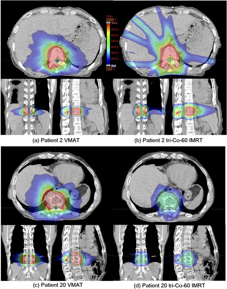
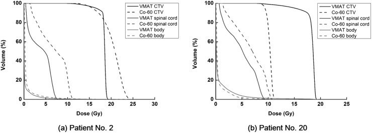
In terms of OARs, all plans, including both VMAT and tri-Co-60 IMRT plans, were clinically acceptable, showing lower irradiation of OARs than the tolerance doses set forth by the RTOG 0631 study.15 This was reasonable because we lowered the prescription doses to meet the spinal cord tolerance level, which is the most challenging part in spine SABR. With regard to the differences in the dose–volumetric parameters between VMAT plans and tri-Co-60 IMRT plans, no statistically significant differences were observed except D10% of the spinal cord (8.2 Gy for VMAT plans vs 10.1 Gy for tri-Co-60 IMRT plans with p = 0.002). However, the differences were not clinically significant. Representative dose distributions of a clinically appropriate case to use the ViewRay system for spine SABR (Patient 2) as well as a clinically inappropriate case to use the ViewRay system for spine SABR (Patient 20) are shown in Figure 2. Their dose–volume histograms are plotted in Figure 3. For the appropriate patient case (Patient 2), the target volume did not wrap around the spinal cord completely, while the target volume completely wrapped the spinal cord for the inappropriate patient case (Patient 20). The appropriate patient could receive the initial prescription dose of 18 Gy while maintaining a dose lower than the tolerance level to the spinal cord. On the contrary, the inappropriate patient could receive only 10 Gy as a prescription dose while maintaining a dose to the spinal cord below the tolerance level. In this case, no steep dose gradient was generated between the target volume and spinal cord; instead, a uniform dose was delivered to both the target volume and spinal cord.
Figure 2.
Representative dose distributions of a clinically appropriate spine stereotactic ablative radiotherapy (SABR) case to use the ViewRay™ system (ViewRay inc., Cleveland, OH) (Patient 2) as well as a clinically inappropriate case to use the ViewRay system (Patient 20) to perform spine SABR are shown. Dose distributions of a volumetric-modulated arc therapy (VMAT) plan (a) and a tri-Co-60 intensity-modulated radiation therapy (IMRT) plan (b) for Patient 2 are shown. For Patient 20, dose distributions of a VMAT plan (c) and a tri-Co-60 IMRT plan (d) are also shown.
Figure 3.
Representative dose–volume histograms (DVHs) of a clinically appropriate spine stereotactic ablative radiotherapy (SABR) case to use the ViewRay™ system (ViewRay inc., Cleveland, OH) (Patient 2) as well as a clinically inappropriate case to use the ViewRay system (Patient 20) to perform spine SABR are shown. The DVHs of Patient 2 (a) are shown for the target volume (black line), spinal cord (dark grey line) and body (grey line). The DVHs of Patient 20 (b) are also shown for the target volume (black line), spinal cord (dark grey line) and body (grey line). The DVHs of the volumetric-modulated arc therapy (VMAT) plans are shown with solid lines while those of the tri-Co-60 intensity-modulated radiation therapy plans are shown with dashed lines. CTV, clinical target volume.
The mean value of the body integral dose of the VMAT plans was lower than that of the tri-Co-60 IMRT plans with statistical significance (16,570 ± 1768 vs 22,087 ± 2986 Gy cm3 with p = 0.044). In the same vein, the mean values of GM and GI of the VMAT plans were lower than those of the tri-Co-60 IMRT plans with statistical significance (1.4 cm vs 2.2 cm with p < 0.001 for GM and 4.5 vs 7.4 with p < 0.001 for GI). The dose–volumetric quality of the VMAT plans was superior to the quality of the tri-Co-60 IMRT plans in terms of sparing dose to normal tissues.
Beam-on time, dose gradient and penumbra
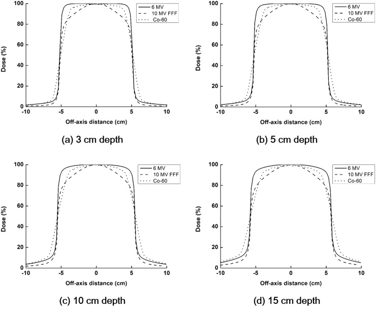
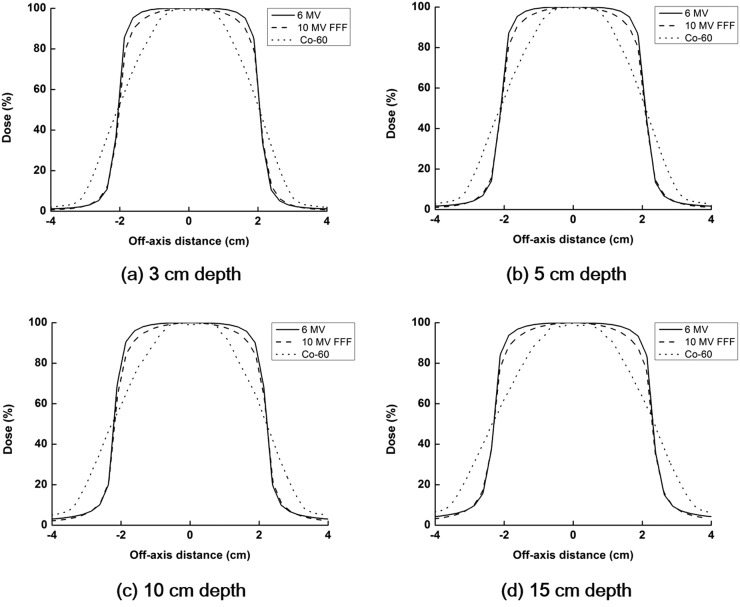
The average calculated beam-on time with the TPS was 3.95 min for the VMAT, while it was 48.82 min for the tri-Co-60 IMRT (p < 0.001). The beam-on time of the ViewRay system was calculated under the assumption that the activities of each Co-60 source were 15,000 Ci. The average values of the dose gradient of the VMAT plans and the tri-Co-60 IMRT plans were 10.0%/mm (1.9–15.8%/mm) and 2.6%/mm (0.1–9.3%/mm), respectively. The calculated penumbrae using the TPS of the 6- and 10-MV FFF photon beams by a linear accelerator and Co-60 gamma ray by the ViewRay system with field sizes of 10 × 10 cm2 and 4 × 4 cm2 are shown in Figures 4 and 5, respectively. The values of penumbrae at various depths are summarized in Table 2. The penumbrae of the Co-60 source with a field size of 10 × 10 cm2 were approximately two times larger than those of 10-MV FFF photon beams. Compared with the penumbrae of 6-MV photon beams, the penumbrae of the Co-60 source were approximately four times as large. In the case of 4 × 4 cm2, the penumbrae of the Co-60 source were approximately three and four times larger than those of 10-MV FFF and 6-MV photon beams, respectively.
Figure 4.
The calculated penumbrae using the treatment planning system in a water phantom with a dimension of 30 × 30 × 30 cm are shown. The field sizes are 10 × 10 cm and the source-to-surface distance is 100 cm. Penumbrae of 6- and 10-MV flattening filter-free (FFF) photon beams by a linear accelerator and Co-60 gamma ray by the ViewRay™ (ViewRay inc., Cleveland, OH) system are shown with solid lines, dashed lines and dotted lines, respectively. Penumbrae at depths of 3 (a), 5 (b), 10 (c) and 15 cm (d) are shown.
Figure 5.
The calculated penumbrae using the treatment planning system in a water phantom with a dimension of 30 × 30 × 30 cm are shown. The field sizes are 4 × 4 cm and the source-to-surface distance is 100 cm. Penumbrae of 6- and 10-MV flattening filter-free (FFF) photon beams by a linear accelerator and Co-60 gamma ray by the ViewRay™ system (ViewRay inc., Cleveland, OH) are shown with solid lines, dashed lines and dotted lines, respectively. Penumbrae at depths of 3 (a), 5 (b), 10 (c) and 15 cm (d) are shown.
Table 2.
Penumbrae of the photon beams generated with a linear accelerator and Co-60 radiation source
| Depth | 6-MV FFF | 10-MV FFF | Co-60 |
|---|---|---|---|
| Field size of 10 × 10 cm2 | |||
| 3-cm depth (cm) | 0.35 | 0.85 | 1.52 |
| 5-cm depth (cm) | 0.40 | 0.85 | 1.62 |
| 10-cm depth (cm) | 0.48 | 1.03 | 1.88 |
| 15-cm depth (cm) | 0.55 | 1.18 | 2.13 |
| Field size of 4 × 4 cm2 | |||
| 3-cm depth (cm) | 0.38 | 0.45 | 1.41 |
| 5-cm depth (cm) | 0.40 | 0.45 | 1.46 |
| 10-cm depth (cm) | 0.41 | 0.48 | 1.58 |
| 15-cm depth (cm) | 0.43 | 0.53 | 1.76 |
FFF, flattening filter free.
DISCUSSION
In this study, the quality of tri-Co-60 IMRT plans for spine SABR was compared with that of VMAT plans with an HD 120 MLC and 10-MV FFF photon beams. Although the volumes of the spinal cords of the tri-Co-60 IMRT were smaller than those of the VMAT plans (1.8 vs 4.4 cm3 with p = 0.001), VMAT plans showed consistently better plan quality in terms of both the target volume and OARs. The VMAT plans could deliver higher prescription doses than the tri-Co-60 IMRT plans while maintaining spinal cord irradiation below the tolerance level (17.5 ± 0.9 vs 12.6 ± 3.3 Gy for prescription dose). The values of CI and HI also indicated that the VMAT plans had better plan quality than the tri-Co-60 IMRT plans (0.85 vs 1.58 with p = 0.020 for CI and 0.09 vs 0.24 with p = 0.003 for HI). Normal tissue irradiation of the VMAT plans was consistently lower than that of the tri-Co-60 IMRT plans, showing lower integral doses as well as lower values of GM in the VMAT plans than those in the tri-Co-60 IMRT plans (16,570 vs 22,087 Gy cm3 with p = 0.044 for integral dose and 1.4 vs 2.2 cm with p < 0.001 for GM). This was due to the steep dose gradients of the VMAT plans around the target volume, which was 10%/mm on an average, compared with only 2.6%/mm for tri-Co-IMRT plans. This discrepancy was mainly caused by the large penumbra of the Co-60 source of the ViewRay system. Table 2 shows the penumbra of the ViewRay system, which is nearly two times larger than that of the 10-MV FFF photon beam generated with a linear accelerator. In addition, the large leaf width of the ViewRay system (1-cm leaf width at 100-cm SSD) likely contributed to the worse reduced target conformity and lower dose gradients near the target volume in the tri-Co-60 IMRT plans than that in the VMAT plans with TrueBeam STx (0.25-cm inner leaf width at 100-cm SSD), as several studies have already shown decreased target conformity when using MLCs with large leaf widths.21–23 Reviewing the results on normal tissue irradiation, the VMAT plans were also superior to the tri-Co-60 IMRT plans because of the lower penetrating power of the photon beams of the ViewRay system in comparison with a linear accelerator. Previous studies have demonstrated that low-energy photon beams increased body integral doses owing to the low penetrating power of the photon beams when treating thicker body parts.19,24 The body volume irradiated by doses higher than 50% of the prescription dose in the tri-Co-60 IMRT plans was larger than that of VMAT plans, showing higher values of GM. Comprehensively reviewing the dose–volumetric results, tri-Co-60 IMRT with the ViewRay system seems inappropriate for spine SABR in comparison with VMAT, since the dose–volumetric parameters of both the target volume and normal tissue indicated that the ViewRay system had a lower plan quality.
The ViewRay system was able to reduce the spinal cord radius by 1 mm owing to the use of MR images; however, the advantage gained from this extra precision was offset by the reduced ability to decrease dose around the target volume, as the ViewRay system was able to achieve dose gradients of only 2.6%/mm, in comparison with 10%/mm for the VMAT system. Therefore, the superior capability of the ViewRay system in terms of image guidance did not contribute to the improvement of the plan quality for spine SABR owing to its inferior beam delivery capability compared with VMAT.
The treatment time per fraction is another important consideration for spine SABR, since the patients with spine metastasis generally have difficulty holding the treatment position for a long period of time owing to pain.4 As shown in the results, the beam-on time of the ViewRay system was much longer than that of VMAT because the delivery technique of the ViewRay system is static IMRT, which takes much longer than VMAT.25,26 The long treatment time was also attributed to the low dose rate of the ViewRay system in comparison with the high dose rate of FFF photon beams (550 vs 2400 cGy min−1 at the depth of dose maximum).27 Thus, the increased treatment time further indicated that the ViewRay system is inappropriate for spine SABR.
The ViewRay system provides some novel features which are mainly based on MRI, such as the ability to achieve adaptive radiation therapy, no extra imaging dose to a patient and gating with almost real-time cine MR images considering internal organ motions. Several previous studies showed clinical acceptability of the treatment plans generated with the ViewRay system for tumours at various sites in the body.9–14 However, for spine SABR, the results in this study showed that no advantage is to be gained by utilizing the ViewRay system. When the target volumes are wrapped around the spinal cord completely, it is particularly inappropriate to use the ViewRay system. The ViewRay system was able to reduce the radius of the spinal cord by 1 mm by virtue of MRI, resulting in dose sparing of the spinal cord; however, this was not enough to offset the poor capability in generating steep dose gradients around the target volume. Moreover, one of the novel features of the ViewRay system, the gating capability based on real-time cine MR images, would not be advantageous for spine SABR, since the critical structures of spine SABR, including the target volume, are minimally affected by the respiratory motion of internal organs. For the above reasons, we conclude that tri-Co-60 IMRT with the ViewRay system is inappropriate for spine SABR.
Acknowledgments
ACKNOWLEDGMENTS
This research was supported by a grant of the Korea Health Technology R&D Project through the Korea Health Industry Development Institute (KHIDI), funded by the Ministry of Health & Welfare, Republic of Korea (grant number : HI16C0059).
Contributor Information
Chang Heon Choi, Email: dm140@naver.com.
So-Yeon Park, Email: vsoyounv@gmail.com.
Jung-in Kim, Email: madangin@gmail.com.
Jin Ho Kim, Email: jinho.kim.md@gmail.com.
Kyubo Kim, Email: kyubokim.ro@gmail.com.
Joel Carlson, Email: joelcarlsonesl@gmail.com.
Jong Min Park, Email: leodavinci@naver.com.
REFERENCES
- 1.Benedict SH, Yenice KM, Followill D, Galvin JM, Hinson W, Kavanagh B, et al. Stereotactic body radiation therapy: the report of AAPM Task Group 101. Med Phys 2010; 37: 4078–101. doi: https://doi.org/10.1118/1.3438081 [DOI] [PubMed] [Google Scholar]
- 2.Lee S, Chun M, Lee M. Stereotactic body radiotherapy for solitary spine metastasis. Radiat Oncol J 2013; 31: 260–6. doi: https://doi.org/10.3857/roj.2013.31.4.260 [DOI] [PMC free article] [PubMed] [Google Scholar]
- 3.Ryu S, Yoon H, Stessin A, Gutman F, Rosiello A, Davis R. Contemporary treatment with radiosurgery for spine metastasis and spinal cord compression in 2015. Radiat Oncol J 2015; 33: 1–11. doi: https://doi.org/10.3857/roj.2015.33.1.1 [DOI] [PMC free article] [PubMed] [Google Scholar]
- 4.Wu QJ, Yoo S, Kirkpatrick JP, Thongphiew D, Yin FF. Volumetric arc intensity-modulated therapy for spine body radiotherapy: comparison with static intensity-modulated treatment. Int J Radiat Oncol Biol Phys 2009; 75: 1596–604. doi: https://doi.org/10.1016/j.ijrobp.2009.05.005 [DOI] [PubMed] [Google Scholar]
- 5.Bhattacharya IS, Woolf DK, Hughes RJ, Shah N, Harrison M, Ostler PJ, et al. Stereotactic body radiotherapy (SBRT) in the management of extracranial oligometastatic (OM) disease. Br J Radiol 2015; 88: 20140712. doi: https://doi.org/10.1259/bjr.20140712 [DOI] [PMC free article] [PubMed] [Google Scholar]
- 6.Lee KN, Lee DJ, Suh TS. Three-dimensional verification of intracranial target point deviation using MRI-based polymer-gel dosimetry for conventional and fractionated stereotactic radiosurgery. J Radiat Prot Res 2011; 36: 107–18. [Google Scholar]
- 7.Park JM, Park S, Wu H, Kim J. Commissioning experience of tri-cobalt-60 MRI-guided radiation therapy system. Prog Med Phys 2015; 26: 193–200. doi: https://doi.org/10.14316/pmp.2015.26.4.193 [Google Scholar]
- 8.Ezzell GA, Burmeister JW, Dogan N, LoSasso TJ, Mechalakos JG, Mihailidis D, et al. IMRT commissioning: multiple institution planning and dosimetry comparisons, a report from AAPM Task Group 119. Med Phys 2009; 36: 5359–73. doi: https://doi.org/10.1118/1.3238104 [DOI] [PubMed] [Google Scholar]
- 9.Wooten HO, Rodriguez V, Green O, Kashani R, Santanam L, Tanderup K, et al. Benchmark IMRT evaluation of a Co-60 MRI-guided radiation therapy system. Radiother Oncol 2015; 114: 402–5. doi: https://doi.org/10.1016/j.radonc.2015.01.015 [DOI] [PubMed] [Google Scholar]
- 10.Wooten HO, Green O, Yang M, DeWees T, Kashani R, Olsen J, et al. Quality of intensity modulated radiation therapy treatment plans using a 60Co magnetic resonance image guidance radiation therapy system. Int J Radiat Oncol Biol Phys 2015; 92: 771–8. doi: https://doi.org/10.1016/j.ijrobp.2015.02.057 [DOI] [PubMed] [Google Scholar]
- 11.Kishan AU, Cao M, Wang PC, Mikaeilian AG, Tenn S, Rwigema JC, et al. Feasibility of magnetic resonance imaging-guided liver stereotactic body radiation therapy: a comparison between modulated tri-cobalt-60 teletherapy and linear accelerator-based intensity modulated radiation therapy. Pract Radiat Oncol 2015; 5: 330–7. doi: https://doi.org/10.1016/j.prro.2015.02.014 [DOI] [PubMed] [Google Scholar]
- 12.Kim J, Park S, Lee YH, Shin KH, Wu H, Park JM. Effect of low magnetic field on dose distribution in the partial-breast irradiation. Prog Med Phys 2015; 26: 208–14. doi: https://doi.org/10.14316/pmp.2015.26.4.208 [Google Scholar]
- 13.Merna C, Rwigema JC, Cao M, Wang PC, Kishan AU, Michailian A, et al. A treatment planning comparison between modulated tri-cobalt-60 teletherapy and linear accelerator-based stereotactic body radiotherapy for central early-stage non-small cell lung cancer. Med Dosim 2016; 41: 87–91. doi: https://doi.org/10.1016/j.meddos.2015.09.002 [DOI] [PubMed] [Google Scholar]
- 14.Park JM, Park SY, Kim HJ, Wu HG, Carlson J, Kim JI. A comparative planning study for lung SABR between tri-Co-60 magnetic resonance image guided radiation therapy system and volumetric modulated arc therapy. Radiother Oncol 2016; 120: 279–85. doi: https://doi.org/10.1016/j.radonc.2016.06.013 [DOI] [PubMed] [Google Scholar]
- 15.Ryu S, Pugh SL, Gerszten PC, Yin FF, Timmerman RD, Hitchcock YJ, et al. RTOG 0631 phase 2/3 study of image guided stereotactic radiosurgery for localized (1–3) spine metastases: phase 2 results. Pract Radiat Oncol 2014; 4: 76–81. doi: https://doi.org/10.1016/j.prro.2013.05.001 [DOI] [PMC free article] [PubMed] [Google Scholar]
- 16.Matuszak MM, Yan D, Grills I, Martinez A. Clinical applications of volumetric modulated arc therapy. Int J Radiat Oncol Biol Phys 2010; 77: 608–16. doi: https://doi.org/10.1016/j.ijrobp.2009.08.032 [DOI] [PubMed] [Google Scholar]
- 17.Shaw E, Kline R, Gillin M, Souhami L, Hirschfeld A, Dinapoli R, et al. Radiation Therapy Oncology Group: radiosurgery quality assurance guidelines. Int J Radiat Oncol Biol Phys 1993; 27: 1231–9. doi: https://doi.org/10.1016/0360-3016(93)90548-A [DOI] [PubMed] [Google Scholar]
- 18.Zhang P, Happersett L, Hunt M, Jackson A, Zelefsky M, Mageras G. Volumetric modulated arc therapy: planning and evaluation for prostate cancer cases. Int J Radiat Oncol Biol Phys 2010; 76: 1456–62. doi: https://doi.org/10.1016/j.ijrobp.2009.03.033 [DOI] [PubMed] [Google Scholar]
- 19.Park JM, Kim JI, Heon Choi C, Chie EK, Kim IH, Ye SJ. Photon energy-modulated radiotherapy: Monte Carlo simulation and treatment planning study. Med Phys 2012; 39: 1265–77. doi: https://doi.org/10.1118/1.3682172 [DOI] [PubMed] [Google Scholar]
- 20.Paddick I, Lippitz B. A simple dose gradient measurement tool to complement the conformity index. J Neurosurg 2006; 105: 194–201. doi: https://doi.org/10.3171/sup.2006.105.7.194 [DOI] [PubMed] [Google Scholar]
- 21.Lafond C, Chajon E, Devillers A, Louvel G, Toublanc S, Olivier M, et al. Impact of MLC leaf width on volumetric-modulated arc therapy planning for head and neck cancers. J Appl Clin Med Phys 2013; 14: 4074. doi: https://doi.org/10.1120/jacmp.v14i6.4074 [DOI] [PMC free article] [PubMed] [Google Scholar]
- 22.Park JM, Park SY, Kim JH, Carlson J, Kim JI. The effect of extremely narrow MLC leaf width on the plan quality of VMAT for prostate cancer. Radiat Oncol 2016; 11: 85. doi: https://doi.org/10.1186/s13014-016-0664-0 [DOI] [PMC free article] [PubMed] [Google Scholar]
- 23.Shang Q, Qi P, Ferjani S, Xia P. Effect of MLC leaf width on treatment adaptation and accuracy for concurrent irradiation of prostate and pelvic lymph nodes. Med Phys 2013; 40: 061701. doi: https://doi.org/10.1118/1.4803499 [DOI] [PubMed] [Google Scholar]
- 24.Park JM, Choi CH, Ha SW, Ye SJ. The dosimetric effect of mixed-energy IMRT plans for prostate cancer. J Appl Clin Med Phys 2011; 12: 3563. doi: https://doi.org/10.1120/jacmp.v12i4.3563 [DOI] [PMC free article] [PubMed] [Google Scholar]
- 25.Kim YS, Lee J, Park JI, Sung W, Lee SM, Kim GE. Volumetric modulated arc therapy for carotid sparing in the management of early glottic cancer. Radiat Oncol J 2016; 34: 18–25. doi: https://doi.org/10.3857/roj.2016.34.1.18 [DOI] [PMC free article] [PubMed] [Google Scholar]
- 26.Park JM, Kim K, Chie EK, Choi CH, Ye SJ, Ha SW. RapidArc vs intensity-modulated radiation therapy for hepatocellular carcinoma: a comparative planning study. Br J Radiol 2012; 85: e323–9. doi: https://doi.org/10.1259/bjr/19088580 [DOI] [PMC free article] [PubMed] [Google Scholar]
- 27.Kim JI, Park SY, Kim HJ, Kim JH, Ye SJ, Park JM. The sensitivity of gamma-index method to the positioning errors of high-definition MLC in patient-specific VMAT QA for SBRT. Radiat Oncol 2014; 9: 167. doi: https://doi.org/10.1186/1748-717X-9-167 [DOI] [PMC free article] [PubMed] [Google Scholar]


