Abstract
Purpose/Background
Several studies have examined the effect of hamstring strength exercises upon hamstring strains in team sports that involve many sprints. However, there has been no cross comparison among muscle activation of these hamstring training exercises with actual sprinting. Therefore, the aim of this study was to examine different hamstring exercises and compare the muscle activity in the hamstring muscle group during various exercises with the muscular activity produced during maximal sprints.
Methods
Twelve male sports students (age 25 ± 6.2 years, 1.80 ± 7.1 m, body mass 81.1 ± 15.6 kg) participated in this study. Surface EMG electrodes were placed on semimembranosus, semitendinosus and biceps femoris to measure muscle activity during seven hamstrings exercises and sprinting together with 3D motion capture to establish at what hip and knee angles maximal muscle activation (EMG) occurs. Maximal EMG activity during sprints for each muscle was used in order to express each exercise as a percentage of max activation during sprinting.
Results
The main findings were that maximal EMG activity of the different hamstring exercises were on average between 40-65% (Semitendinosus), 18-40% (biceps femoris) and 40-75% (Semimembranosus) compared with the max EMG activity in sprints, which were considered as 100%. The laying kick together with the Nordic hamstring exercises and its variations had the highest muscle activations, while the cranes showed the lowest muscle activation (in all muscles) together with the standing kick for the semimembranosus. In addition, angles at which the peak EMG activity of the hamstring muscle occurs were similar for the Nordic hamstring exercises and different for the two crane exercises (hip angle), standing kick (hip angle) and the laying kick (knee angle) compared with the sprint.
Conclusions
Nordic hamstring exercises with its variation together with the laying kick activates the hamstrings at high levels and at angles similar to the joint angles at which peak hamstring activation occurs during sprinting, while cranes did not reach high levels of hamstring activation compared with sprinting.
Level of Evidence
1b
Keywords: Electromyography, muscle activity, hamstring, sprint
INTRODUCTION
Hamstring strain injuries are one of the most frequently occurring injuries in sports, representing approximately 12-24% of all athletic injuries.1-3 There is a high prevalence of hamstring strain injuries in many sports, including soccer, 4,5 Australian football, 6 American football7 and sprinting. High-speed running is a common denominator among these activities and is the activity accounting for the majority of hamstring strains.8
The hamstring muscles are mostly active during the late swing phase and the start of the stance phase.9 During sprinting the hamstring muscles contract eccentrically during the late swing and late stance phases to control knee and hip extension, which makes the risk of hamstring injury greatest during those phases.10,11 Yu, et al.10 argued that hamstring strain injuries may be most likely to occur at the muscle tendon junction during the late stance phase, and in the muscle belly during the late swing phase.
Different risk factors for these hamstring strains were identified, which can be categorized in two categories: 1) unmodifiable factors like age,12 previous hamstring injury2,12-14 and 2) modifiable factors: hamstring weakness,14,15, muscle fatigue,16 decreased flexibility,17 poor running technique and altered neuromuscular function.18 Since it is not possible to affect the unmodifiable factors, only the modifiable factors are of interest when aiming to avoid a future hamstring strain.
Hamstring weakness is one of the most common risk factors associated with hamstring injuries. It has been suggested that hamstrings can produce sufficient force to counter the force produced by the quadriceps during various movements.15 Thereby, a stronger muscle could provide adequate protection from stretching and tearing the muscle fibers.8
Several training studies were conducted to investigate how to decrease weakness and increase neurological control. Guex, et al.8 presented a conceptual framework for strengthening the hamstring and for developing specific exercises. They proposed six key parameters to be considered when developing exercises: contraction type, load, range of motion, angular velocity, uni/bilateral exercises, and the kinetic chain. Guex, et al.8 concluded that the hamstring strength exercises used should be specific to simulate the power developed by the hamstring during the late swing phase of sprinting. However, none of the existing training studies directly compared the hamstring activity during the different strength training exercises and the activity during the sprint phase, which makes it difficult to determine whether the muscle training is specific enough for high speed running. Furthermore, it is not known if the angles at which the different exercises are conducted are similar to the position of the limb during late swing phase and thereby the hamstring length at peak tension. At present, information is limited regarding what resistance training maximally activates the hamstring, which exercise is the most specific related to sprint and targets the hamstrings strength in the most vulnerable position that occurs in high speed running: the angles seen in late swing phase, that occur with a fast eccentric contraction. Training the hamstring muscle group is critical for performance and plays an important role in hamstring injury prevention. The Nordic hamstring exercises is commonly used for prevention of hamstring strains. It is an exercise that is suggested to target the hamstrings effectively and has been shown to prevent hamstring strains.19 However, whether the angles at which the exercise is conducted are similar to the late swing phase has not been studied, and it is not known if hamstring muscle activity is high enough during this exercise to elicit a strength training stimulus, which has been suggested to be at least 70% of a maximal voluntary contraction.20-22 There may be more effective strength exercises like some explosive exercises that may better target the hamstring muscles, and at more specific angles and higher movement velocities that resemble the demands of the late swing and early stance phase in high speed running.
Therefore, the aim of this study was to examine different hamstring exercises and compare the muscle activity in the hamstring muscle group during various exercises with the muscular activity produced during maximal sprints. The gained information regarding muscle activity during the different hamstring strength exercises could help trainers, physiotherapists, and athletes to develop strength training programs that could target the hamstrings in an effective way to gain hamstring strength and potentially reduce the chance of hamstring strain during high-speed running.
MATERIALS AND METHODS
To compare maximal electromyography (EMG) activity for the hamstring muscle group, during a maximal sprint with several types of hamstring strength exercises, seven popular hamstring exercises were chosen. The exercises were able to be performed without much specific strength equipment. Some of the chosen exercises, such as the Nordic hamstring exercise, were selected from existing research papers.27,28 In addition, some modifications to the Nordic hamstring exercise that have been suggested to target the hamstrings even more than the standard Nordic hamstring exercise, were included as well as some less well investigated exercises that simulate higher movement velocities seen in high speed running. All the exercises focused on targeting the hamstrings in lengthened conditions, which represents the risk situation the hamstrings undergo during the running cycle.
SUBJECTS
Twelve male sports students (age 25 ± 6.2 years, 1.80 ± 7.1 m, body mass 81.1 ± 15.6 kg) participated in the study. Participants were excluded from the study if they had a former hamstring strain (the previous year), or if they had muscular pain or illness that could reduce their effort under each exercise. All participants were familiar with resistance training of the lower extremity. The participants were asked to refrain from any heavy strength training targeting the lower body during the 48 hours before testing, in order to ensure that they were free from strains and well trained. Before testing, a written consent was contained from the participants. The study was conducted with approval of the Regional Committee for Medical Research Ethics and conformed to the latest revision of the Declaration of Helsinki.
Procedures
One to two weeks before testing day each subject had a familiarization session with the different exercises and the sprint on a non-motorized treadmill. On testing day, the subjects were briefed about the exercise order. The sprint was always conducted first (after warm-up) followed by the seven hamstring exercises, which were performed in a randomized order for each subject to avoid the order effect due to fatigue. Before the warm-up, the hamstring area of the right leg was shaved, making the electrode adherence faster. After shaving the leg, the subjects performed a general warm up: 15 min of running on a non-motorized treadmill. Following the warm-up, the subjects were allowed five minutes of rest, where they were allowed to drink water. During this rest, electrodes and 21 retroreflective markers were placed on the hamstring muscles and anatomical landmarks.
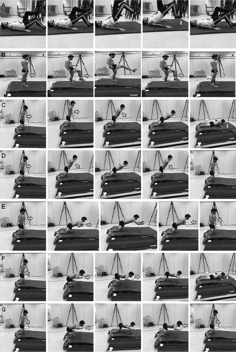
The test started with a sprint wearing their regular running shoes on the Woodway Curve 2.0 (Woodway Inc., Waukesha, USA). This is a non-motorized treadmill with curved running surface, which makes it possible for the subjects to run upright and at their own controlled pace. Furthermore, it made it possible to easily measure maximal peak velocity together with 3D kinematics and muscle activity. After the sprint the subject had five minutes rest before performing one of the seven hamstring strength exercises. The different exercises were: a) the laying hamstring kick, b) the standing hamstring kick, c) Nordic hamstring, d) Nordic hamstring with return, e) Nordic hamstring + bump, f) Hamstring cranes without return, and the g) Hamstring cranes with return exercise. See Figure 1 for the description and performance of each exercise. Limiting the test to three repetitions per exercise, randomization of the exercise order, and five minutes of recovery1 provided between the exercises were employed to reduce fatigue and an order effect.
Figure 1.
Performance of the different hamstring exercises. (A) Laying kick: lay on back with on shoulders, hips, and right heel on the ground, kick left heel up, in order to lift right foot off the ground and land on it again (B) Standing kick: stand on left foot, lift right knee and then kick out rapidly with the right foot. (C) Nordic hamstrings: with the feet held by a belt, lean forward with straight hips and back, until unable to hold, release and absorb forces with the hands in an eccentric push up motion. (D) Nordic with return: Same set up as (C), lean forwards with straight hips until unable to hold any longer, and return to the upright starting position. (E) Nordic with bump: lean forwards at the limit of your ability to hold, then move the 5kg weight straight forwards and back as fast as possible. (F) Cranes: flex hips to 90 degrees. Then extend knees until unable to hold it anymore, absorb forces with the hands in an eccentric push up motion. (G) Cranes with return: flex hips to 90 degrees. Then extend knees until unable to hold any longer, and return to the starting position.
Measurements
Electromyography (EMG) was used to quantify muscle activity during the sprint and the various exercises. Wireless EMG was recorder by using a Musclelab 6000 system and analyzed by Musclelab™ v10.73 software (Ergotest Technology AS, Langesund, Norway). Before placing the gel coated self-adhesive electrodes (Dri-Stick Silver circular sEMG Electrodes AE-131, NeuroDyne Medical, USA), the skin was shaved, abraded and washed with alcohol. The electrodes (11 mm contact diameter and a 2 cm center-to-center distance) were placed along the presumed direction of the underlying muscle fiber according to the recommendations by SENIAM 23. The electrodes were placed on the right leg on the muscle belly of the biceps femoris, semitendinosus and semimembranosus. The raw EMG signals, sampled at 1000 Hz were amplified and filtered using a preamplifier located as close to the pickup point as possible directly connected to the electrodes. The signals were bandpass filtered (fourth-order Butterworth filter) with cut-off frequencies of 20 Hz and 500 Hz. The preamplifier had a common mode rejection ratio of 100 dB. The EMG signals were converted to root mean square (RMS) EMG signals using a hardware circuit network (frequency response 20 - 500 kHz, integrating moving average filter with 100 ms width, total error ± 0.5%). In this study, a comparison of muscle activation during the various exercises was investigated in relation to the maximal EMG activity that occurred during a maximal sprint. The peak RMS converted and filtered data was obtained from the hamstring exercises and presented as percentage of the maximum activation. The peak RMS converted and filtered data from the muscles during the sprint was used as reference (100%).
A three-dimensional (3D) motion capture system (Qualysis, Gothenburg, Sweden) with eight cameras operating with a frequency of 500 Hz was used to track reflective markers, creating a 3D positional measurement. The markers were placed on spinous processes of the fifth lumbar vertebrae, one on each side of the body on lateral tip of the acromion, the iliac crests, greater trochanters, on the lateral and medial condyles of the knee, on the lateral and medial malleolei, the distal ends of metatarsals I and V. Segments of the feet, lower and upper leg, pelvis and trunk were made in Visual 3D v5 software (C-Motion, Germantown, MD, USA). Joint angles were measured during the sprint and all seven exercises. 3D motion capture data was synchronized with the wireless EMG recordings. The hip and knee joint angles at which maximal muscle activation of the biceps femoris, semitendinosus and semimembranosus occurred were recorded, and were used for further analysis.
STATISTICAL ANALYSIS
To assess differences in kinematics and EMG activity during the sprint and the seven exercises, a One-way ANOVA with repeated measures for each of three muscles was used. In addition, to compare the kinematics (knee and hip joint angles) at which maximal muscle activity for each muscle occurred in the sprint and the seven exercise a 3 (muscles: semitendinosus, semimembranosus and biceps) × 8 (exercise) with repeated measures performed. If the sphericity assumption was violated the Greenhouse-Geisser adjustments of the p-values were reported in the results. Post hoc test using Holm-Bonferroni probabilities adjustment was used to locate significant differences. The level of significance was set at p≤0.05. For statistical analysis purposes SPSS Statistics v21 (SPSS, Inc., Chicago, IL) was used. All results are presented as means ± standard deviations and effect size was evaluated with (Eta partial squared) where 0.01<η2<0.06 constitutes a small effect, a medium effect when 0.06<η2<0.14 and a large effect when η2>0.14 24.
RESULTS
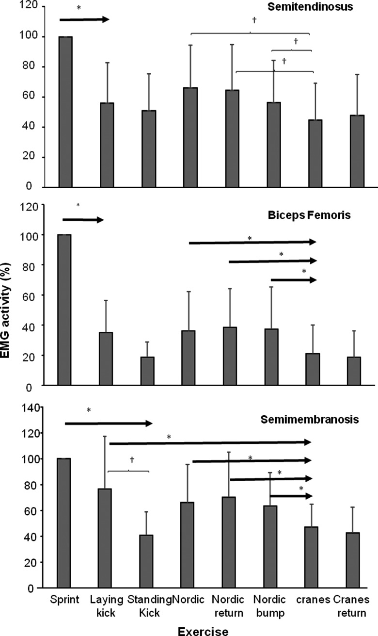
The maximal sprint velocity on the non-motorized treadmill was 22.5 ± 2.0 km/t, Significant differences in EMG activity for the Semitendinosus (F = 9.28, p<0.001, η2 = 0.48), Semimembranosus (F = 14.1 p < 0.001, η2 = 0,56) and Biceps femoris (F = 47.45, p < 0.001, η2 = 0.81) were found between the sprint and the seven exercises. Post hoc comparison showed that the maximum EMG activity during sprinting was significantly higher compared with all other exercises for the semitendinosus and biceps femoris, while for the semimembranosus the laying kick was not significantly different from the sprint (Figure 2). In addition, the semimembranosus EMG activity was also significantly higher during the laying kick compared with the standing kick, and the two cranes exercises (Figure 2). For the semitendinosus and biceps femoris the maximal EMG activity during the crane and the crane with return were significantly lower than the three different Nordic hamstrings exercises. For the semimembranosus only the EMG activity of the cranes were significantly lower than the three Nordic hamstring exercises (Figure 2).
Figure 2.
Maximum EMG activity for semitendinosus, semimembranosus, and biceps femoris during the different exercises (%) related to the sprint (100%).
*Indicates a significant difference between this EMG activity and to the right of the arrow on a (p<0.05)
†Indicates a significant difference in EMG activity between these two exercises (p<0.05)
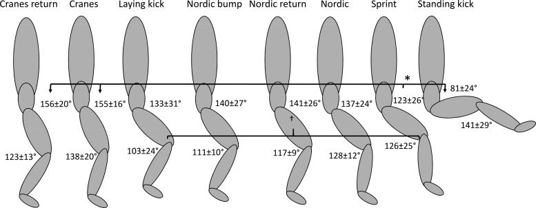
No significant effect between the muscles were found for the hip (F = 0.79, p = 0.47, η2=0.14), and knee angle (F = 0.46, p = 0.64, η2=0.07) at which the maximal EMG activity occurred. However, the hip (F = 9.1, p<0.001, η2=0.65) and knee joint angles (F = 13.0, p < 0.001, η2=0.69) at which the maximal EMG activity occurred was significantly different between the exercises (Figure 3 and Table 1). Post hoc comparison showed that the knee angle at which maximal EMG occurred was significantly lower (more knee flexion) in the laying kick for all three muscles compared with the knee joint angle during the sprints. In addition, the knee angle at which maximal EMG of the biceps occurred during the standing kick was significantly greater (less knee flexion) than the angle during the sprints (Table 1). The hip angle of maximal EMG was significantly greater (less hip flexion) for the cranes and cranes with return and lower (more hip flexion) for the standing kick compared with the sprints for all three muscles (Table 1).
Figure 3.
The angles (+/-SD) at maximal EMG activity for the hip and knee joints, averaged over all three muscles for each hamstring exercise and sprint.
*Indicates a significant difference for hip joint angle for this exercise with the sprints (p<0.05)
†Indicates a significant difference for knee joint angle between this exercise and the sprints on a (p<0.05)
Table 1.
Hip and knee joint angles at which maximal EMG activity was recorded of the three muscles during sprint and the seven hamstring strength exercises. All values are reported as degrees, +/− standard deviations.
| Sprint | Laying kick | Standing kick | Nordic | Nordic with return | Nordic with bump | Cranes | Cranes with return | |
|---|---|---|---|---|---|---|---|---|
| Hip joint angle | ||||||||
| Semimembranosus | 123 ± 25 | 134 ± 31 | 82 ± 27 | 140 ± 24 | 141 ± 26 | 140 ± 29 | 150 ± 18 | 160 ± 21 |
| Semitendinosus | 120 ± 25 | 132 ± 30 | 80 ± 27 | 135 ± 22 | 141 ± 26 | 138 ± 25 | 154 ± 18 | 153 ± 22 |
| Biceps Femoris | 126 ± 28 | 134 ± 33 | 81 ± 30 | 138 ± 24 | 141 ± 27 | 141 ± 27 | 160 ± 13 | 156 ± 19 |
| Knee joint angle | ||||||||
| Semimembranosus | 123 ± 27 | 107 ± 24 | 133 ± 38 | 128 ± 15 | 119 ± 7 | 111 ± 9 | 141 ± 15 | 119 ± 15 |
| Semitendinosus | 128 ± 27 | 103 ± 29 | 137 ± 30 | 128 ± 11 | 120 ± 9 | 112 ± 9 | 133 ± 24 | 125 ± 12 |
| Biceps Femoris | 127 ± 23 | 98 ± 19 | 153 ± 20 | 129 ± 10 | 111 ± 12 | 111 ± 12 | 139 ± 23 | 125 ± 12 |
→ sign indicates a significant difference between sprints with this exercise on a p<0.05 level.
No significant interaction effect was found for the occurrence at which the maximal EMG activity occurred for the hip (F = 0.23, p = 0.99, η2=0.04) and knee joint angles (F = .52, p = 0.98, η2=0.08).
DISCUSSION
The aim of this study was to compare hamstring muscles activity during different hamstring strengthening exercises with the hamstring muscles activation during a maximal sprint. The main findings were that maximal EMG activity of the different hamstring exercises were on average between 40-65% (Semitendinosus), 18-40% (biceps femoris) and 40-75% (Semimembranosus) of the max EMG activity produced by the hamstrings during a sprint. Cranes showed the lowest muscle activation (in all muscles) together with the standing kick for the semimembranosus (Figure 2). In addition, angles at which the peak EMG activity of the hamstring muscles occurred differed between the two crane exercises (hip angle), standing kick (hip angle) and the laying kick (knee angle) when compared with the sprint.
Analyses of hamstring activation reveal that the maximal sprint resulted in the highest muscle activity for the semitendinosus and biceps femoris when compared to all of the hamstring exercises. For the semimembranosus only the laying kick had no significant difference in level of activation with the sprint, while maximal activation during the other exercises was lower than activation during the sprint.
It was expected that hamstring activation during the sprints would be the highest since it involves rapid hip and knee joint movements that utilizes the hamstring to a large degree. Previous authors 25-27 have found that hamstring muscles are most active during the late swing phase of the sprint, in order to slow the forward moving limb. During this part of the running gait cycle, the hamstring muscles work mostly eccentrically, therefore the authors’ chose different hamstring strength exercises also suggested to eccentrically stimulate the hamstrings. However, most of the studied exercises reported an EMG activity lower than 70% of maximal EMG activity during sprint. Only the semitendinosus during the three Nordic exercises the and the laying kick demonstrated an average activation of respectively around 70% and 80% and was not significantly different from the sprint. It has been suggested that in order to gain strength in a muscle, an activity has to be at least 70% of a maximal voluntary contraction. 20,22 In the present study, no maximal voluntary contractions were measured, rather activity during exercises were related to the maximal contractions demonstrated during sprints. In support of this methodology, Jönhagen, et al. 25 showed that maximal voluntary contractions in the hamstring were equal to maximal muscle activation during maximal sprints. None of the investigated exercises in the present study reached the level of activation needed for strength gain (70%) for the biceps femoris (Figure 2). Muscle activity during the two crane exercises and the standing kick were also not of a high enough intensity to be a strengthening stimulus for any of the muscles.
Despite the relatively low levels of muscle activation (≤70%) during the Nordic hamstrings exercises several recent studies have shown that training the Nordic hamstring exercise can increase strength and muscle activation of the hamstrings.28,29 Bourne, et al.28 showed that Nordic hamstring exercise training promoted the elongation of the fascicles of the biceps femoris long head while it did not promote hypertrophy of the biceps femoris long head. They found that it preferentially developed hypertrophy of the semitendinosus. This is in accordance with the present study that shows activation levels in the semitendinosus that reached almost 70% compared with the biceps femoris of only 40% when compared to values during sprinting. Delahunt, et al.29 found similar average percentage EMG levels for the semitendinosus (65% of eccentric MVC) in the Nordic hamstring exercise, as in the present study. However, after six weeks of Nordic hamstring exercise training the subjects increased this percentage to 80% of eccentric MVC. In the present study some subjects had much training experience with the chosen hamstring exercises, while others did not have as much experience. This may have influenced the variation in EMG activity percentage between subjects during these exercises as shown by the large standard deviations (Figure 2).
It was expected that the hamstring activity would be higher in the modifications of the Nordic hamstring exercise (Nordic with return and Nordic with bump) than the traditional Nordic hamstring exercise, especially the Nordic with bump. By leaning forwards as far as possible the subject could hold their body with the hamstring muscles and then add the movement of pushing and pulling a 5 kg weight (Figure 1), which was projected to increase the activity of the hamstring muscles even more. However, this was not the case. An explanation for these findings may be that the subjects did not lean as far forward during the modified Nordic hamstrings exercises as shown in the lower knee angles achieved during the exercise (Table 1 and Figure 3). This causes lower initial work for hamstrings, but the extra weight that has to be pushed, or the return movement compensates for this decrease in leaning. The same was found between the cranes and the cranes with return i.e. less shifting forward in the cranes with return (lower knee angle) so that the subjects could be sure that they had the strength to return to starting position. This resulted in the same maximal EMG activation during these two crane exercises. It seems that the level of maximal activation determines the degree of forward lean during these variations of Nordic Hamstring exercise, as the joint angles are similar, and thus these modified Nordic hamstring exercises may be used as variations of the traditional Nordic hamstring during training.
The lower activity in the cranes exercises were probably caused by the different hip angles (more extended) at which maximal hamstring activity was measured as compared to the sprints (Table 1, Figure 3). During the crane exercises subjects first have to flex their hips. This increases the load on the hamstring due to the weight of the trunk that is moving toward horizontal. From there subjects move forwards, while the trunk is maintained horizontally. This probably causes that the hip angle at which the maximal hamstring EMG activity was measured to occur at another hip angle, while the knee angles were comparable with the Nordic exercises (Table 1, Figure 3). The alterations in hip angle in the presence of the same knee angle likely resulted in a different effective hamstring muscle length and thereby lowered muscle activity that was measured (Figure 2).
The standing kick did cause similar, low-level muscle activation as the cranes (Figure 2), which was probably caused by the absence of an added weight that could load the hamstrings. In all other exercises the trunk causes extra load to the hamstrings, as compared with the sprint, during which the load on the hamstrings is caused by the lower and upper limb movements. The standing kick was included to investigate whether kicking in a position of higher knee extension and flexion than in sprinting would cause an extra load to the hamstrings. The present data suggests that this was not the case. However, adding extra weights around the ankle joint may potentially have increased the hamstrings activity to a level comparable to the other exercises, but this was not investigated.
The maximal hamstring activity during sprint was measured for all three muscles at around the same time in the late swing phase, which was comparable with earlier studies 25-27,30. The angles of Nordic hamstrings and modified hamstring exercises at which maximal hamstring activity occurred was similar to the maximal activity seen during sprints indicating that these exercises target the hamstring muscles at the right part of the running cycle when following the principle of specificity. The laying kick exercise produced similar EMG activity to the Nordic hamstring exercises, and for the semitendinosus reached almost the same activity level as in the sprints indicating that this also could be a good hamstring strength exercise. However, in the present study the knee flexion angle was much more flexed for this exercise compared to the sprints, which could target the hamstring muscle at the wrong angle. Yet, subjects were not familiar with this exercise and were perhaps therefore a bit reluctant to put a potentially greater load on the hamstrings by putting the foot more distally on the ground and thereby decreasing knee flexion angle to a degree comparable to the sprint knee angles. With this variation, the exercise could target the hamstring muscle closer angles to those used during sprinting and possibly increase maximal EMG hamstring activity.
When comparing EMG hamstring activity between the laying kick and the Nordic hamstring exercise there were no differences found, while in the laying kick only one leg is trained and in the Nordic hamstring exercises both legs were used. To increase maximal EMG hamstring activity during the Nordic exercises it is possible to perform them with only one leg fastened. This would probably increase the EMG activity to reach a level (>70% of maximal voluntary contraction) and a greater potential for strength gain of these muscles may be possible. Future studies should investigate hamstring muscle activity in these modified Nordic hamstring exercises with one leg fastened as well as the laying kick, with a lower knee flexion to see if they reach high enough muscle activity levels. Furthermore, training studies with the laying kick exercise should be conducted to investigate if this exercise could have the same or even better results in strengthening the hamstring muscles and to reduce the occurrences of hamstrings strains in sports involving sprints.
CONCLUSION
The results of the current study indicate that among the selected exercises the Nordic hamstring exercise (and its variations) activates the hamstrings at high levels and at angles similar to the joint angles at which peak hamstring activation occurred during sprinting, which may in part explain the great potential for hamstring injury prevention shown in other studies. Furthermore, the results show that the explosive laying kick exercise yielded similarly high hamstring activation levels but the knee joint angle was not specific to the angle at which highest activation occurred during the sprint. This exercise must be studied more before the potential as a prophylactic exercise can be determined.
References
- 1.Ebben WP Feldmann CR Dayne A, et al. Muscle activation during lower body resistance training. Int J Sports Med. 2009;30(1):1-8. [DOI] [PubMed] [Google Scholar]
- 2.Schmitt B Tim T McHugh M. Hamstring injury rehabilitation and prevention of reinjury using lengthened state eccentric training: a new concept. Int J Sports Phys Ther. 2012;7(3):333-341. [PMC free article] [PubMed] [Google Scholar]
- 3.Woods C Hawkins RD Maltby S, et al. The Football Association Medical Research Programme: an audit of injuries in professional football--analysis of hamstring injuries. Br J Sports Med. 2004;38(1):36-41. [DOI] [PMC free article] [PubMed] [Google Scholar]
- 4.Ekstrand J Gillquist J. Soccer injuries and their mechanisms: a prospective study. Med Sci Sports Exerc. 1983;15(3):267-270. [DOI] [PubMed] [Google Scholar]
- 5.Inklaar H. Soccer injuries. I: Incidence and severity. Sports Med. 1994;18(1):55-73. [DOI] [PubMed] [Google Scholar]
- 6.Orchard JW Seward H Orchard JJ. Results of 2 decades of injury surveillance and public release of data in the Australian Football League. Am J Sports Med. 2013;41(4):734-741. [DOI] [PubMed] [Google Scholar]
- 7.Elliott MC Zarins B Powell JW, et al. Hamstring muscle strains in professional football players: a 10-year review. Am J Sports Med. 2011;39(4):843-850. [DOI] [PubMed] [Google Scholar]
- 8.Guex K Millet GP. Conceptual framework for strengthening exercises to prevent hamstring strains. Sports Med. 2013;43(12):1207-1215. [DOI] [PubMed] [Google Scholar]
- 9.Mero A Komi PV. Electromyographic activity in sprinting at speeds ranging from sub-maximal to supra-maximal. Med Sci Sports Exerc. 1987;19(3):266-274. [PubMed] [Google Scholar]
- 10.Yu B Queen RM Abbey AN, et al. Hamstring muscle kinematics and activation during overground sprinting. J Biomech. 2008;41(15):3121-3126. [DOI] [PubMed] [Google Scholar]
- 11.Bahr R Holme I. Risk factors for sports injuries--a methodological approach. Br J Sports Med. 2003;37(5):384-392. [DOI] [PMC free article] [PubMed] [Google Scholar]
- 12.Daly C. Sprint-related hamstring injuries. SportEX Med. 2013(58):20-25. [Google Scholar]
- 13.Engebretsen AH Myklebust G Holme I, et al. Intrinsic risk factors for hamstring injuries among male soccer players: a prospective cohort study. Am J Sports Med. 2010;38(6):1147-1153. [DOI] [PubMed] [Google Scholar]
- 14.Agre JC. Hamstring injuries. Proposed aetiological factors, prevention, and treatment. Sports Med. 1985;2(1):21-33. [DOI] [PubMed] [Google Scholar]
- 15.Foreman TK Addy T Baker S, et al. Prospective studies into the causation of hamstring injuries in sport: A systematic review. Phys Ther Sport. 2006;7(2):101-109. [Google Scholar]
- 16.Makaruk B Makaruk H. Changes to flexibility of the hamstring in sprinters in the context of prevention. Pol J Sport Tourism. 2009;16(3):152-154. [Google Scholar]
- 17.Jönhagen S Ackermann P Saartok T. Forward lunge: a training study of eccentric exercises of the lower limbs. J Strength Cond Res. 2009;23(3):972-978. [DOI] [PubMed] [Google Scholar]
- 18.Devlin L. Recurrent posterior thigh symptoms detrimental to performance in rugby union: predisposing factors. Sports Med. 2000;29(4):273-287. [DOI] [PubMed] [Google Scholar]
- 19.van der Horst N Smits D-W Petersen J, et al. The Preventive Effect of the Nordic Hamstring Exercise on Hamstring Injuries in Amateur Soccer Players: A Randomized Controlled Trial. Am J Sports Med. 2015;43(6):1316-1323. [DOI] [PubMed] [Google Scholar]
- 20.Andersen LL Magnusson SP Nielsen M, et al. Neuromuscular activation in conventional therapeutic exercises and heavy resistance exercises: implications for rehabilitation. Phys Ther. 2006;86(5):683-697. [PubMed] [Google Scholar]
- 21.Fry AC. The role of resistance exercise intensity on muscle fibre adaptations. Sports Med. 2004;34(10):663-679. [DOI] [PubMed] [Google Scholar]
- 22.Boren K Conrey C Le Coguic J, et al. Electromyographic analysis of gluteus medius and gluteus maximus during rehabilitation exercises. Int J Sports Phys Ther. 2011;6(3):206-223. [PMC free article] [PubMed] [Google Scholar]
- 23.Hermens HJ Freriks B Disselhorst-Klug C, et al. Development of recommendations for SEMG sensors and sensor placement procedures. J electromyogr Kinesiol. 2000;10(5):361-374. [DOI] [PubMed] [Google Scholar]
- 24.Cohen J. Statistical Power Analysis for the Behavioral Sciences. Hillsdale, NJ, England: Lawrence Erlbaum Associates; 1988. [Google Scholar]
- 25.Jönhagen S Ericson MO Nemeth G, et al. Amplitude and timing of electromyographic activity during sprinting. Scand J Med Sci Sports. 1996;6(1):15-21. [DOI] [PubMed] [Google Scholar]
- 26.Chumanov ES Heiderscheit BC Thelen DG. Hamstring musculotendon dynamics during stance and swing phases of high-speed running. Med Sci Sports Exerc. 2011;43(3):525-532. [DOI] [PMC free article] [PubMed] [Google Scholar]
- 27.Higashihara A Ono T Kubota JUN, et al. Functional differences in the activity of the hamstring muscles with increasing running speed. J Sports Sci. 2010;28(10):1085-1092. [DOI] [PubMed] [Google Scholar]
- 28.Bourne MN Duhig SJ Timmins RG, et al. Impact of the Nordic hamstring and hip extension exercises on hamstring architecture and morphology: implications for injury prevention. Br J Sports Med. 2016. [DOI] [PubMed] [Google Scholar]
- 29.Delahunt E McGroarty M De Vito G, et al. Nordic hamstring exercise training alters knee joint kinematics and hamstring activation patterns in young men. Eur J Appl Physiol. 2016;116(4):663-672. [DOI] [PubMed] [Google Scholar]
- 30.Higashihara A Nagano Y Ono T, et al. Relationship between the peak time of hamstring stretch and activation during sprinting. Eur J Sport Sci. 2016;16(1):36-41. [DOI] [PubMed] [Google Scholar]


