Abstract
Background
The surveillance of hip injuries and risk factors have become an emerging focus in sports medicine due to the increased recognition of hip pathologies. Researchers suggest that decreased hip range of motion (ROM) is a risk factor for injury in various athletic activities. One under reported population that has potential for hip injuries is recreational weight training (WT) participants. Currently, no studies have reported hip ROM values in WT participants which creates a knowledge gap in this population.
Purpose
The purpose of this study was to report hip passive ROM values of WT participants to develop reference data for future research on injury patterns and prevention strategies for this population.
Study Design
Descriptive cross sectional study
Methods
Two-hundred healthy recreational adult WT participants (age = 27.18 ± 9.3 years, height = 174.84 ± 9.8 cm, mass = 91.0 ± 17.9 kg, body mass index = 29.6 ± 4.5 kg/m2) were recruited. Bilateral hip passive ROM was assessed for flexion, extension, internal rotation, external rotation, and abduction. Statistical analysis included subject demographics (means and SD) and a two-tailed independent t-test to compare mean passive hip ROM values between sexes and hips. Statistical significance was considered p < .05.
Results
A total of 400 hundred hips (right + left) were measured for this analysis. When comparing hip ROM values within sexes, men had no significant difference (p≥.28) between the right and left hip for all motions. Women did have a significant difference (p≤.05) between the right and left hip for all motions. The right hip had lower values for all motions than the left hip suggesting a more global decrease in right hip ROM. When comparing hip ROM values between men and women, there was a significant difference (p≤.05) between men and women for all motions. Men had lower ROM values for all hip motions when compared to women.
Conclusion
This is the first investigation to provide a descriptive analysis of hip ROM in healthy recreational WT participants. These data provide a starting point for clinicians and researchers to further study this population for injury prevention.
Evidence Level
2
Keywords: Exercise, hip joint, injury, mobility, prevention
INTRODUCTION
It has been estimated that approximately 45 million Americans participate in some form of resistance training two or more times a week.1 From a benefit perspective, resistance training improves both health and fitness attributes. Specifically, researchers suggest that resistance training may have a positive effect on muscle performance, bone mineral density, and function.2-4 Although the benefits of resistance training have been well documented, participation is not without risk as a significant number of injuries have been reported in the literature.
Approximately 25% of those that participate in weight-training (WT), a form of resistance training, report injuries severe enough for which they sought medical attention.5 It has been reported that WT participants sustained, on average, 2.4-3.3 injuries per 1000 hours of activity.6,7 Injuries to the shoulder, low back, and knee are the most reported injuries among this population.7,8 Recently, injuries to the hip have received more attention due to improved recognition of pathology and the advent of hip arthroscopy.9 Reports of hip injuries among individuals who weight train are lacking in the literature. Among the available studies, Jonasson et al10 reported that 31% of WT injuries were hip related in a sample of 21 male weightlifters and Kulund et al11 reported a 3% hip injury rate among 80 male weightlifters. Unfortunately, the WT population has not been studied in detail to determine the future risk for chronic musculoskeletal condition such as hip osteoarthritis. Nevertheless, many of the occupational risk factors identified for hip Osteoarthritis (OA) seemingly resemble WT activities (e.g. climbing, squatting, lifting).12
Of interest to sports medicine professionals, is the association between hip range of motion (ROM) and the potential for injury in the WT population. The connection between hip ROM deficits and injury has been reported for other athletic activities. For baseball, a higher risk of shoulder,13 elbow,14 and groin injuries15 have been found in players with hip ROM deficits. Hip ROM deficits have also been associated with hip, groin, and knee injuries in soccer,16-19 tennis20 and ice hockey.21 These data provide insight into a potential risk factor for hip injury among these athletic activities which may also be a risk factor in the WT population. To date, a paucity of data has been directly reported in the WT population which creates a gap in the knowledge regarding this potential connection.
Furthermore, clinicians must rely on previously published normative data on hip ROM and attempt to apply the values when treating clients who WT.22 Other sports such as soccer,19 baseball,23,24 tennis, 25 dance26 and golf27 have published reference ROM values. The WT population lacks adequate reporting of hip ROM values and it is not unreasonable to postulate that a difference may exist when compared to the general population as a result of training patterns. Published studies on individuals who participate in WT have focused on hip motion for specific movements such as the squat,28,29 lunge30 or the step down movement.31 To date, no studies have reported hip ROM values for these individuals. Thus, the purpose of this study was to report passive hip ROM values of WT participants to develop reference data for future research on injury patterns and prevention strategies for this population.
METHODS
This descriptive cross sectional study involved the measurement of passive hip ROM in recreational WT participants. This study was approved by the University of Central Florida institutional review board (IRB # IRB00001138).
Participants
A convenience sample of 200 recreational WT participants (400 hips) 18-59 years were recruited via flyers and word of mouth from the university campus, local health clubs, and gymnasiums (Table 1). The inclusion criteria included: history of WT for at least one year and current participation in recreational WT at least two times per week. Exclusion criteria included: current complaint of hip injury or pain, prior surgery to hip joint, and any medical or musculoskeletal condition that would prevent testing. Additionally, participants were excluded if they were currently participating in competitive sports or had a current or past history of competitive bodybuilding or power-lifting. All participants who qualified received detailed information of the study requirements and were required to speak and read English to complete the university approved consent process prior to participation.
Table 1.
Subject Demographics.
| Age (years) | Height (cm) | Mass (kg) | BMI (kg/m2) | Training Experience (years) | Weekly training (days) | |
|---|---|---|---|---|---|---|
|
All subjects (N=200) (mean ± SD) (M-139, F-61) |
27.2 ± 9.3 | 174.8 ± 9.8 | 91.0 ± 17.9 | 29.6 ± 4.5 | 6.4 ± 1.6 | 3.4 ± 1.2 |
| Men (N=139) | 27.0 ± 9.1 | 179.6 ± 6.9 | 99.1 ± 14.4 | 30.7 ± 4.7 | 7.8 ± 1.2 | 3.6 ± 1.4 |
| Women (N=61) | 27.6 ± 9.9 | 163.9 ± 6.7 | 72.6 ± 10.1 | 27.1 ± 4.1 | 5.9 ± 1.8 | 3.3 ± 1.1 |
cm=centimeters; BMI, Body mass index; kg, kilograms
Instrumentation
Measurement of bilateral passive hip flexion, extension, abduction, internal rotation (IR) and external rotation (ER) ROM was performed and measured with a standard goniometer. Standard goniometry has shown to be a valid and reliable instrument for measuring hip ROM.32-35
Pilot Study
Prior to data collection, a pilot study was conducted to determine intersession reliability. Two examiners participated in data collection for this study. The goniometric measurements were performed on 20 independent participants chosen for this portion of the study. The Intraclass Correlation Coefficient (ICC) was used to calculate intersession (ICC model 2, k (95% CI)) reliability.36 For the reliability analysis, a single measurement of the right and left hip were taken and the mean of the two values were used. For passive hip ROM, there was good intersession reliability for IR ICC=0.90 (.87-.92), ER ICC=0.89 (.84-.91), flexion ICC = 0.84 (0.78–0.88), abduction ICC=0.90 (0.86-0.92), and extension ICC=0.81 (.75-.85) ROM. The standard error of measurement (SEM) ranged from 3-degrees for abduction, ER, and IR to 4 degrees for flexion and extension. SEM values were rounded to the nearest degree to reflect the smallest unit available on a goniometer.
Procedures
All measurement were performed in a climate controlled environment and performed based on previously described measurement procedures.37,38 All subjects underwent the same testing procedures by two examiners. Subjects were blinded to the results and from other subjects participating in the study. No practice or warm-up was performed prior to testing. The following procedures for each motion is described below.
Hip Flexion ROM
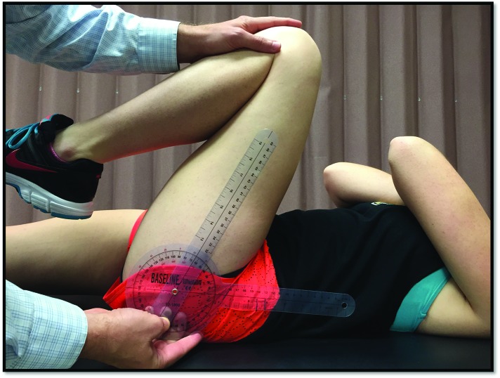
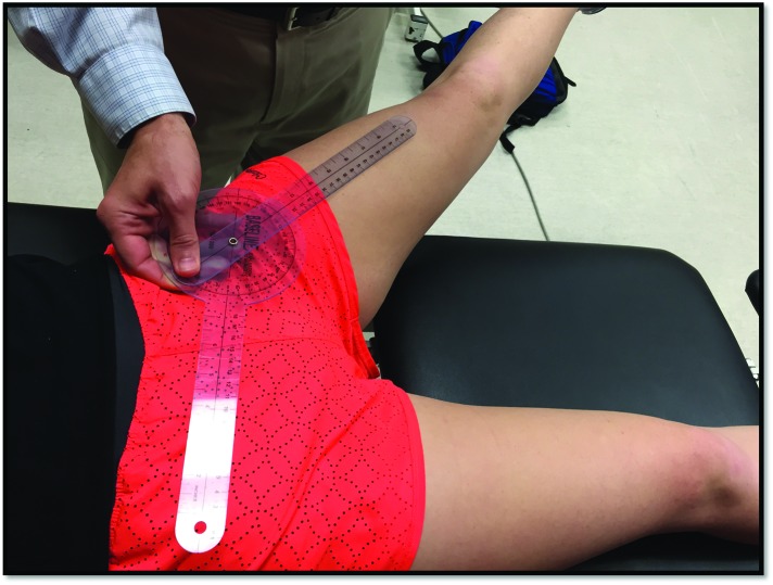
The subject was positioned supine on an examination table. The examiner passively flexed the subject's hip as far as possible with the opposite leg extended. The goniometer was centered at the greater trochanter, aligning one arm along the center of the thigh and the other arm aligned horizontally as illustrated in Figure 1. The examiner monitored for any aberrant pelvic motion prior to taking measurement.37
Figure 1.
Goniometric measurement of supine hip flexion.
Hip Extension ROM
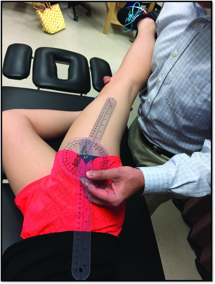
The subject was positioned in the sidelying position on the examination table with the test extremity facing upward. The lowermost extremity was flexed at the hip to 45 degrees and at knee to 90 degrees. The examiner passively extended the hip with knee straight as far as possible. The goniometer was centered at the greater trochanter aligning one arm of goniometer over the center of the thigh and the other arm along a zero-degree position as illustrated in Figure 2. The examiner monitored for any aberrant lumbopelvic motion prior to and during the measurement.37
Figure 2.
Goniometric measurement of side lying hip extension.
Hip IR ROM
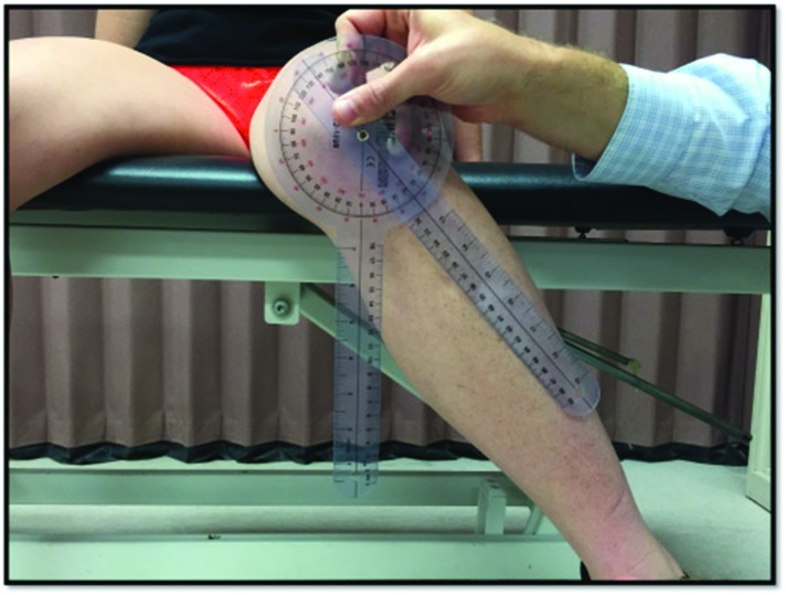
The subject was sitting on an examination table with their knees flexed to 90 ° and feet unsupported. The examiner stood in front of the test leg and centered the goniometer at the lower border of the patella with the arm of the goniometer aligned along the patellar tendon and the other arm aligned vertically. The examiner passively moved the participant's hip into IR, keeping their leg in neutral, to the end of the available range until an “unyielding” end-feel was felt and then took the measurementas illustrated in Figure 3. 37,38 . The examiner provided verbal cues if the participant compensated in any way to ensure no substitute movements occurred during testing.
Figure 3.
Goniometric measurement of seated hip internal rotation.
Hip ER ROM
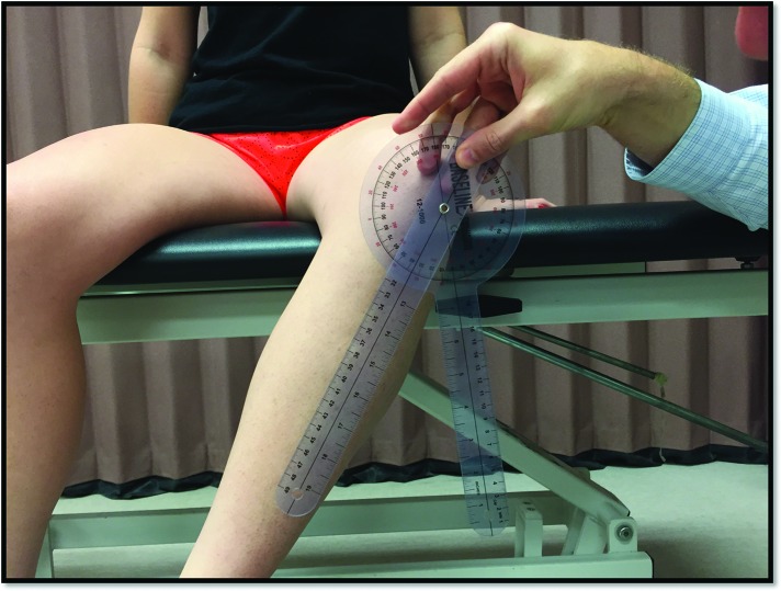
The subject was sitting on an examination table with their knees flexed to 90 ° and feet unsupported. The examiner stood in front of the test leg and centered the goniometer at the lower border of the patella with the arm of the goniometer aligned along the patellar tendon and the other arm aligned vertically. The examiner passively moved the participant's hip into ER, keeping their leg in neutral, to the end of the available range until an “unyielding” end-feel was felt and then took the measurement as illustrated in Figure 4.37,38 The examiner provided verbal cues if participant compensated in any way to ensure no substitute movements occurred during testing.
Figure 4.
Goniometric measurement of seated hip external rotation.
Hip Abduction ROM
The subject was positioned supine on the examination table with legs extended. The examiner stood on the side of the test leg. The goniometer was centered midway between the subject's anterior superior iliac spine and pubic symphysis, aligning one arm centrally over their thigh. The examiner passively abducted the subject's leg as far as possible, without causing any aberrant pelvic motion, and then took the measurement as illustrated in Figure 5. 37 The examiner monitored for any aberrant lumbopelvic motion prior to and during the measurement.37
Figure 5.
Goniometric measurement of supine hip abduction.
Statistical Analysis
Statistical analysis was performed using SPSS version 24.0 (IBM SPSS, Chicago, IL, USA). Participant descriptives were calculated and reported as the mean and standard deviation (SD) for age, height, mass, body mass index, and ROM values. A two-tailed independent t-test was used to compare mean passive hip ROM values between the right and left leg to determine asymmetries as well as to compare men and women. Statistical significance was considered as p < 0.05. The SEM was calculated for the reliability pilot study using a previously established formula SEM = standard deviation multiplied by the square root of 1-ICC value.39
RESULTS
Participant demographic data is presented in Table 1. Both men and women reported participation in WT a mean 3.4 times per week with no significant differences between men and women (p = .88). Training experience was reported at a mean of 5.9 years for women and 7.8 years for men. Significant differences for training experience were not found (p = .22). Tables 2 and 3 present mean passive ROM values. When comparing hip ROM values among sexes, men had no significant differences (p≥.28) between the right and left hip for all motions. Women did have a significant differences (p≤.05) between the right and left hip for all motions. The right hip had lower values for all motions than the left hip suggesting a more global decrease in right hip PROM (Table 3). When comparing hip ROM values between men and women, there was a significant difference (p≤.05) between men and women for all motions. Men had lower ROM values for all right and left hip motions when compared to women (Table 4).
Table 2.
Hip ROM Values for Recreational Weight Training Participants.
| Right Hip | Left Hip | p-value | |
|---|---|---|---|
| Flexion | 120.4 ± 14.5 ° | 121.3 ± 13.8 ° | .50 |
| Extension | 12.6 ± 5.9 ° | 12.6 ± 7.6 ° | .95 |
| Internal Rotation | 36.4 ± 9.5 ° | 36.1 ± 8.7 ° | .82 |
| External Rotation | 32.2 ± 8.7 ° | 32.0 ± 9.4 ° | .78 |
| Abduction | 42.6 ± 11.3 ° | 43.2 ± 12.3 ° | .64 |
* = statistically significant difference
Table 3.
Comparison between Right and Left Hip ROM among Sexes.
| Men | Women | |||||
|---|---|---|---|---|---|---|
| Right | Left | P-value | Right | Left | p-value | |
| Flexion | 117.0 ± 14.9 ° | 118.0 ± 14.4 ° | .28 | 118.0 ± 14.4 ° | 128.9 ± 8.2 ° | <.001* |
| Extension | 11.2 ± 5.4 ° | 10.8 ± 5.5 ° | .72 | 10.8 ± 5.5 ° | 16.7 ± 9.9 ° | <.001* |
| Internal Rotation | 34.6 ± 9.2 ° | 34.4 ± 8.0 ° | .58 | 34.4 ± 8.0 ° | 40.1 ± 8.8 ° | <.001* |
| External Rotation | 30.3 ± 8.5 ° | 29.8 ± 9.1 ° | .68 | 29.8 ± 9.1 ° | 37.0 ± 8.1 ° | <.001* |
| Abduction | 41.9 ± 12.0 ° | 42.1 ± 11.4 ° | .44 | 42.1 ± 11.4 ° | 45.8 ± 13.8 ° | .05* |
* = statistically significant difference
Table 4.
Comparison between Male and Female Recreational Weight Training Participants.
| Right Hip | Left Hip | |||||
|---|---|---|---|---|---|---|
| Men (N=139) | Women (N=61) | P-value | Men (N=139) | Women (N=61) | p-value | |
| Flexion | 117.0 ± 14.9 ° | 127.9 ± 9.9 ° | .001* | 118.0 ± 14.4 ° | 128.9 ± 8.2 ° | <.001* |
| Extension | 11.2 ± 5.4 ° | 15.7 ± 5.7 ° | .001* | 10.8 ± 5.5 ° | 16.7 ± 9.9 ° | <.001* |
| Internal Rotation | 34.6 ± 9.2 ° | 40.4 ± 9.1 ° | .001* | 34.4 ± 8.0 ° | 40.1 ± 8.8 ° | <.001* |
| External Rotation | 30.3 ± 8.5 ° | 36.7 ± 7.7 ° | .001* | 29.8 ± 9.1 ° | 37.0 ± 8.1 ° | <.001* |
| Abduction | 41.9 ± 12.0 ° | 44.4 ± 9.1 ° | .05* | 42.1 ± 11.4 ° | 45.8 ± 13.8 ° | .04* |
* = statistically significant difference
DISCUSSION
This is the first investigation to report hip passive ROM values in recreational WT participants. This group has been understudied compared to other athletic groups which leaves a gap in the knowledge regarding hip ROM and the potential risk for injury. These results provide reference hip ROM values that may help to further classify these individuals for future research on injury surveillance and prevention strategies.
The results of the study suggest that among recreational WT participants, women have greater hip ROM in all motions (p≤.05) than men. This is consistent with prior research reporting greater hip ROM values in adult women when compared to adult men.22,40 However, it is often difficult to make a direct comparison among populations due to the variation in study methodology and the procedure by which ROM may be tested. Currently, there is no standard method for measuring hip ROM since many researchers measure both active and passive ROM in the supine, prone, sidelying, and seated positions.41,42 With this being stated, the passive ROM findings of this investigation are limited to the specific procedures used. This is a necessary consideration as prone hip ER and IR may have produced different values.
When comparing results from this study to published reference values, only one comparable study was found that used similar methods for measuring passive hip IR and ER in adults.40 The WT men and women in the current study had lower seated passive hip IR (right + left) (31.1 ° versus 37.9 °) and ER (right + left) (36.2 ° versus 40.7 °) when compared to the published adult values of Kouyoumdjian et al.40 Potential reasons for the differences reside in measurement technique (e.g. positioning), procedure, and age. In the Kouyoumdjian et al study subjects were older (mean age 39.1 years), measurements were performed in supine and prone, and a digital camera with software was used to quantify ROM.40
Injuries to the lower extremities have been reported among elite competitive weightlifters and powerlifters but not in recreational WT participants.6,8,43 Researchers are just beginning to report injuries specific to the hip among general WT participants. Polesello et al44 reported on 47 individuals who underwent arthroscopic surgery for hip labral tears and chondral lesions after developing painful symptoms associated with the leg press and squat which are common WT movements. The researchers reported the post-surgical outcomes but did not provide any insight regarding the correlation between the WT movements, hip ROM, and the diagnosed hip injuries. Other researchers have evaluated pre-surgical and post-surgical unilateral and bilateral squat performance in individuals with femoral acetabular impingement (FAI). The researchers observed decreased squat depth, hip internal rotation, and decreased posterior pelvic tilt in individuals with CAM-type FAI.9,45,46 Researchers have also observed that squat performance improved post-surgically with subjects having a greater squat depth and pelvic motion.9 Despite these reported finding, the researchers did not discuss if the squat movement was a risk factor for injury which leaves a gap in our understanding of this common exercise.9,45,46 Future research is necessary to examine the correlation between common WT movements, the required hip ROM, and risk of hip injury.
The data from this study provides a beginning for clinicians to understand common hip ROM values in the WT population. Impaired hip ROM may be a relative factor needing to be considered for injury prevention and athletic performance, thus should be considered for inclusion when prescribing exercises for these individuals.14,19,23 These data are the first to be reported among WT participants, thus should be considered for clinical practice when managing such patients. WT participants may have different values based upon the types of WT activities, performed, thus general population normative values may not be relevant.
When interpreting differences in ROM values between men and women as well as side-to-side differences it should be recognized that a statistically significant difference does not necessarily mean a clinically important difference nor does it mean error in the measurement is accounted for. Moreover, it is not unreasonable for mean and women to have differences given the potential for training differences as well as body morphology.
One way to determine the error in a measurement is to consider the SEM. The SEM is an index of the expected variation of a score due to measurement error. The SEM is reported in terms of specific value and as a confidence interval around a mean. One SEM value represents 68% of the population. For example, the results of this study suggest that women have statistically greater bilateral hip ROM when compared to men. As an example, when comparing the mean angle of right hip abduction for men (41.9 degrees) to women (44.4 degrees) a difference of 2.5 degrees is present. While this difference is statistically significantly different, the SEM for hip abduction is 3 degrees. This suggests that the angles reported will vary +/- 3 degrees (68% of the time) from the mean for men and women, thus the difference may reflect error.
Limitations
When considering the methodology of this descriptive study, several limitations need to be discussed. First, this investigation reported values in healthy subjects which limits the generalizability of these results to this population. However, no reference data has been reported in this population, thus the information provided may guide practice or be used for reference values. Second, weight-training participants comprise a heterogeneous population and variability within training patterns and styles may indeed influence mobility. The effort to use only recreational participants was an attempt to capture a homogenous subgroup, however, subjects were not grouped according to a specific intensity level of exercise which may have influenced hip ROM values. Perhaps a further classification based on such variables may help guide injury prevention strategies. Third, passive hip IR and ER ROM were measured in the seated position where other investigations have measured hip ROM in different positions.47-49 This must be considered when interpreting these results or comparing to other values to inform clinical practice. Fourth, hip adduction measurements were omitted which limits the understanding of the complete range of hip mobility in these participants. Lastly, standard goniometry was used in lieu of a digital device, as the standard goniometer is a common tool used in the clinical setting.32-35
Future Research
Future research should focus on prospective injury surveillance among recreational WT participants. Given the recent evidence associating hip ROM deficits and athletic injuries, future research in warranted in this population.13-15,50-52,20,21 Also, attempts to further classify the recreational WT participant according to the type of weight training and level of training may assist in providing a better understating of subgroups based on WT activities. This may provide insight into common mechanisms of injury related to specific weight training activities and help guide injury prevention strategies. Finally, it would be beneficial for future studies to capture hip adduction range of motion and limited mobility in this plane may have consequences in terms of function and sport participation.
CONCLUSION
This study reported passive hip ROM values in recreational WT participants. Women WT participants had asymmetrical passive hip ROM whereas men had symmetrical measurements. With regard to sex, men had lower overall hip ROM compared to women. Implications for these findings may include the use of clinical efforts to increase global ROM in men, whereas women should focus on symmetry. Lastly the right hip had grossly lower ROM values among all participants, which may suggest a participation type dominance which could be addressed with efforts to achieve symmetry. This is the first study to report reference data for recreational WT participants which provides a starting point for future research. Future investigations should focus on injury surveillance and injury prevention strategies in this population.
References
- 1.Trends in strength training--United States, 1998-2004. MMWR Morb Mortal Wkly Rep. 2006;55(28):769-772. [PubMed] [Google Scholar]
- 2.Tonnesen R Schwarz P Hovind PH, et al. Physical exercise associated with improved BMD independently of sex and vitamin D levels in young adults. Eur J Appl Physiol. 2016;116(7):1297-1304. [DOI] [PMC free article] [PubMed] [Google Scholar]
- 3.Mosti MP Carlsen T Aas E, et al. Maximal strength training improves bone mineral density and neuromuscular performance in young adult women. J Strength Cond Res. 2014;28(10):2935-2945. [DOI] [PubMed] [Google Scholar]
- 4.Garber CE Blissmer B Deschenes MR, et al. American College of Sports Medicine position stand. Quantity and quality of exercise for developing and maintaining cardiorespiratory, musculoskeletal, and neuromotor fitness in apparently healthy adults: guidance for prescribing exercise. Med Sci Sports Exerc. 2011;43(7):1334-1359. [DOI] [PubMed] [Google Scholar]
- 5.Powell KE Heath GW Kresnow MJ, et al. Injury rates from walking, gardening, weightlifting, outdoor bicycling, and aerobics. Med Sci Sports Exerc. 1998;30(8):1246-1249. [DOI] [PubMed] [Google Scholar]
- 6.Raske A Norlin R. Injury incidence and prevalence among elite weight and power lifters. Am J Sports Med. 2002;30(2):248-256. [DOI] [PubMed] [Google Scholar]
- 7.Aasa U Svartholm I Andersson F, et al. Injuries among weightlifters and powerlifters: a systematic review. Br J Sports Med. 2016. [DOI] [PubMed] [Google Scholar]
- 8.Calhoon G Fry AC. Injury rates and profiles of elite competitive weightlifters. J Athl Train. 1999;34(3):232-238. [PMC free article] [PubMed] [Google Scholar]
- 9.Lamontagne M Brisson N Kennedy MJ, et al. Preoperative and postoperative lower-extremity joint and pelvic kinematics during maximal squatting of patients with cam femoro-acetabular impingement. J Bone Joint Surg Am. 2011;93 Suppl 2:40-45. [DOI] [PubMed] [Google Scholar]
- 10.Jonasson P Halldin K Karlsson J, et al. Prevalence of joint-related pain in the extremities and spine in five groups of top athletes. Knee Surg Sports Traumatol Arthrosc. 2011;19(9):1540-1546. [DOI] [PubMed] [Google Scholar]
- 11.Hanson PG Angevine M Juhl JH. Osteitis Pubis in Sports Activities. Physician sports med. 1978;6(10):111-114. [DOI] [PubMed] [Google Scholar]
- 12.Cheatham SW Kolber MJ. Orthopedic Management of the Hip and Pelvis. Elsevier: - Health Sciences Division; 2015. [Google Scholar]
- 13.Scher S Anderson K Weber N, et al. Associations among hip and shoulder range of motion and shoulder injury in professional baseball players. J Athl Train. 2010;45(2):191-197. [DOI] [PMC free article] [PubMed] [Google Scholar]
- 14.Saito M Kenmoku T Kameyama K, et al. Relationship Between Tightness of the Hip Joint and Elbow Pain in Adolescent Baseball Players. Orthop J Sports Med. 2014;2(5):2325967114532424. [DOI] [PMC free article] [PubMed] [Google Scholar]
- 15.Li X Ma R Zhou H, et al. Evaluation of Hip Internal and External Rotation Range of Motion as an Injury Risk Factor for Hip, Abdominal and Groin Injuries in Professional Baseball Players. Orthop Rev (Pavia). 2015;7(4):6142. [DOI] [PMC free article] [PubMed] [Google Scholar]
- 16.Tak I Glasgow P Langhout R, et al. Hip Range of Motion Is Lower in Professional Soccer Players With Hip and Groin Symptoms or Previous Injuries, Independent of Cam Deformities. Am J Sports Med. 2016;44(3):682-688. [DOI] [PubMed] [Google Scholar]
- 17.Ellera Gomes JL Palma HM Ruthner R. Influence of hip restriction on noncontact ACL rerupture. Knee Surg Sports Traumatol Arthrosc. 2014;22(1):188-191. [DOI] [PubMed] [Google Scholar]
- 18.Gomes JL de Castro JV Becker R. Decreased hip range of motion and noncontact injuries of the anterior cruciate ligament. Arthroscopy. 2008;24(9):1034-1037. [DOI] [PubMed] [Google Scholar]
- 19.Nguyen AD Zuk EF Baellow AL, et al. Longitudinal Changes in Hip Strength and Range of Motion in Female Youth Soccer Players: Implications for ACL Injury. A Pilot Study. J Sport Rehabil. 2016. [DOI] [PubMed] [Google Scholar]
- 20.Young SW Dakic J Stroia K, et al. Hip range of motion and association with injury in female professional tennis players. Am J Sports Med. 2014;42(11):2654-2658. [DOI] [PubMed] [Google Scholar]
- 21.Wilcox CR Osgood CT White HS, et al. Investigating Strength and Range of Motion of the Hip Complex in Ice Hockey Athletes. J Sport Rehabil. 2015;24(3):300-306. [DOI] [PubMed] [Google Scholar]
- 22.Simoneau GG Hoenig KJ Lepley JE, et al. Influence of hip position and gender on active hip internal and external rotation. J Orthop Sports Phys Ther. 1998;28(3):158-164. [DOI] [PubMed] [Google Scholar]
- 23.Picha KJ Harding JL Bliven KC. Glenohumeral and Hip Range-of-Motion and Strength Measures in Youth Baseball Athletes. J Athl Train. 2016;51(6):466-473. [DOI] [PMC free article] [PubMed] [Google Scholar]
- 24.Sauers EL Huxel Bliven KC Johnson MP, et al. Hip and glenohumeral rotational range of motion in healthy professional baseball pitchers and position players. Am J Sports Med. 2014;42(2):430-436. [DOI] [PubMed] [Google Scholar]
- 25.Moreno-Perez V Ayala F Fernandez-Fernandez J, et al. Descriptive profile of hip range of motion in elite tennis players. Phys Ther Sport. 2016;19:43-48. [DOI] [PubMed] [Google Scholar]
- 26.Bennell KL Khan KM Matthews BL, et al. Changes in hip and ankle range of motion and hip muscle strength in 8-11 year old novice female ballet dancers and controls: a 12 month follow up study. Br J Sports Med. 2001;35(1):54-59. [DOI] [PMC free article] [PubMed] [Google Scholar]
- 27.Gulgin H Armstrong C Gribble P. Weight-bearing hip rotation range of motion in female golfers. N Am J Sports Phys Ther. 2010;5(2):55-62. [PMC free article] [PubMed] [Google Scholar]
- 28.Kim SH Kwon OY Park KN, et al. Lower extremity strength and the range of motion in relation to squat depth. J Hum Kinet. 2015;45:59-69. [DOI] [PMC free article] [PubMed] [Google Scholar]
- 29.Schutz P List R Zemp R, et al. Joint angles of the ankle, knee, and hip and loading conditions during split squats. J Appl Biomech. 2014;30(3):373-380. [DOI] [PubMed] [Google Scholar]
- 30.Riemann BL Lapinski S Smith L, et al. Biomechanical analysis of the anterior lunge during 4 external-load conditions. J Athl Train. 2012;47(4):372-378. [DOI] [PMC free article] [PubMed] [Google Scholar]
- 31.Bell-Jenje T Olivier B Wood W, et al. The association between loss of ankle dorsiflexion range of movement, and hip adduction and internal rotation during a step down test. Man Ther. 2016;21:256-261. [DOI] [PubMed] [Google Scholar]
- 32.Holm I Bolstad B Lutken T, et al. Reliability of goniometric measurements and visual estimates of hip ROM in patients with osteoarthrosis. Physiother Res Int. 2000;5(4):241-248. [DOI] [PubMed] [Google Scholar]
- 33.Nussbaumer S Leunig M Glatthorn JF, et al. Validity and test-retest reliability of manual goniometers for measuring passive hip range of motion in femoroacetabular impingement patients. BMC Musculoskelet Disord. 2010;11:194. [DOI] [PMC free article] [PubMed] [Google Scholar]
- 34.Pua YH Wrigley TV Cowan SM, et al. Intrarater test-retest reliability of hip range of motion and hip muscle strength measurements in persons with hip osteoarthritis. Arch Phys Med Rehabil. 2008;89(6):1146-1154. [DOI] [PubMed] [Google Scholar]
- 35.Roach S San Juan JG Suprak DN, et al. Concurrent validity of digital inclinometer and universal goniometer in assessing passive hip mobility in healthy subjects. Int J Sports Phys Ther. 2013;8(5):680-688. [PMC free article] [PubMed] [Google Scholar]
- 36.Portney LG Watkins MP. Foundations of Clinical Research: Applications to Practice. Pearson/Prentice Hall; 2009. [Google Scholar]
- 37.Cibere J Thorne A Bellamy N, et al. Reliability of the hip examination in osteoarthritis: effect of standardization. Arthritis Rheum. 2008;59(3):373-381. [DOI] [PubMed] [Google Scholar]
- 38.Shimamura KK Cheatham S Chung W, et al. Regional interdependence of the hip and lumbo-pelvic region in divison ii collegiate level baseball pitchers: a preliminary study. Int J Sports Phys Ther. 2015;10(1):1-12. [PMC free article] [PubMed] [Google Scholar]
- 39.Kolber MJ Hanney WJ. The reliability, minimal detectable change and construct validity of a clinical measurement for identifying posterior shoulder tightness. N Am J Sports Phys Ther. 2010;5(4):208-219. [PMC free article] [PubMed] [Google Scholar]
- 40.Kouyoumdjian P Coulomb R Sanchez T, et al. Clinical evaluation of hip joint rotation range of motion in adults. Orthop Traumatol Surg Res. 2012;98(1):17-23. [DOI] [PubMed] [Google Scholar]
- 41.Martin HD Kelly BT Leunig M, et al. The pattern and technique in the clinical evaluation of the adult hip: the common physical examination tests of hip specialists. Arthroscopy. 2010;26(2):161-172. [DOI] [PubMed] [Google Scholar]
- 42.Kouyoumdjian P Coulomb R Sanchez T, et al. Clinical evaluation of hip joint rotation range of motion in adults. Orthop Traumalol Surg Res. 2012;98(1):17-23. [DOI] [PubMed] [Google Scholar]
- 43.Keogh J Hume PA Pearson S. Retrospective injury epidemiology of one hundred one competitive Oceania power lifters: the effects of age, body mass, competitive standard, and gender. J Strength Cond Res. 2006;20(3):672-681. [DOI] [PubMed] [Google Scholar]
- 44.Polesello GC Cinagawa EH Cruz PD, et al. Surgical treatment for femoroacetabular impingement in a group that performs squats. Rev Bras Ortop. 2012;47(4):488-492. [DOI] [PMC free article] [PubMed] [Google Scholar]
- 45.Bagwell JJ Snibbe J Gerhardt M, et al. Hip kinematics and kinetics in persons with and without cam femoroacetabular impingement during a deep squat task. Clin Biomech (Bristol, Avon). 2016;31:87-92. [DOI] [PubMed] [Google Scholar]
- 46.Lamontagne M Kennedy MJ Beaule PE. The effect of cam FAI on hip and pelvic motion during maximum squat. Clin Orthop Relat Res. 2009;467(3):645-650. [DOI] [PMC free article] [PubMed] [Google Scholar]
- 47.Robb AJ Fleisig G Wilk K, et al. Passive Ranges of Motion of the Hips and Their Relationship With Pitching Biomechanics and Ball Velocity in Professional Baseball Pitchers. Am J Sports Med. 2010;38(12):2487-2493. [DOI] [PubMed] [Google Scholar]
- 48.Ellenbecker TS Ellenbecker GA Roetert EP, et al. Descriptive Profile of Hip Rotation Range of Motion in Elite Tennis Players and Professional Baseball Pitchers. Am J Sports Med. 2007;35(8):1371-1376. [DOI] [PubMed] [Google Scholar]
- 49.Laudner K Wong R Onuki T, et al. The relationship between clinically measured hip rotational motion and shoulder biomechanics during the pitching motion. J Sci Med Sport. 2014. [DOI] [PubMed] [Google Scholar]
- 50.Sadeghisani M Manshadi FD Kalantari KK, et al. Correlation between Hip Rotation Range-of-Motion Impairment and Low Back Pain. A Literature Review. Ortop Traumatol Rehabil. 2015;17(5):455-462. [DOI] [PubMed] [Google Scholar]
- 51.Van Dillen LR Bloom NJ Gombatto SP, et al. Hip rotation range of motion in people with and without low back pain who participate in rotation-related sports. Phys Ther Sport. 2008;9(2):72-81. [DOI] [PMC free article] [PubMed] [Google Scholar]
- 52.Roach SM San Juan JG Suprak DN, et al. Passive hip range of motion is reduced in active subjects with chronic low back pain compared to controls. Int J Sports Phys Ther. 2015;10(1):13-20. [PMC free article] [PubMed] [Google Scholar]


