Abstract
Purpose
To characterize eye movements made by patients with intermittent exotropia when fusion loss occurs spontaneously and to compare them with those induced by covering 1 eye and with strategies used to recover fusion.
Design
Prospective study of a patient cohort referred to our laboratory.
Participants
Thirteen patients with typical findings of intermittent exotropia who experienced frequent spontaneous loss of fusion.
Methods
The position of each eye was recorded with a video eye tracker under infrared illumination while fixating on a small central near target.
Main Outcome Measures
Eye position and peak velocity measured during spontaneous loss of fusion, shutter-induced loss of fusion, and recovery of fusion.
Results
In 10 of 13 subjects, the eye movement made after spontaneous loss of fusion was indistinguishable from that induced by covering 1 eye. It reached 90% of full amplitude in a mean of 1.75 seconds. Peak velocity of the deviating eye’s movement was highly correlated for spontaneous and shutter-induced events. Peak velocity was also proportional to exotropia amplitude. Recovery of fusion was more rapid than loss of fusion, and often was accompanied by interjection of a disconjugate saccade.
Conclusions
Loss of fusion in intermittent exotropia is not influenced by visual feedback. Excessive divergence tone may be responsible, but breakdown of alignment occurs via a unique, pathological type of eye movement that differs from a normal, physiological divergence eye movement.
The most extraordinary feature of intermittent exotropia is that it is intermittent.1 Subjects usually have normal visual acuity in each eye. They are able to fuse on targets, especially at near, and enjoy stereopsis.2–9 Occasionally, this intact state is interrupted by the abrupt outward deviation of 1 eye. Once fusion is broken, 2 sensory adaptations become engaged immediately. First, the peripheral temporal retina in each eye is suppressed to prevent diplopia.10–12 Second, the perceived location of images falling on the deviated eye’s retina is shifted to avoid visual confusion.12–14 These compensatory mechanisms are so efficient that some patients are unaware when they lose fusion.15
Other forms of strabismus, such as infantile esotropia or accommodative esotropia, generally carry a worse prognosis for binocular vision than does intermittent exotropia. In these conditions, amblyopia is more common and the ability to fuse is often absent.2,16 In contrast, the neural substrate for binocularity remains intact in intermittent exotropia. A cure would be possible if one could devise an effective strategy to prevent periodic interruption of fusion. Unfortunately, we remain ignorant of the mechanism that triggers the loss of fusion in intermittent exotropia. No eye movement recordings have been published that capture the moments when the eye breaks fusion and drifts outward. It is not even clear how the outward rotation of the deviating globe should be classified among the repertoire of eye movements made by primates.17
While investigating how patients with exotropia locate and fixate visual targets, we occasionally recorded epochs when the eyes were straight, followed by loss of fusion and the subsequent outward movement of 1 eye.18 Such transitions were sometimes involuntary, or even occult, made while subjects were attempting to maintain alignment on a visual target. In other cases, subjects were able to exert some control over the process, and in fact were encouraged to release ocular fusion so that the resulting eye movement could be documented. All such episodes were classified as “spontaneous,” to distinguish them from exodeviation triggered by shutter occlusion. Recordings were also made of the recovery of ocular alignment from an exotropic state. These findings are presented in the hope that they may yield insight into the neural mechanisms that underlie intermittent exotropia.
Methods
The 13 subjects (6 male, 7 female) with a history of intermittent exotropia who contributed to this study had a mean age of 30 years (range, 11–61 years). They were referred by local ophthalmologists or recruited from the Neuro-Ophthalmology/Pediatric Ophthalmology Service at the University of California, San Francisco. The study was approved by the Institutional Review Boards at the University of California, San Francisco, and at Kaiser Permanente Northern California. Adult subjects provided informed consent to participate; minors gave their assent and a parent gave informed consent.
The goal of this study was to record the eye movements made by patients during episodes of spontaneous loss of fusion. A comparison was made with the eye movements induced by occluding 1 eye with a shutter. Most patients manifested a spontaneous exotropia of either eye. However, in 5 of 13 subjects, eye dominance was so strong that a spontaneous exotropia occurred only in the nondominant eye. In graphs of spontaneous exotropia data, these 5 patients’ data are based on 1 eye only, rather than the mean of both eyes. Eye movements were also recorded during recovery of eye alignment, occurring either spontaneously or after the shutter was removed.
Each subject received an ophthalmologic examination, which included measurement of the best-corrected visual acuity in each eye and a cycloplegic refraction. Assessment was made of the pupils, eye movements, alignment, and stereopsis (Randot circles and stereo butterfly). Slit-lamp and fundus examinations were also performed. Inclusion criteria were as follows: (1) intermittent exotropia since early childhood, (2) 20/20 Snellen acuity in each eye with refractive correction, (3) no eye disease except strabismus, (4) ability to alternate ocular fixation freely, (5) no pathologic nystagmus, (6) absence of diplopia, (7) normal stereopsis, and (8) no history of strabismus surgery. Subjects with more than 4 diopters of myopia, hyperopia, or astigmatism were excluded. Testing was performed with no refractive correction. After clinical evaluation for eligibility, patients were scheduled on a different day for recording of eye movements.
Subjects were seated in a dimly lit room, with their head relatively immobile in a chin/forehead rest, facing a translucent tangent screen at a distance of 57 cm. Their task was simply to fixate a spot of light, 0.5 degrees in diameter, presented at the center of the screen. Stimuli were projected onto the tangent screen with a digital light processing projector or a laser. Infrared filter shutters, controlled by pneumatic pistons, were positioned to descend rapidly over either eye. The shutters blocked visible light, but passed infrared wavelengths, allowing one to continue recording eye movements without interruption. An infrared video-based eye tracker (iView X, SensoMotoric Instruments), sampling at 60 Hz, recorded the position of each eye. The trackers were calibrated independently. Under ideal circumstances, each eye tracker has a resolution of better than ±0.50 degrees.19,20 Optimal performance was obtained in subjects with wide palpebral fissures, prominent globes, few blinks, high iris/pupil contrast, light eyelashes, and a relatively small pupil. For practical purposes, in a mixed cohort of untrained subjects, each tracker had a potential position error of up to ±1 degree.21 Eye, stimulus, and shutter positions were digitized and acquired for later analysis. Blinks caused interruption of eye tracking. Blinks were removed from traces and replaced by linear interpolation. Events that were marred by excessive blinking or loss of eye tracking were not analyzed. Further details regarding the equipment, experimental design, and tracker resolution have been published.18,20 An excerpt from a typical data recording session is available for review (Supplemental Video 1; available at www.aaojournal.org).
Results
Similarity of Spontaneous and Cover-Induced Exotropia
Figure 1 shows examples of eye movement recordings made from 2 patients with intermittent exotropia. After descent of the shutter, the occluded eye began to move outward (Fig 1A, C). The mean latency for initiation of globe movement for all subjects was 240±60 milliseconds (range, 140–320 milliseconds, n = 13). In these 2 patients, the globe position reached its maximum excursion within 2 seconds. One subject had an exotropia of 10 to 11 degrees and the other subject had an exotropia of 19 degrees. When in a deviated state, each subject exhibited a strong eye fixation preference. However, it made no difference which eye was occluded: The right exotropia and the left exotropia were equal in both individuals.
Figure 1.
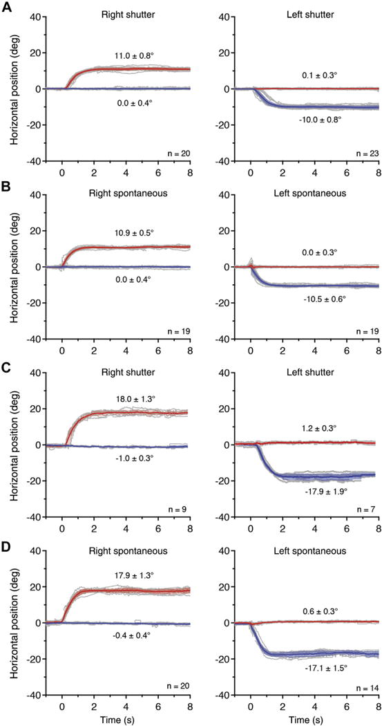
Cover-induced exotropia is equal to spontaneous exotropia. A, Recordings from a 26-year-old woman (refraction: plano both eyes [OU]) during fixation on a 0.5-degree target at 57 cm. The mean position (solid line) and standard deviation (shading) are shown for the right eye (red) and left eye (blue). At t ¼ 0 seconds, a shutter occluded either the right eye or the left eye, inducing an exodeviation. n = number of events. Positive and negative values for horizontal deviation denote right and left gaze, respectively. B, Spontaneous exotropia occurring intermittently between cover-induced episodes has an amplitude nearly equal to shutter-induced exotropia, with a difference of 0.1 degree for right exotropia and 0.4 degree for left exotropia. The shapes of the mean position traces and the variability in individual position traces are also similar. C: Recordings from a 36-year-old woman (refraction: −1.00 OU), showing a larger exotropia. D: Interspersed episodes of spontaneous fusion loss are similar to those from shutter occlusion, with an amplitude difference of 0.7 degrees for right exotropia and 1.4 degrees for left exotropia.
During the cover test recording session, while fixating a central target binocularly, both subjects experienced frequent bouts of spontaneous exotropia (Fig 1B, D). The cardinal finding was that the eye movement made during such events closely resembled the eye movement induced by shutter occlusion, both in velocity and in amplitude. Each eye tracker has a potential position error of ±1 degree, so only exotropia differences exceeding 2 degrees could be detected reliably. Of the 13 subjects recorded during spontaneous loss of fusion, only 3 showed more than 2 degrees of difference between spontaneous exotropia and shutter-induced exotropia. Hence, spontaneous and shutter-induced events were not measurably different in 10 of 13 subjects. In 7 of 13 subjects, spontaneous and shutter exotropia differed by less than 1 degree. Data from all subjects are provided in the Supplementary Figure S1 (available at www.aaojournal.org).
An example of a subject exhibiting a difference between shutter and spontaneous exotropia is shown in Figure 2 (see also Supplementary Video 1, available at www.aaojournal.org). The subject had a strong left eye fixation preference, and manifested spontaneous exotropia of only the right eye. Right shutter exotropia was less than right spontaneous exotropia, by 3.8 degrees. The left shutter exotropia was highly variable in time course, because on some trials the dominant left eye remained fairly straight transiently, even after its view of the target was blocked by the shutter. Ultimately, it reached nearly the same amplitude as the right exotropia. There were 2 other patients who showed a difference between shutter and spontaneous exotropia. One patient (Supplementary Figure S1, subject E) had a larger right (16.6 degrees) and left (16.4 degrees) spontaneous exotropia compared with the corresponding right (13.0 degrees) and left (12.1 degrees) shutter exotropia. The other patient (Supplementary Figure S1, subject G) had a highly asymmetrical exotropia. It was essentially equal in the right eye for spontaneous (10.9 degrees) and shutter exotropia (12.2 degrees), but smaller in the left eye for spontaneous exotropia (2.1 degrees), compared with shutter exotropia (4.8 degrees). We have no explanation for these discrepancies, but they highlight the diversity of patient findings in intermittent exotropia.
Figure 2.
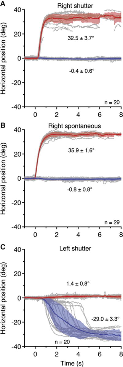
Cover-induced exotropia different from spontaneous exotropia. Recordings from a 35-year-old man (refraction: −1.25 +0.75 right eye; −1.25 left eye), showing that A, right exotropia induced by occlusion was smaller and more variable than B, spontaneous right exotropia. C, Covering the dominant left eye produced a highly variable outward eye movement; spontaneous left exotropia never occurred (Supplementary Video 1, available at www.aaojournal.org).
In exotropia, the deviated eye is more variable in position than the fixating eye.19 It is unknown, however, if occlusion of the deviating eye makes any difference to its stability. In Figure 2, the deviated right eye had a standard deviation of ±3.7 degrees in position during shutter-induced exotropia, but only ±1.6 degrees during spontaneous right exotropia. This difference implies that being uncovered (and thus potentially able to perceive the visual scene) may stabilize the deviating eye. To test this idea, the standard deviation of the deviating eye’s position was compared for all subjects during shutter exotropia (n = 26) and spontaneous exotropia (n = 18). The mean standard deviation in position for the deviated eye was 2.3±1.4 degrees (95% confidence interval [CI], 1.7–2.8 degrees) during shutter exotropia and 1.7±0.7 degrees (95% CI, 1.3–2.0 degrees) during spontaneous exotropia. Overlap in the 95% CIs indicates that it made no difference whether the deviated eye was covered or not. A larger population would need to be tested, however, to exclude the possibility that the exotropic eye is more stable when uncovered than covered.
Peak Velocity of Eye Movement in Exotropia
Saccadic eye movements exhibit a predictable correlation between amplitude and peak velocity, known as the main sequence relationship.22,23 To see if a relationship exists in exotropia, the peak velocity of the deviating eye’s outward movement was calculated for each subject (Supplementary Figure S2, available at www.aaojournal.org). A plot of the amplitude of spontaneous exotropia versus peak eye velocity (Fig 3) showed that subjects with a larger exotropia moved their eye outward more rapidly (r = 0.81). A linear fit yielded the following: peak velocity 1.36 × exotropia amplitude/second.
Figure 3.
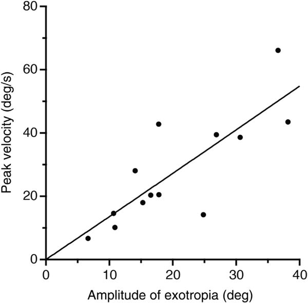
Deviation amplitude is correlated with peak velocity of the deviating eye’s movement during onset of spontaneous exotropia.
The spontaneous deviation reached 90% of its full amplitude within a mean of 1.75±1.01 seconds, n = 13. There was little correlation (r = −0.18) between the exotropia magnitude and the duration of the outward eye movement, because the globe deviated faster in patients with a large exotropia. In most patients (8/13) the full exotropic amplitude was reached within 3 seconds, but in some patients the exotropic movement was quite prolonged, and even took 10 seconds in 1 subject.
The peak velocity of the deviating eye’s movement was highly correlated (r = 0.91) for spontaneous and shutter-induced events (Fig 4). This finding indicates that exotropic eye movements are similar in both amplitude and velocity, whether they occur via occlusion or spontaneously.
Figure 4.
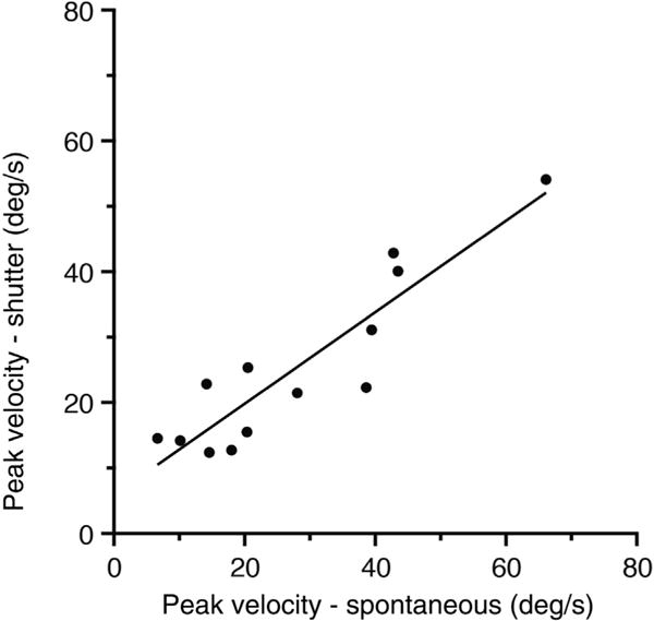
Peak velocity of the deviating eye’s movement is similar for spontaneous exotropia compared with shutter-induced exotropia.
Recovery of Ocular Alignment
After shutter removal the timing of fusion recovery was highly variable, and indeed, on some trials the exotropic eye remained deviated. The shortest latency for the initiation of an adducting eye movement after shutter removal was 100 milliseconds, recorded in 2 patients. Recovery of alignment was often accompanied by frequent blinking. In 2 of 13 patients, the blinking was so severe that eye tracking was lost, making it impossible to determine the strategy used to realign the eyes. In the remaining subjects, ocular alignment was regained through 1 of 4 stereotypic strategies (Fig 5). All strategies favored the dominant eye, either because (1) it showed little or no displacement from the target when the nondominant eye was uncovered, or (2) an alternating saccade was used to bring the dominant eye onto the target immediately upon uncovering. In nearly every case, one could predict which eye was dominant simply by inspecting the recordings that showed how ocular alignment was reacquired.
Figure 5.
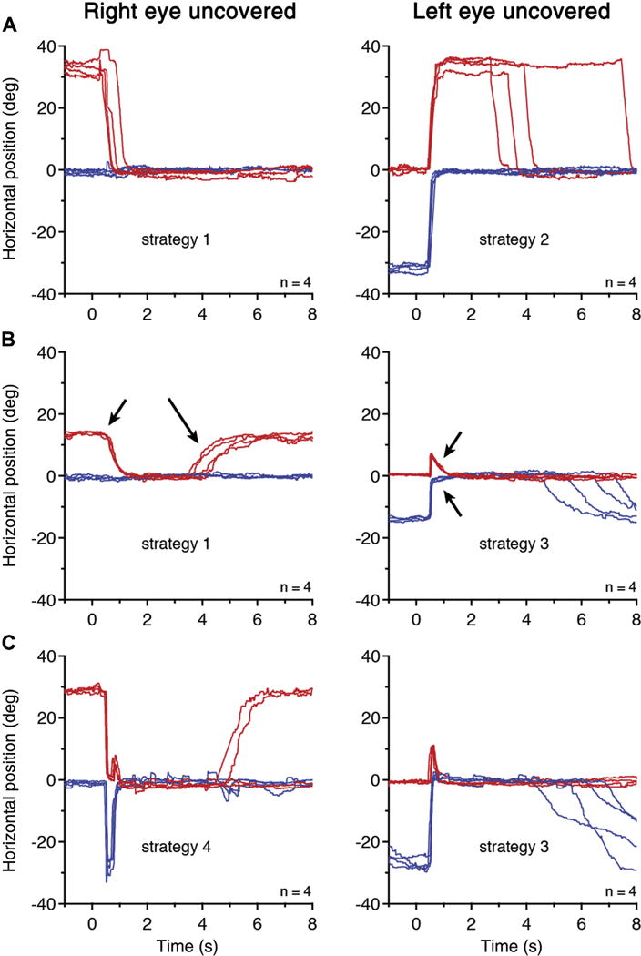
Four strategies for ocular realignment. A, A 35-year-old man with left eye dominance. Strategy 1: After the nondominant right eye was uncovered at t = 0 seconds,it made a vergence-like adducting movement. Strategy 2: After the dominant left eye was uncovered, an alternating saccade was made, followed at variable intervals by a vergence-like movement of the right eye. Blinks have been removed (Supplementary Video 1, available at www.aaojournal.org). B, A 31-year-old man with left eye dominance. After the right eye was uncovered, strategy 1 was employed. Convergence (small arrow) was faster than divergence (large arrow), which occurred with subsequent episodes of fusion loss. Strategy 3: After the left eye was uncovered, a disconjugate saccade (much larger in the adducting, left eye) was made, followed by vergence movements (arrows). The vergence movement was larger in the nondominant right eye. C, An 11-year-old girl with right eye dominance. Strategy 4: After the dominant eye was uncovered, an alternating saccade was made to bring it onto the target (first stage of strategy 2). This was followed by a disconjugate saccade and asymmetrical vergence movement (strategy 3). After the left eye was uncovered, this child used strategy 3 to recover fusion.
Strategy 1 was the simplest realignment technique (Fig 5). After the nondominant eye was uncovered, it made a vergence-type adducting movement while the dominant eye stayed riveted on the target. In strategy 2, when the dominant eye was uncovered, an alternating saccade was made to bring it onto the target. This saccade was followed by strategy 1 to unite the eyes. Altogether, 10 of 11 patients adducted their nondominant eye with a unilateral vergence-type movement, by using strategy 1 or 2. The adducting movement had a mean peak velocity of 51.9 26.5 degrees/second. This was far more rapid than the abducting movement that occurred with fusion loss, which had a mean peak velocity of 27.9±17.1 degrees/second (Fig 4).
Strategy 3 was another common way to regain fusion, occurring in 8 eyes of 11 patients. Either eye could be the dominant eye. As the eyes began to move toward each other, a disconjugate saccade was made to bring the deviated eye close to the target. This occurred at the expense of the fixating eye, which moved the “wrong” way, bringing it off the target. The saccade had a low peak velocity, compared with saccades of the same magnitude made in isolation.24 The eyes then continued to move slowly and asymmetrically toward each other until fusion was achieved. Strategy 4 used an alternating saccade that first brought the dominant eye onto the target, and was then followed by strategy 3.
Type of Eye Movement Made in Intermittent Exotropia
After spontaneous loss of fusion, 1 eye drifts outward in a fashion that resembles a divergence eye movement (Fig 1). The globe rotation is slow, disjunctive (eyes moving in opposite directions), and accompanied by dilation of the pupil. It has not been established, however, that it is a physiological divergence eye movement. In one respect it cannot be, given that in normal subjects the eyes never diverge beyond parallel. The following experiments attempted to replicate, with a normal divergence eye movement, the same eye motion made after loss of fusion. A single example is illustrated, but similar results were obtained in 4 subjects.
In the first task, the subject fixated a target at distance with both eyes. Spontaneous loss of fusion resulted in an outward movement of 1 eye, in this example the right eye (Fig 6A). In the second task, the subject fixated a crosshair at near with both eyes. He aligned the crosshair with a distant laser spot along the visual axis of the left eye.25 While this eye continued to fixate the crosshair, a shutter descended over the right eye. It deviated out immediately, mimicking the exodeviation movement made at distance, but within a physiological range (Fig 6B). In the third task, the subject was asked to fixate the near crosshair with both eyes, and then to shift his gaze to the distant laser spot. The goal was to generate a unilateral divergence movement of the right eye, matching the eye movement made in the previous task, but without resorting to occlusion. Instead, the subject always made a mixed vergence–saccade movement, even when instructed to fixate the laser spot assiduously (Fig 6C). This behavior thwarted our attempt to match the exodeviatory eye movement of strabismus with a normal divergence eye movement. Interestingly, after the gaze shift, the subject maintained fusion at distance. The act of shifting vergence angle seemed to transiently inhibit the tendency to manifest a spontaneous exotropia. To generate an exotropia with this maneuver, a fourth task was devised. The subject fixated on the near crosshair with both eyes. At the moment the laser spot appeared, signaling to the subject to shift his gaze from near to far, a shutter descended over the right eye. This almost always eliminated the unwanted saccade (Fig 6D). The occluded eye made a unilateral divergence movement, taking it to the same position assumed in spontaneous exotropia after loss of fusion at distance (Fig 6A). The velocity of the movement was much faster, however, because of its large amplitude. It fell along the “main sequence” for exodeviatory eye movement velocities (Fig 3).
Figure 6.
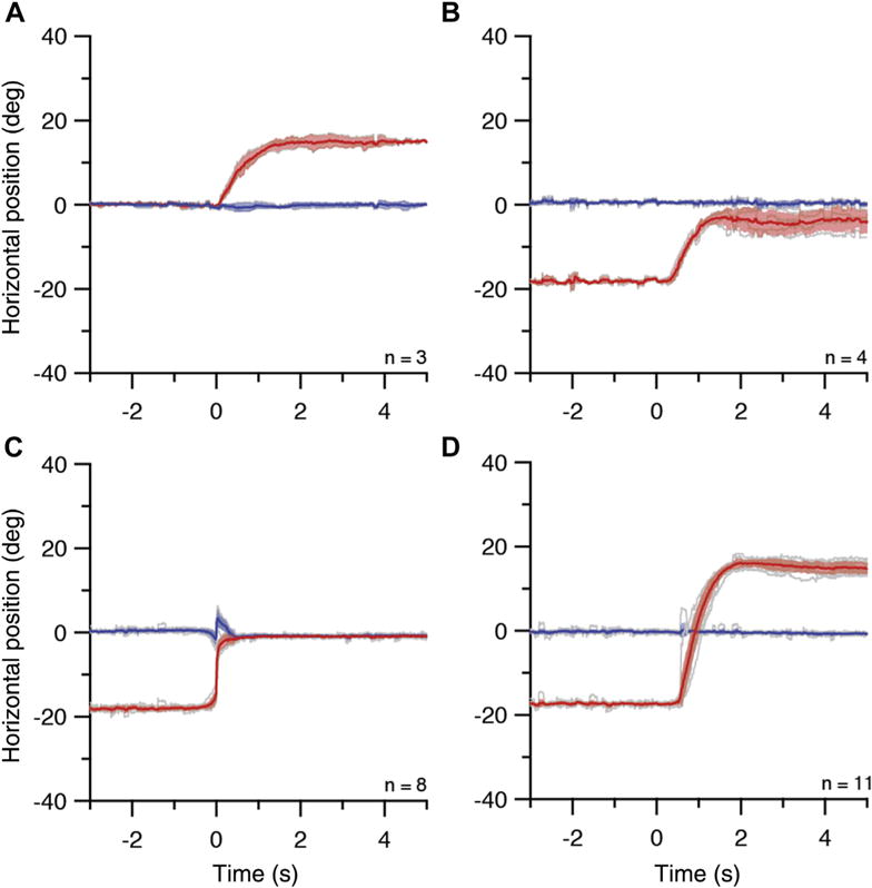
Exotropia at near and distance. The eye trackers were calibrated at distance; 0 degrees refers to fixation at 305 cm. A, Task 1: Spontaneous episodes of right exotropia (amplitude 15.0 degrees, peak velocity 20.1 degrees/second) in a 46-year-old man during fixation at 305 cm. B, Task 2: Exotropia (amplitude 14.2 degrees, peak velocity 24.5 degrees/second) after shutter occlusion of the right eye, while fixating on a crosshair at ~21 cm from the left eye along its line of sight with a distant laser spot. The intent was to mimic the eye movement in (A), by moving the right eye from −15.0 degrees to 0 degrees. The crosshair may have been placed a few centimeters closer than intended, resulting in a movement of the right eye from −18.1 degrees to −3.9 degrees. C, Task 3: Gaze shift along left eye’s line of sight from a crosshair nominally at 21 cm to a laser spot at 305 cm resulted in an outward movement of the right eye (amplitude 18.1 degrees, peak velocity 142.2 degrees/second) that combined divergence with a disconjugate saccade. Replication of the right eye’s movement in (B) was intended, but was foiled by the occurrence of a saccade. D, Task 4: Same task as (C), except that when the distant laser spot appeared, a shutter simultaneously covered the right eye. The right eye moved outward (amplitude 33.0 degrees, peak velocity 43.4 degrees/second) from −17.4 degrees to 15.3 degrees, to assume the same final position as in (A).
Discussion
Doubtless our observations are familiar to experienced clinicians engaged in daily care of patients with stra-bismus, but there is some value to quantitative measurement given our primitive understanding of intermittent exotropia. The use of an infrared camera is especially useful for documenting movement of the eye hidden behind the occluder. In the future, data from eye-movement recordings may provide the basis for confirming or refuting various theories about the neural basis of intermittent exotropia.
When a subject loses fusion and becomes exotropic, how should the eye movement be classified? The globe rotates outward: In that sense it is a divergence eye movement. Recall, however, that when normal subjects shift their gaze from near to far, they usually do not make a pure divergence eye movement. Instead, they combine a disconjugate saccade with a divergence eye movement, allowing them to acquire a new target more rapidly and efficiently.24,26,27 Exotropic subjects display the same behavior when shifting gaze to a distant target, even when it is located along the line of sight of 1 eye.28 Under such conditions (Fig 6C), the interjected saccade actually takes the fixating eye off the target momentarily. This ocular motor behavior is quite different from the exotropic eye movement that occurs with spontaneous loss of fusion (Fig 1, Fig 6A). The latter eye movement is slow, is unilateral, and contains no embedded saccade.
It is possible, albeit less common, for normal subjects to execute a step shift in gaze angle from near to far by making a pure divergence movement with no saccade.26,29 There are several reasons, however, for rejecting the idea that such a movement is tantamount to the eye movement accompanying fusion loss in exotropia. First, pure divergence is more likely when the target location is midway between each eye’s line of sight, requiring a relatively equal divergence movement in each eye. This is different from exotropia, in which only 1 eye moves outward. Second, the main sequence relationship for normal divergence is peak velocity = 5.0 × vergence step amplitude/second.30 The corresponding main sequence relationship for the abducting eye movement in exotropia is peak velocity = 1.36 × exotropia amplitude/second (Fig 3). Therefore, the peak velocity of a physiological divergence movement is faster than the diverging eye movement made in exotropia.
It appears, therefore, that when exotropic subjects lose fusion, the movement of the deviating eye does not represent an inadvertent attempt to shift gaze from near to far. As we have shown, exotropic subjects have the capacity to execute such normal gaze shifts (with an interjected saccade) while in a normal binocular state. In fact, when they do, loss of fusion is transiently suppressed (Fig 6C). It would be worthwhile to document what type of ocular motor behavior provokes fusion loss in exotropic patients. This could be done by tracking their eye movements during long periods while they are ambulatory and engaged in normal exploration of real-life visual scenes. We predict that fusion loss typically occurs during episodes of steady fixation on targets, and when it follows a gaze shift to a distant target, there is a transient delay before onset.
To summarize, the outward movement of the eye in exotropia shares some features with normal divergence, but it should be classified as a unique, pathologic type of human eye movement. As mentioned previously, when normal subjects shift their gaze from near to far, they usually make a combined saccadeevergence movement.26,31,32 This type of hybrid eye movement does not occur when fusion is lost spontaneously in exotropic subjects (Fig 1). A normal subject could, in principle, mimic the slow outward eye movement made by an exotropic subject by tracking a receding target that was moving slowly along the line of sight of 1 eye. However, that type of slow divergence pursuit movement requires a visible, moving target. In exotropia, the subject has no target to pursue as the eye moves outward. Finally, divergence of the ocular axes never occurs beyond parallel in normal subjects.
In normal divergence, eye velocity depends on initial stimulus position.33 Our data showed an interesting and unexpected correlation between the velocity of the outward eye movement and its amplitude (Fig 3). This feature is usually associated with saccadic eye movements. It is reminiscent of the expansion of a spring upon release.
The eye movement made to recover fusion is much faster than the movement that occurs with loss of fusion (Fig 5). Four different strategies were observed in our patients. Several were described previously in an elegant study of cover test dynamics in exophoric subjects.34 Although fusion is often regained by making an inward movement of 1 eye only, a combined saccade–vergence eye movement is sometimes employed, as if the subject were attempting to shift gaze from far to near. In this respect, the realignment of the eyes more closely resembles a physiological vergence movement than does the movement that occurs with the loss of fusion.
In most patients, the eye movement that occurs during spontaneous loss of fusion is identical to the movement made when the eye is covered. In exotropes, divergence tone is excessive relative to convergence tone,35 explaining why adducting saccades are hypometric relative to abducting saccades.18 The eyes are prevented from drifting apart by cortical binocular mechanisms, which maintain fusion. When 1 eye is occluded, cortical drive is interrupted, and the covered eye drifts outward. This can occur remarkably fast, in as little as 140 milliseconds. It requires 80 milliseconds for erasure of cortical images after loss of retinal input.36 That leaves only 60 milliseconds for initiation of an eye movement in subjects with the shortest latency. Why does exotropia occur on a spontaneous basis, given that both eyes are open and actively engaged in perception? The cortical drive for binocular vision may be weak or abnormal, creating permissive conditions for fusion loss. For example, it has been reported that children with intermittent exotropia exhibit suppression of their temporal retinae even while their eyes are still aligned.37 Apparently, they are primed to suppress, which could lower the threshold that normally blocks a transition from fusion to exotropia.
Alternatively, cortical binocular drive may be normal in intermittent exotropia, but the imbalance in vergence tone too powerful to permit maintenance of constant fusion. Our observation that spontaneous and occluder-induced outward eye movements are equal does not enable us to rule out either theory: weak cortical binocular drive and/or excessive divergence tone. We can conclude, however, that once the ocular axes separate, visual feedback has no impact on the subsequent outward movement of the deviating eye to its final position, at least in most patients. After fusion loss, the deviating eye moves outward in a stereotypic, reflexive fashion. Otherwise, there would be a difference between shutter-induced and spontaneous exotropia.
Supplementary Material
Acknowledgments
Jessica Wong and Brittany C. Rapone participated in the testing of subjects. The authors thank the ophthalmologists who referred subjects, and Anthony T. Moore and Arthur Jampolsky for reading the manuscript.
Financial Disclosure(s):
Supported by grants EY10217 (J.C.H.), EY02162 (Beckman Vision Center) from the National Eye Institute and a Physician-Scientist Award from Research to Prevent Blindness (J.C.H.).
Abbreviations and Acronyms
- CI
confidence interval
- OU
both eyes.
Footnotes
Author Contributions:
Conception and design: Economides, Adams, Horton
Analysis and interpretation: Economides, Adams, Horton
Data collection: Economides, Adams, Horton
Obtained funding: Horton
Overall responsibility: Economides, Adams, Horton
References
- 1.Hatt SR, Mohney BG, Leske DA, Holmes JM. Variability of control in intermittent exotropia. Ophthalmology. 2008;115(2):371–376. doi: 10.1016/j.ophtha.2007.03.084. [DOI] [PMC free article] [PubMed] [Google Scholar]
- 2.Chia A, Roy L, Seenyen L. Comitant horizontal strabismus: an Asian perspective. Br J Ophthalmol. 2007;91(10):1337–1340. doi: 10.1136/bjo.2007.116905. [DOI] [PMC free article] [PubMed] [Google Scholar]
- 3.Kushner BJ, Morton GV. Distance/near differences in intermittent exotropia. Arch Ophthalmol. 1998;116(4):478–486. doi: 10.1001/archopht.116.4.478. [DOI] [PubMed] [Google Scholar]
- 4.Buck D, Powell C, Cumberland P, et al. Presenting features and early management of childhood intermittent exotropia in the UK: inception cohort study. Br J Ophthalmol. 2009;93(12):1620–1624. doi: 10.1136/bjo.2008.152975. [DOI] [PubMed] [Google Scholar]
- 5.Nusz KJ, Mohney BG, Diehl NN. The course of intermittent exotropia in a population-based cohort. Ophthalmology. 2006;113(7):1154–1158. doi: 10.1016/j.ophtha.2006.01.033. [DOI] [PubMed] [Google Scholar]
- 6.Romanchuk KG, Dotchin SA, Zurevinsky J. The natural history of surgically untreated intermittent exotropia-looking into the distant future. J AAPOS. 2006;10(3):225–231. doi: 10.1016/j.jaapos.2006.02.006. [DOI] [PubMed] [Google Scholar]
- 7.Hoyt CS, Pesic A. The many enigmas of intermittent exotropia. Br J Ophthalmol. 2012;96(10):1280–1282. doi: 10.1136/bjophthalmol-2012-302345. [DOI] [PubMed] [Google Scholar]
- 8.Brodsky MC, Jung J. Intermittent exotropia and accommodative esotropia: distinct disorders or two ends of a spectrum? Ophthalmology. 2015;122(8):1543–1546. doi: 10.1016/j.ophtha.2015.03.004. [DOI] [PubMed] [Google Scholar]
- 9.Jampolsky A. Physiology of intermittent exotropia. Am Orthopt J. 1963;13:5–13. [PubMed] [Google Scholar]
- 10.Joosse MV, Simonsz HJ, van Minderhout EM, et al. Quantitative visual fields under binocular viewing conditions in primary and consecutive divergent strabismus. Graefes Arch Clin Exp Ophthalmol. 1999;237(7):535–545. doi: 10.1007/s004170050276. [DOI] [PubMed] [Google Scholar]
- 11.Cooper J, Feldman J. Panoramic viewing, visual acuity of the deviating eye, and anomalous retinal correspondence in the intermittent exotrope of the divergence excess type. Am J Optom Physiol Opt. 1979;56(7):422–429. doi: 10.1097/00006324-197907000-00003. [DOI] [PubMed] [Google Scholar]
- 12.Economides JR, Adams DL, Horton JC. Perception via the deviated eye in strabismus. J Neurosci. 2012;32(30):10286–10295. doi: 10.1523/JNEUROSCI.1435-12.2012. [DOI] [PMC free article] [PubMed] [Google Scholar]
- 13.Herzau V. How useful is anomalous correspondence? Eye (Lond) 1996;10(Pt 2):266–269. doi: 10.1038/eye.1996.56. [DOI] [PubMed] [Google Scholar]
- 14.Cooper J, Record CD. Suppression and retinal correspondence in intermittent exotropia. Br J Ophthalmol. 1986;70:673–676. doi: 10.1136/bjo.70.9.673. [DOI] [PMC free article] [PubMed] [Google Scholar]
- 15.Hatt SR, Leske DA, Holmes JM. Awareness of exodeviation in children with intermittent exotropia. Strabismus. 2009;17(3):101–106. doi: 10.1080/09273970903107972. [DOI] [PMC free article] [PubMed] [Google Scholar]
- 16.Birch EE, Holmes JM. The clinical profile of amblyopia in children younger than 3 years of age. J AAPOS. 2010;14(6):494–497. doi: 10.1016/j.jaapos.2010.10.004. [DOI] [PMC free article] [PubMed] [Google Scholar]
- 17.Leigh R, Zee D. The Neurology of Eye Movements. 4th. New York: Oxford University Press; 2006. [Google Scholar]
- 18.Economides JR, Adams DL, Horton JC. Eye choice for acquisition of targets in alternating strabismus. J Neurosci. 2014;34(44):14578–14588. doi: 10.1523/JNEUROSCI.3278-14.2014. [DOI] [PMC free article] [PubMed] [Google Scholar]
- 19.Economides JR, Adams DL, Horton JC. Variability of ocular deviation in strabismus. JAMA Ophthalmol. 2016;134(1):63–69. doi: 10.1001/jamaophthalmol.2015.4486. [DOI] [PMC free article] [PubMed] [Google Scholar]
- 20.Economides JR, Adams DL, Jocson CM, Horton JC. Ocular motor behavior in macaques with surgical exotropia. J Neurophysiol. 2007;98(6):3411–3422. doi: 10.1152/jn.00839.2007. [DOI] [PubMed] [Google Scholar]
- 21.Hvelplund KT. Eye tracking and the translation process: reflections on the analysis and interpretation of eye-tracking data. In: Munoz Martin R., editor. Minding Translation – MonTI Special Issue. 2014. pp. 201–223. [Google Scholar]
- 22.Bahill A, Clark M, Stark L. The main sequence, a tool for studying human eye movements. Math Biosci. 1975;24:191–204. [Google Scholar]
- 23.Boghen D, Troost BT, Daroff RB, et al. Velocity characteristics of normal human saccades. Invest Ophthalmol. 1974;13(8):619–623. [PubMed] [Google Scholar]
- 24.Collewijn H, Erkelens CJ, Steinman RM. Voluntary binocular gaze-shifts in the plane of regard: dynamics of version and vergence. Vision Res. 1995;35(23–24):3335–3358. doi: 10.1016/0042-6989(95)00082-p. [DOI] [PubMed] [Google Scholar]
- 25.Müller J. Zur Vergleichenden Physiologie des Gesichtssinnes des Menschen und der Thiere. Leipzig: Cnobloch; 1826. [Google Scholar]
- 26.Zee DS, Fitzgibbon EJ, Optican LM. Saccade-vergence interactions in humans. J Neurophysiol. 1992;68(5):1624–1641. doi: 10.1152/jn.1992.68.5.1624. [DOI] [PubMed] [Google Scholar]
- 27.Enright JT. Changes in vergence mediated by saccades. J Physiol. 1984;350:9–31. doi: 10.1113/jphysiol.1984.sp015186. [DOI] [PMC free article] [PubMed] [Google Scholar]
- 28.Kenyon RV, Ciuffreda KJ, Stark L. Dynamic vergence eye movements in strabismus and amblyopia: asymmetric vergence. Br J Ophthalmol. 1981;65(3):167–176. doi: 10.1136/bjo.65.3.167. [DOI] [PMC free article] [PubMed] [Google Scholar]
- 29.Rashbass C, Westheimer G. Disjunctive eye movements. J Physiol (Lond) 1961;159:339–360. doi: 10.1113/jphysiol.1961.sp006812. [DOI] [PMC free article] [PubMed] [Google Scholar]
- 30.Hung GK, Ciuffreda KJ, Semmlow JL, Horng JL. Vergence eye movements under natural viewing conditions. Invest Ophthalmol Vis Sci. 1994;35(9):3486–3492. [PubMed] [Google Scholar]
- 31.Erkelens CJ, Steinman RM, Collewijn H. Ocular vergence under natural conditions. II. Gaze shifts between real targets differing in distance and direction. Proc R Soc Lond B Biol Sci. 1989;236(1285):441–465. doi: 10.1098/rspb.1989.0031. [DOI] [PubMed] [Google Scholar]
- 32.Erkelens CJ, Van der Steen J, Steinman RM, Collewijn H. Ocular vergence under natural conditions. I. Continuous changes of target distance along the median plane. Proc R Soc Lond B Biol Sci. 1989;236(1285):417–440. doi: 10.1098/rspb.1989.0030. [DOI] [PubMed] [Google Scholar]
- 33.Alvarez TL, Semmlow JL, Pedrono C. Divergence eye movements are dependent on initial stimulus position. Vision Res. 2005;45(14):1847–1855. doi: 10.1016/j.visres.2005.01.017. [DOI] [PubMed] [Google Scholar]
- 34.Peli E, McCormack G. Dynamics of cover test eye movements. Am J Optom Physiol Opt. 1983;60(8):712–724. doi: 10.1097/00006324-198308000-00010. [DOI] [PubMed] [Google Scholar]
- 35.Burian HM. Exodeviations: their classification, diagnosis and treatment. Am J Ophthalmol. 1966;62(6):1161–1166. doi: 10.1016/0002-9394(66)92570-0. [DOI] [PubMed] [Google Scholar]
- 36.Coppola D, Purves D. The extraordinarily rapid disappearance of entoptic images. Proc Natl Acad Sci U S A. 1996;93(15):8001–8004. doi: 10.1073/pnas.93.15.8001. [DOI] [PMC free article] [PubMed] [Google Scholar]
- 37.Serrano-Pedraza I, Manjunath V, Osunkunle O, et al. Visual suppression in intermittent exotropia during binocular alignment. Invest Ophthalmol Vis Sci. 2011;52(5):2352–2364. doi: 10.1167/iovs.10-6144. [DOI] [PubMed] [Google Scholar]
Associated Data
This section collects any data citations, data availability statements, or supplementary materials included in this article.


