Abstract
Matrix-assisted laser desorption ionization time-of-flight mass spectrometry (MALDI-TOF MS) is a reliable and rapid technique applied widely in the identification and classification of microbes. MALDI-TOF MS has been used to identify many endospore-forming Bacillus species; however, endospores affect the identification accuracy when using MALDI-TOF MS because they change the protein composition of samples. Since culture conditions directly influence endospore formation and Bacillus growth, in this study we clarified how culture conditions influence the classification of Bacillus species by using MALDI-TOF MS. We analyzed members of the Bacillus subtilis group and Bacillus cereus group using different incubation periods, temperatures and media. Incubation period was found to affect mass spectra due to endospores which were observed mixing with vegetative cells after 24 hours. Culture temperature also resulted in different mass spectra profiles depending on the temperature best suited growth and sporulation. Conversely, the four common media for Bacillus incubation, Luria-Bertani agar, nutrient agar, plate count agar and brain-heart infusion agar did not result in any significant differences in mass spectra profiles. Profiles in the range m/z 1000–3000 were found to provide additional data to the standard ribosomal peptide/protein region m/z 3000–15000 profiles to enable easier differentiation of some highly similar species and the identification of new strains under fresh culture conditions. In summary, control of culture conditions is vital for Bacillus identification and classification by MALDI-TOF MS.
Introduction
The genus Bacillus is ubiquitous in the environment, including in terrestrial and aquatic systems, and in the atmosphere. Members of the Bacillus genus are rod-shaped, endospore-forming, Gram positive bacteria that widely affect human life and the ecological environment1. Over 200 species of Bacillus have been identified and differentiated by genetic approaches and also through biological assays according to their activities in the environment or their pathogenicities2,3. One method of identifying bacteria is 16S rRNA gene-based taxonomy, but some Bacillus species are highly similar according to 16S rRNA sequence4. The Bacillus subtilis group, Bacillus cereus group and Bacillus pumilus group have multiple highly-related species within their groups. In the B. subtilis group, B. amyloliquifaciens, B. atrophaeus, B. licheniformis, B. mojavensis, B. subtilis subsp. spizizenii, B. subtilis subsp. subtilis and B. vallismortis are included but share different features5. The B. cereus group comprises B. anthracis, B. cereus, B. mycoides, B. pseumycoides, B. thuringiensis and B. weihenstephaniensis, and they are differentiated mainly according to their pathogenicities to humans, insects or others and free-living2. The B. pumilus group contains B. altitudinis, B. aerophilus, B. safiensis, B. pumilus and B. stratosphaericus which are differentiated through the gyrB (β-subunit of DNA gyrase) sequence and bacterial phenotypic features, chemotaxonomic characteristics and DNA-DNA connectedness5. Multi-locus sequence typing and whole genome sequencing6 can be used to differentiate close Bacillus species and even strains, but no high-throughput approach is commonly applied for classifying and identifying the many Bacillus isolates found in environmental samples.
Matrix assisted-laser desorption ionization time-of-flight mass spectrometry (MALDI-TOF MS) has been used for microbial identification and commercialized for more than a decade7,8. Several companies along with homemade hardware and software platforms have supported the identification of clinical microorganisms and an abundance of datasets have been compiled9. The ribosomal peptides/proteins extracted from microbial samples are the targets of MALDI-TOF MS analysis, and can provide overall and unique spectra of the samples10. The mass spectral profiles are compared with microbial protein profile databases through different algorithms which give possible classifications between sample and references. The technique is highly efficient and reliable and results are obtained within an hour11.
Identification of Bacillus species through MALDI-TOF MS has been conducted for several years and is often applied to important clinical pathogens like B. anthracis and B. cereus 12–15. However, pretreatment for Bacillus identification is not as simple as for other bacteria because of endospore formation. Endospores have different protein expression compared to vegetative cells and small acid soluble proteins (SASPs) have a crucial role in endospore formation16,17. In anthrax diagnosis, for example, endospore protein composition was shown to differentiate between pathogenic and non-pathogenic B. cereus group species18,19. If Bacillus are classified through general pretreatment of culture, extraction and matrix deposition for MALDI-TOF MS analysis, endospore protein signals cannot be ignored20. In addition, incubating Bacillus under different conditions influences the protein percentages due to different extents of endospore-formation21. Furthermore, different extents of endospore-formation affect the comparison of spectra when Bacillus is grown under different culture conditions. Despite culture conditions being critical for accurate identification, a systematic protocol has not been established for the classification of most Bacillus species from the environment.
In this study, we evaluated the influence of culture conditions including incubation time, temperature and culture medium and identified the factors that crucially affect accurate classification when Bacillus is isolated from different environmental origins. Furthermore, we offer new insights that may strengthen the classification within the B. cereus group and the B. subtilis group by including different molecular weight regions observed by MALDI-TOF MS.
Results
16S rRNA analysis of Bacillus strains
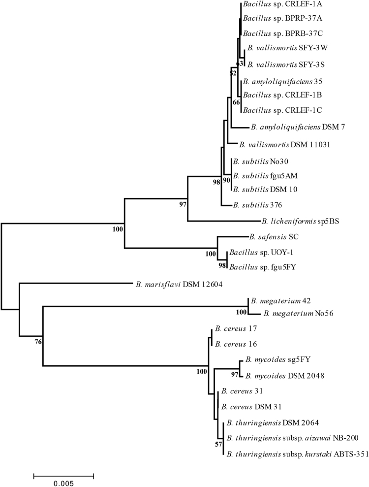
We sequenced and compared 16S rRNA of 30 Bacillus strains which were collected from multiple origins and isolated through different media by ourselves and the Bioresource Collection and Research Center (BCRC) in Taiwan (Table 1). In our collections, the B. subtilis group, containing three species, B. amyloliquefaciens, B. subtilis subsp. subtilis and B. vallimortis, and the B. cereus group including type strains B. cereus, B. mycoides and B. thuringiensis were the two main groups according to the 16S rRNA phylogenetic tree which revealed high similarity within each group of strains (Fig. 1). In each group, the Bacillus strains that shared 100% 16S rRNA sequence similarity were grouped together. For example, in Figure S1, the B. vallimortis strain SFY-3S and the SFY-3W strain showed entirely different morphology on Luria-Bertani (LB) agar, but could not be differentiated by their 16S rRNA sequences.
Table 1.
List of Bacillus used in this study as well as 16S rRNA accession number, the source of isolation or order, the original cultivation medium, and culture temperature.
| Organism | 16S rRNA accession Number | Source | Original Medium | Original Culture Temperature |
|---|---|---|---|---|
| B. thuringiensis DSM 2046 | D16281 | Type strain, BCRC | Nutrient agar | 30 °C |
| B. mycoides DSM 2048 | AB021192 | Type strain, BCRC | Nutrient agar | 25 °C |
| B. subtilis subsp. subtilis DSM 10 | AJ276351 | Type strain, BCRC | Nutrient agar | 30 °C |
| B. amyloliquefaciens DSM 7 | AB006920 | Type strain, BCRC | Nutrient agar | 30 °C |
| B. cereus DSM 31 | AE016877 | Type strain, BCRC | Nutrient agar | 30 °C |
| B. vallismortis DSM 11031 | AB021198 | Type strain, BCRC | Nutrient agar | 30 °C |
| B. marisflavi DSM 16204 | AF483624 | Type strain, BCRC | Bacto marine broth | 30 °C |
| B. cereus 31 | KY819017 | Terrestrial soil | Potato dextrose agar | 25 °C |
| B. cereus 17 | KY819023 | Terrestrial soil | Brain-heart infusion | 37 °C |
| B. cereus 16 | KY819024 | Terrestrial soil | Brain-heart infusion | 37 °C |
| B. thuringiensis subsp. kurstaki ABTS-351 | KY819035 | Commercial biopesticide | Nutrient agar | 30 °C |
| B. thuringiensis subsp. aizawai NB-200 | KY819034 | Commercial biopesticide | Nutrient agar | 30 °C |
| B. mycoides sg5CY | KY819039 | Marine shrimp gut | Yeast malt agar | 25 °C |
| B. subtilis 376 | KY819033 | Terrestrial soil | Luria-Bertani agar | 28 °C |
| B. subtilis No30 | KY819027 | Marine sediment | Luria-Bertani agar | 25 °C |
| B. subtilis fgu5AM | KY819036 | Fish gut | Marine agar | 25 °C |
| B. vallismortis SFY-3W | KY819019 | Sea fern | Yeast malt agar | 25 °C |
| B. vallismortis SFY-3S | KY819018 | Sea fern | Yeast malt agar | 25 °C |
| B. amyloliquefaciens 35 | KY819021 | Terrestrial soil | Potato dextrose agar | 37 °C |
| Bacillus sp. CRLEF-1A | KY819028 | Catharanthus roseus root | Luria-Bertani agar | 30 °C |
| Bacillus sp. CRLEF-1B | KY819029 | Catharanthus roseus root | Luria-Bertani agar | 30 °C |
| Bacillus sp. CRLEF-1C | KY819030 | Catharanthus roseus root | Luria-Bertani agar | 30 °C |
| Bacillus sp. BPRB37-A | KY819031 | Banana root | Luria-Bertani agar | 30 °C |
| Bacillus sp. BPRB37-C | KY819032 | Banana root | Luria-Bertani agar | 30 °C |
| B. licheniformis sp5BS | KY819038 | Sea pig | Yeast malt agar | 25 °C |
| B. megaterium No56 | KY819025 | Marine sediment | Luria-Bertani agar | 25 °C |
| B. megaterium 42 | KY819022 | Terrestrial soil | Potato dextrose agar | 30 °C |
| Bacillus sp. fgu5FY | KY819037 | Fish gut | Yeast malt agar | 25 °C |
| B. safensis SC | KY819026 | Bee gut | Luria-Bertani agar | 30 °C |
| Bacillus sp. UOY-1 | KY819020 | Sea urchin | Yeast malt agar | 25 °C |
Figure 1.
Phylogenic analysis of the Bacillus species 16S rRNA sequences using the neighbor-joining method. The sequences were aligned and analyzed using the neighbor-joining method with 1000 bootstrap replications using MEGA 5. The numbers over 50% on the branches represent the bootstrap value and the bar indicates a phylogenetic tree distance of 0.5%.
MALDI-TOF MS analysis of Bacillus with different incubation temperatures and periods
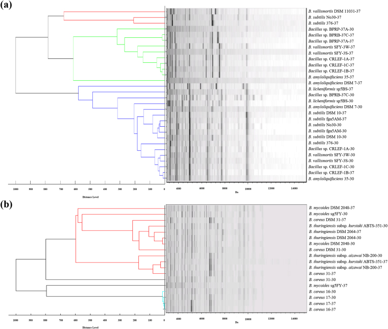
The inconsistencies in Bacillus identification through MALDI-TOF MS are mostly because of endospore formation and culture conditions. In addition, different species of Bacillus isolated from different environments have a variety of optimal growth temperatures which further influences the endospore formation rate. The B. subtilis group revealed a consistent main spectrum (MSP) with strains incubated at 30 °C or 37 °C mainly separated into two clusters (Fig. 2a). But the B. cereus group did not share similar MSP patterns to the B. subtilis group. For example, B. cereus 16 and B. cereus 17 had similar MSPs at two different temperatures (Fig. 2b).
Figure 2.
MSP dendrograms and spectra gel view of (a) Bacillus subtilis and (b) Bacillus cereus group species incubated at 30 °C and 37 °C for 24 hours. Numbers behind the species and strain name indicate the culture temperature.
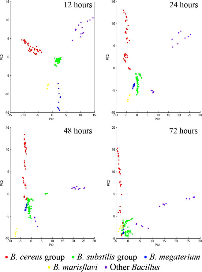
Different incubation times for Bacillus ranging from 12 hours to 72 hours had a dramatic effect on the endospore populations (Table S1 and Fig. S2). Principal component analysis plots were used to illustrate the MSP variances in the Bacillus groups cultured for different times (Fig. 3 and Fig. S4). At 12 hours, each Bacillus group was clearly separated; however, the B. cereus group was divided into two sections after incubation for 24 hours. After 48 to 72 hours, several isolates from each Bacillus group were not significantly distinguished from other groups in principal component analysis. The MSP dendrograms and spectral gel views are provided in Fig. S3. We then identified SASPs, biomarkers of endospores, and found they were accumulated in both B. cereus and B. subtilis samples cultured over 48 and 24 hours, respectively (Fig. S5). These results demonstrated that the uncertainty of endospore formation affects classification using MALDI-TOF MS and fresh Bacillus is an ideal sample for classification and identification. But interestingly, the MSPs of a few species such as B. mycoides still showed obvious differences between 12 and 72 hours even though almost no endospores were detected within three days incubation (in the period 12–72 hours) (Table S1).
Figure 3.
Two-dimensional principal component analysis plots of Bacillus cereus group, Bacillus subtilis group, Bacillus megaterium, Bacillus marisflavi and other Bacillus species cultured for 12 hours, 24 hours, 48 hours, and 72 hours. The spot presents one spectrum and each Bacillus isolate includes five spectra. The plots were generated by first two principal components: PC1 and PC2.
MALDI-TOF MS analysis of Bacillus with different incubation agar media
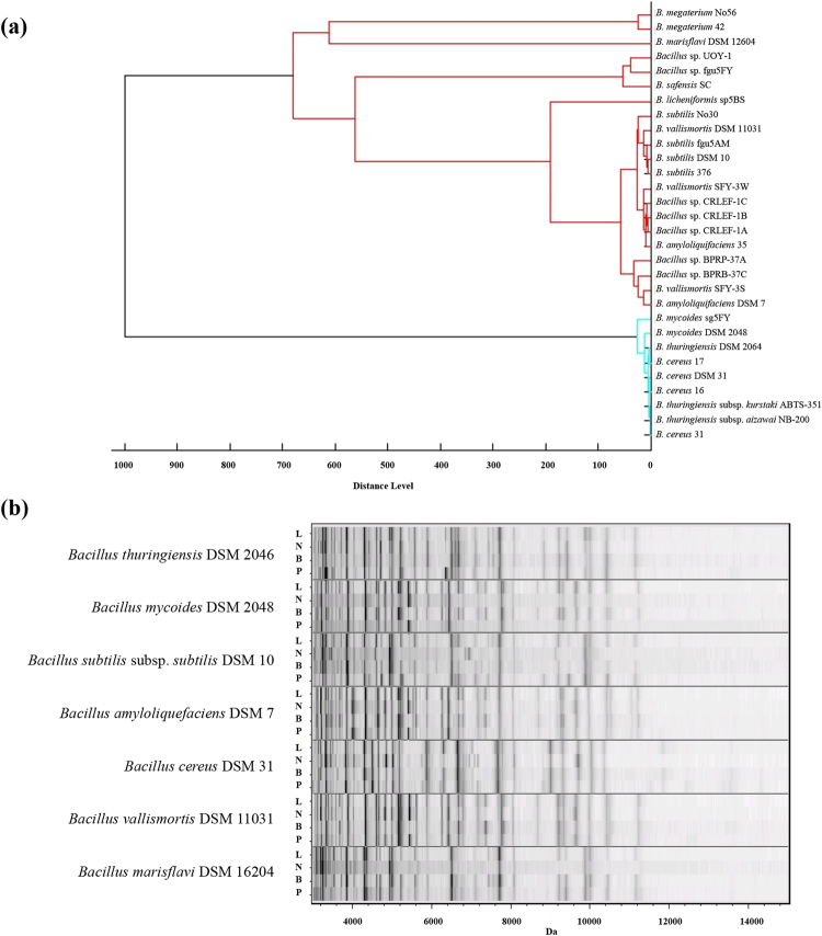
To further determine the stability and reliability of Bacillus classification through MALDI-TOF MS, the samples were cultured and collected 12 hours after incubation and all kept at 30 °C. Then the impact of different agar media was evaluated. LB agar, nutrient agar (NA), plate count agar (PCA) and brain-heart infusion (BHI) agar, which are commonly applied for isolating and culturing Bacillus from clinical and environmental samples, were investigated. The four different agar media had no dramatic effect on Bacillus MSP dendrograms (Fig. 4a: LB and Fig. S6: LB, NA, BHI, PCA with gel views). B. cereus, B. thuringiensis and B. mycoides strains were still clustered in the B. cereus group with high similarity and all the tested strains in the B. subtilis group were firmly linked in the same clade. The MSP of other species like B. megaterium strains and B. marisflavi type strain were also consistently found in the same clades no matter which media were used. The high intensity peaks in MSPs are crucial in the identification and taxonomy of microorganisms. MSP gel view of type strains showed that high intensity peaks in each strain were not significantly different between the Bacillus strains incubated in the four different media (Fig. 4b).
Figure 4.
Biotyper analysis of Bacillus with different culture media. (a) MSP dendrogram of Bacillus cultured on LB agar at 30 °C for 12 hours and (b) spectral gel view of seven Bacillus type strains cultured on LB agar (L), NA (N), BHI agar (B) and PCA (P) at 30 °C for 12 hours.
Range m/z 1000–3000 assisted the standard region (m/z 3000–15000) for differentiation of highly similar strains
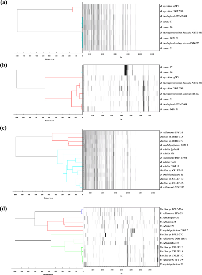
Many isolates in B. subtilis and B. cereus groups are not easily distinguished using single genomic approaches such as 16S rRNA sequencing or MALDI-TOF MS analysis in the range m/z 3000–15000. To investigate how to reinforce the differentiation of strains within these groups using MALDI-TOF MS, we set up the standard identification procedure (LB, 30 °C, 12 hours, m/z 3000–15000) and a modified procedure (LB, 30 °C, 12 hours, m/z 1000–3000) in this study. To eliminate the high intensity peaks in m/z 3000–15000 affecting distinguishability, m/z 1000–3000 was utilized in strain differentiation independently. The B. subtilis and B. cereus groups demonstrated much greater relative distance or different results for classification in MSP dendrogram analysis at m/z 1000–3000, in comparison to those derived from MSP at m/z 3000–15000 (Fig. 5).
Figure 5.
MSP dendrogram and spectra gel view of (a, b) Bacillus cereus and (c, d) Bacillus subtilis group species cultured on LB agar at 30 °C for 12 hours. The analysis and construction of dendrograms (a) and (c) are based on the m/z 3000–15000 range and dendrogram (b) and (d) are based on the m/z 1000–3000 range.
Discussion
The B. subtilis and B. cereus groups, which are highly genetically similar to each other, were the two targets of this study. Although, the strains were gathered from completely different origins, phylogenetic analysis based on 16S rRNA sequences in the species of each group showed no significant differences (Fig. 1). Some even showed 100% similarity in 16S rRNA sequences but had different colony morphologies, highlighting the difficulty of using 16S rRNA sequences to differentiate strains or species in these groups. Previously, 23S rRNA, gyrB gene and other pathogenic gene clusters have been used to support the classification and identification of these groups22,23; however, the results were either efficient or reliable, but not both, suggesting improvements are needed. MALDI-TOF MS-based microbial classification has also been used with some success to identify Bacillus pathogenic and non-pathogenic strains through the MSP24.
Protein profiles collected by MALDI-TOF MS have become an efficient method to rapidly identify microbial samples, especially for clinical diagnosis, and bacterial and fungal protein databases have been comprehensive for a decade25. Many species of Bacillus are collected in the commercial databases; however, because of endospores, which mix together with vegetative cells, classification and identification of Bacillus strains by using MALDI-TOF MS is particularly difficult. The variable vegetative-endospore cell composition directly affects identification of members of the Bacillus genus13. Moreover, the fraction of SASPs which is around 8% to 20% in different species of endospores is variable26, further hindering the accurate identification of several Bacillus species. Therefore, a method that can analyze total proteins of vegetative cells without endospore interference would be ideal for classification of endospore-forming bacteria. In this study, we were able to obtain endospore-free Bacillus through adopting simple culturing methods.
Optimizing the culture conditions is a straightforward way to avoid endospore formation. Growth rate directly influences the transformation from vegetative cells to endospores. Three culture conditions that affect the growth rate of Bacillus, temperature, incubation time and medium, were evaluated in this study. We demonstrated that temperature partially affected the MSP of some Bacillus strains. Of note, it had considerable effects on B. subtilis group at 30 °C and 37 °C within one day incubation (Fig. 2). Moreover, incubation periods of Bacillus on agar media also dramatically influenced the MSP and principal component analysis results of both the B. cereus group and the B. subtilis group, as well as other Bacillus strains (Fig. 3). Endospores were easily observed when Bacillus were mature or overgrown on agar media (Table S1). A previous study indicated that B. subtilis and B. licheniformis incubated for one, two and four days at 30 °C on PCA had no significant effect on protein profiles27. In this study, MALDI-TOF MS, endospore measurement and microscopy observation were used to confirm that some strains are affected to different extents depending on the growth rate of the Bacillus. The same phenomenon was reported by Chambers et al. who showed that endospores directly decreased the identification scores which were evaluated by MALDI-TOF MS, and disturbed identification of B. subtilis 28. Moreover, SASPs, the essential elements for endospore formation, were also detected in reference strains after 24 hours or longer after incubation, but not at 12 hours, and these results also verified that SASPs, including α-SASP, β-SASP and α/β-SASP, are usually observed under the standard conditions (LB, 24 hours, 37 °C)24,29. MSP dendrograms revealed how the vegetative-endospore cell mixture influenced the taxonomy of Bacillus and what conditions can be used to circumvent these situations (Fig. S5). Keeping the Bacillus sample as fresh as possible is a critical step to stabilize the result of identification and classification. Here we recommend the use of LB, 12 hours, 30 °C as standard culture conditions for Bacillus classification by MALDI-TOF MS.
The culture medium is one factor that has physiological effects on bacteria that further influences MALDI-TOF MS-based identification. Culture medium has been studied for the identification of many bacteria such as E. coli 30, Yersinia 30, Lactobacillus 31, Staphylococcus 32 , Pseudomonas 32 , Salmonella 32 , Klebsiella 32 and so on. Some results revealed highly significant differences when bacteria were cultured on different media. To confirm whether the culture media affect the MSP and classification, in this study, we chose four culture media, LB, NA, BHI and PCA, which are commonly used in isolation and incubation of Bacillus. In comparison with 16S rRNA phylogenetic analysis the MSP dendrograms demonstrated no significant differences (Figs 1 and 4). The results also showed no substantial change from one medium to another and the major peaks in mass spectra of all species had only inappreciable differences in intensity. Valentine et al. revealed that B. subtilis cultured on four different media showed slightly different spectral patterns; however, in our study B. subtilis were well identified no matter which culture medium was applied for MALDI-TOF MS analysis30 (Fig. 4 and Fig. S6).
In Bacillus, the major peptides and proteins over m/z 3000 are ribosomal subunit proteins, SASPs or other conserved domain and putative uncharacterized peptides and proteins that give protein fingerprints that are relatively consistent13,33. Nonetheless, analysis of mass signals in this range restricts the discrimination of strains, subspecies and even species like the B. cereus group and B. subtilis group. The mass region from m/z 1000–3000 is much more variable and it contains several important lipopeptide-like antibiotics, like surfactins, iturins and fengycins, which are synthesized by specific strains of Bacillus 34. Therefore, this region has the potential to distinguish the Bacillus strains from non-antibiotics producing strains through MALDI-TOF MS. In Fig. 5, the MSP dendrograms preprocessed at m/z 1000–3000 were compared with those at m/z 3000–15000. The non-canonical mass range showed an increase in distance between strains, and they also revealed different and diverse results of classification which can support classifying a number of isolates and eliminating duplicates in same origins. For example, in the B. subtilis group, Bacillus sp. SFY-3S and SFY-3W shared 100% similarity of 16S rRNA sequence but revealed quite a difference in this mass range. Although the biosynthetic gene expression of non-ribosomal peptides and antibiotics are manipulated by culture conditions, these results offer an alternative strategy for strain level differentiation or dereplication from numerous samples—known as subtyping, and can be efficient and reliable. Previously, a variation of this strategy was used with Trichomonas vaginalis, a parasitic protozoan. The analytical range was modified from m/z 3000–15000 to m/z 6000–10000 to exclude the non-relevant peaks of T. vaginalis and the identification of the protozoa was thus strengthened35.
In summary, here we confirmed that temperature and incubation period, which are critical to the formation of endospores, have considerable effects on Bacillus classification using MSP, regardless of the origin of the Bacillus samples. In addition, we found that culture media had an insignificant effect on MSP under fresh culture conditions (30 °C, 12 hours). Moreover, different mass detection ranges were able to support the classification of infraspecies. Culturing Bacillus with fresh culture conditions is important for classification by MALDI-TOF MS.
Methods
Bacillus strains and identification
A total of 30 strains collected from different sources were first identified as Bacillus by 16S rRNA. Seven strains were purchased from the Bioresource Collection and Research Center (BCRC), Taiwan. Two strains were collected from commercial bio-pesticides. The others were collected and isolated from terrestrial and marine biological material including plants and shrimps, and so on (Table 1). The 16S rRNA accession number of type strains and sequences of isolated strains are also shown in Table 1.
Phylogenic analysis of Bacillus 16S rRNA sequence
The 16S rRNA sequences were amplified with 16S-8F primer [5′AGAGTTTGATCCTGGCTCAG3′] and 16S-1541R primer [5′AAGGAGGTGATCCAGCCGCA3′] by Expand High Fidelity PCR System (Roche Applied Science) following the manufacturer’s instructions36. The sequences were edited and aligned by MEGA 5.037. The phylogenic analysis was constructed by MEGA 5.0 following the neighbor-joining method with 1000 bootstrap replicates.
Culture conditions
The Bacillus in frozen stocks were recovered onto LB medium with 1.5% agar at 30 °C for at least 24 hours and single colonies were transferred to other media for further tests. For assay of incubation time, all Bacillus were cultured on LB 1.5% agar at 30 °C and single colonies were collected at 12, 24, 48 and 72 hours after inoculation. For assay of different culture temperatures, Bacillus was cultured on LB agar for 24 hours at 30 °C or 37 °C. For assay of different media, Bacillus were cultured on LB agar, NA, PCA and BHI agar for 12 hours at 30 °C.
Counting of endospores
For the procedure, please refer to methods of Leuschner et al.38. Briefly, single colonies picked up from LB agar at 12, 24, 48 and 72 hours after inoculation were suspended in 100 μL sterilized water and 50 μL suspension was heated at 80 °C for 10 minutes and the rest was kept at room temperature. The samples were incubated on LB medium with 1.5% agar at 30 °C for at least 12 hours after properly diluting and spread onto agar plates. The total cell counts were obtained from untreated samples and endospore counts were from heated samples. The Bacillus samples, incubated for 72 hours and stained by Schaeffer and Fulton Spore Stain Kit (Sigma-Aldrich), were examined by Eclipse Ci-L microscope (Nikon) with eyepiece lens 10X, objective oil lens 100X and the software NIS Element D 4.5 (Nikon).
Protein/peptide extraction for MALDI-TOF MS
The single colonies picked up from agar were suspended in the 300 μL sterile water and mixed with 900 μL absolute ethanol. The suspension was centrifuged at 17000 g for one minute then the supernatant was discarded. After the pellet was air-dried at room temperature, an appropriate amount (from 5 to 50 μL) of 70% formic acid was added and mixed with the pellet. The sample was added with equal amount of acetonitrile and then centrifuged at 17000 g for one minute to prepare the supernatant for MALDI-TOF MS analysis. The procedure was mainly derived from Sauer et al.39 and Shih et al.40.
MALDI-TOF MS analysis
Peptide/protein extract (1 μL) was dropped on the MTP 384 target plate (Bruker Daltonics) and dried at room temperature. Then 1 μL of matrix solution (10 mg α-cyano-4-hydroxycinnamic acid in 1 mL standard solution: 50% acetonitrile and 2.5% trifluoroacetic acid) was dropped onto the target sample. The targets were detected by Autoflex Speed MALDI-TOF/TOF mass spectrometry (Bruker Daltonics) in linear positive mode in the range of m/z 1000–20000 (Detector Gain 8.1 × 2819 V) and the laser frequency was 500 Hz. The laser pulses of each target were 1500 laser shots from three 500 laser shots per position and five spectra were collected from five targets. The standards of calibration were Protein Calibration Standard for Mass Spectrometry (Bruker Daltonics) and the total protein extracted from Escherichia coli DH5α, and then the DH5α was used as a positive control of identification by Biotyper 3.1 (Bruker Daltonics) with scores higher than 2.3 against the DH5α MSP in the Biotyper database.
Mass spectrum profile analysis
The raw mass spectra were preprocessed by Biotyoer 3.1 before further analysis according to Biotyper Preprocessing Standard Method (smoothing method: Savitski-Golay; baseline subtraction method: multipolygon; normalization method: maximum normalization). The range of mass spectra was restricted in m/z 3000–15000 (default mass range) or m/z 1000–3000 and the method of peak picking was the spectra classification method and the ratio of signal to baseline noise was 3. The maximum of the picked peaks in a spectrum was 100 and the minimum of desired peak frequency was 60%. The MSP dendrograms were generated by Biotyper MSP Dendrogram Creation Standard Method (distance measure: correlation; linkage method: average; score oriented). Principal component analysis was performed by ClinProTools 3.0. The raw spectra (m/z 3000–15000) were also prepared through default setting (smoothing method: Savitski-Golay; baseline subtraction method: top hat baseline) and the method of peak picking was the spectra classification method and the ratio of signal to baseline noise was 5. The scaling method of calculating the principal component analysis was level scaling. Each isolate with different incubation time had 5 spectra. The results of principal component analysis displayed data plotted according to the first three components (PC1, PC2 and PC3) which explained most of the variance in the data set.
Electronic supplementary material
Acknowledgements
This study was supported by Agricultural Biotechnology Research Center of Academia Sinica.
Author Contributions
L.-J.S. and Y.-L.Y. designed the experiments, analyzed data, and wrote the manuscript. L.-J.S. performed the experiments.
Competing Interests
The authors declare that they have no competing interests.
Footnotes
Electronic supplementary material
Supplementary information accompanies this paper at 10.1038/s41598-017-15808-5.
Publisher's note: Springer Nature remains neutral with regard to jurisdictional claims in published maps and institutional affiliations.
References
- 1.Schallmey M, Singh A, Ward OP. Developments in the use of Bacillus species for industrial production. Can. J. Microbiol. 2004;50:1–17. doi: 10.1139/w03-076. [DOI] [PubMed] [Google Scholar]
- 2.Rasko DA, Altherr MR, Han CS, Ravel J. Genomics of the Bacillus cereus group of organisms. Fems Microbiol. Rev. 2005;29:303–329. doi: 10.1016/j.femsre.2004.12.005. [DOI] [PubMed] [Google Scholar]
- 3.Alcaraz LD, et al. Understanding the evolutionary relationships and major traits of Bacillus through comparative genomics. BMC Genomics. 2010;11:332. doi: 10.1186/1471-2164-11-332. [DOI] [PMC free article] [PubMed] [Google Scholar]
- 4.Jeyaram K, et al. Distinct differentiation of closely related species of Bacillus subtilis group with industrial importance. J. Microbio.l Methods. 2011;87:161–164. doi: 10.1016/j.mimet.2011.08.011. [DOI] [PubMed] [Google Scholar]
- 5.Liu Y, et al. Phylogenetic diversity of the Bacillus pumilus group and the marine ecotype revealed by multilocus sequence analysis. PLoS One. 2013;8:e80097. doi: 10.1371/journal.pone.0080097. [DOI] [PMC free article] [PubMed] [Google Scholar]
- 6.Maiden MC, et al. MLST revisited: the gene-by-gene approach to bacterial genomics. Nat. Rev. Microbiol. 2013;11:728–736. doi: 10.1038/nrmicro3093. [DOI] [PMC free article] [PubMed] [Google Scholar]
- 7.Keys CJ, et al. Compilation of a MALDI-TOF mass spectral database for the rapid screening and characterisation of bacteria implicated in human infectious diseases. Infect Genet Evol. 2004;4:221–242. doi: 10.1016/j.meegid.2004.02.004. [DOI] [PubMed] [Google Scholar]
- 8.Wang Z, Russon L, Li L, Roser DC, Long SR. Investigation of spectral reproducibility in direct analysis of bacteria proteins by matrix-assisted laser desorption/ionization time-of-flight mass spectrometry. Rapid Commun. Mass Spectrom. 1998;12:456–464. doi: 10.1002/(SICI)1097-0231(19980430)12:8<456::AID-RCM177>3.0.CO;2-U. [DOI] [PubMed] [Google Scholar]
- 9.Clark AE, Kaleta EJ, Arora A, Wolk DM. Matrix-assisted laser desorption ionization-time of flight mass spectrometry: a fundamental shift in the routine practice of clinical microbiology. Clin. Microbiol. Rev. 2013;26:547–603. doi: 10.1128/CMR.00072-12. [DOI] [PMC free article] [PubMed] [Google Scholar]
- 10.Freiwald A, Sauer S. Phylogenetic classification and identification of bacteria by mass spectrometry. Nat. Protoc. 2009;4:732–742. doi: 10.1038/nprot.2009.37. [DOI] [PubMed] [Google Scholar]
- 11.Luzzatto-Knaan T, Melnik AV, Dorrestein PC. Mass spectrometry tools and workflows for revealing microbial chemistry. Analyst. 2015;140:4949–4966. doi: 10.1039/C5AN00171D. [DOI] [PMC free article] [PubMed] [Google Scholar]
- 12.Jeong Y-S, Lee J, Kim S-J. Discrimination of Bacillus anthracis spores by direct in-situ analysis of matrix-assisted laser desorption/ionization time-of-flight mass spectrometry. Bull. Korean Chem. Soc. 2013;34:2635–2639. doi: 10.5012/bkcs.2013.34.9.2635. [DOI] [Google Scholar]
- 13.Lasch P, et al. Identification of Bacillus anthracis by using matrix-assisted laser desorption ionization-time of flight mass spectrometry and artificial neural networks. Appl. Environ. Microbiol. 2009;75:7229–7242. doi: 10.1128/AEM.00857-09. [DOI] [PMC free article] [PubMed] [Google Scholar]
- 14.Lasch P, et al. MALDI-TOF mass spectrometry compatible inactivation method for highly pathogenic microbial cells and spores. Anal. Chem. 2008;80:2026–2034. doi: 10.1021/ac701822j. [DOI] [PubMed] [Google Scholar]
- 15.Weller SA, Stokes MG, Lukaszewski RA. Observations on the inactivation efficacy of a MALDI-TOF MS chemical extraction method on Bacillus anthracis vegetative cells and spores. PLoS One. 2015;10:e0143870. doi: 10.1371/journal.pone.0143870. [DOI] [PMC free article] [PubMed] [Google Scholar]
- 16.Hathout Y, et al. Identification of Bacillus spores by matrix-assisted laser desorption ionization-mass spectrometry. Appl. Environ. Microbiol. 1999;65:4313–4319. doi: 10.1128/aem.65.10.4313-4319.1999. [DOI] [PMC free article] [PubMed] [Google Scholar]
- 17.Setlow P. Resistance of spores of Bacillus species to ultraviolet light. Environ. Mol. Mutagen. 2001;38:97–104. doi: 10.1002/em.1058. [DOI] [PubMed] [Google Scholar]
- 18.Castanha ER, Fox A, Fox KF. Rapid discrimination of Bacillus anthracis from other members of the B. cereus group by mass and sequence of “intact” small acid soluble proteins (SASPs) using mass spectrometry. J. Microbiol. Methods. 2006;67:230–240. doi: 10.1016/j.mimet.2006.03.024. [DOI] [PubMed] [Google Scholar]
- 19.Elhanany E, Barak R, Fisher M, Kobiler D, Altboum Z. Detection of specific Bacillus anthracis spore biomarkers by matrix-assisted laser desorption/ionization time-of-flight mass spectrometry. Rapid Commun. Mass Spectrom. 2001;15:2110–2116. doi: 10.1002/rcm.491. [DOI] [PubMed] [Google Scholar]
- 20.Elssner, T., Kostrzewa, M., Maier, T. & Kruppa, G. Detection of biological agents for the prevention of bioterrorism (ed. Banoub, J.) 99–113 (Springer, 2011).
- 21.Hagan, N. A. et al. Rapid characterization of microorganisms by mass spectrometry (ed. Fenselau, C. & Demirev, P.) Ch. 8, 211–224 (ACS Publications, 2011).
- 22.Bavykin SG, et al. Use of 16S rRNA, 23S rRNA, and gyrB gene sequence analysis to determine phylogenetic relationships of Bacillus cereus group microorganisms. J. Clin. Microbiol. 2004;42:3711–3730. doi: 10.1128/JCM.42.8.3711-3730.2004. [DOI] [PMC free article] [PubMed] [Google Scholar]
- 23.Kolsto AB, Tourasse NJ, Okstad OA. What sets Bacillus anthracis apart from other Bacillus species? Annu. Rev. Microbiol. 2009;63:451–476. doi: 10.1146/annurev.micro.091208.073255. [DOI] [PubMed] [Google Scholar]
- 24.Dybwad M, van der Laaken AL, Blatny JM, Paauw A. Rapid identification of Bacillus anthracis spores in suspicious powder samples by using matrix-assisted laser desorption ionization-time of flight mass spectrometry (MALDI-TOF MS) Appl. Environ. Microbiol. 2013;79:5372–5383. doi: 10.1128/AEM.01724-13. [DOI] [PMC free article] [PubMed] [Google Scholar]
- 25.Kliem M, Sauer S. The essence on mass spectrometry based microbial diagnostics. Curr. Opin. Microbiol. 2012;15:397–402. doi: 10.1016/j.mib.2012.02.006. [DOI] [PubMed] [Google Scholar]
- 26.Setlow P. Mechanisms for the prevention of damage to DNA in spores of Bacillus species. Annu. Rev. Microbiol. 1995;49:29–54. doi: 10.1146/annurev.mi.49.100195.000333. [DOI] [PubMed] [Google Scholar]
- 27.Fernandez-No, I. C. et al. Characterisation and profiling of Bacillus subtilis, Bacillus cereus and Bacillus licheniformis by MALDI-TOF mass fingerprinting. Food Microbiol. (2013). [DOI] [PubMed]
- 28.Chambers, T., Culak, R., Gharbia, S. E. & Shah, H. N. Minor differences in the proteome of Bacillus subtilis and Bacillus mojavensis based upon high abundance/conserved protein mass spectra; implications for rapid, improved identification of two pathogen genetically closely related. J. Proteomics Enzymol. 2015 (2016).
- 29.Hathout Y, Setlow B, Cabrera-Martinez R-M, Fenselau C, Setlow P. Small, acid-soluble proteins as biomarkers in mass spectrometry analysis of Bacillus spores. Appl. Environ. Microbiol. 2003;69:1100–1107. doi: 10.1128/AEM.69.2.1100-1107.2003. [DOI] [PMC free article] [PubMed] [Google Scholar]
- 30.Valentine N, Wunschel S, Wunschel D, Petersen C, Wahl K. Effect of culture conditions on microorganism identification by matrix-assisted laser desorption ionization mass spectrometry. Appl. Environ. Microbiol. 2005;71:58–64. doi: 10.1128/AEM.71.1.58-64.2005. [DOI] [PMC free article] [PubMed] [Google Scholar]
- 31.Sedo O, Vavrova A, Vad’urova M, Tvrzova L, Zdrahal Z. The influence of growth conditions on strain differentiation within the Lactobacillus acidophilus group using matrix-assisted laser desorption/ionization time-of-flight mass spectrometry profiling. Rapid Commun. Mass Spectrom. 2013;27:2729–2736. doi: 10.1002/rcm.6741. [DOI] [PubMed] [Google Scholar]
- 32.Anderson NW, et al. Effects of solid-medium type on routine identification of bacterial isolates by use of matrix-assisted laser desorption ionization-time of flight mass spectrometry. J. Clin. Microbiol. 2012;50:1008–1013. doi: 10.1128/JCM.05209-11. [DOI] [PMC free article] [PubMed] [Google Scholar]
- 33.Ryzhov V, Fenselau C. Characterization of the protein subset desorbed by MALDI from whole bacterial cells. Anal. Chem. 2001;73:746–750. doi: 10.1021/ac0008791. [DOI] [PubMed] [Google Scholar]
- 34.Sumi CD, Yang BW, Yeo IC, Hahm YT. Antimicrobial peptides of the genus Bacillus: a new era for antibiotics. Can. J. Microbiol. 2015;61:93–103. doi: 10.1139/cjm-2014-0613. [DOI] [PubMed] [Google Scholar]
- 35.Calderaro A, et al. MALDI-TOF mass spectrometry as a potential tool for Trichomonas vaginalis identification. BMC Infect. Dis. 2016;16:261. doi: 10.1186/s12879-016-1594-z. [DOI] [PMC free article] [PubMed] [Google Scholar]
- 36.Loffler FE, Sun Q, Li J, Tiedje JM. 16S rRNA gene-based detection of tetrachloroethene-dechlorinating Desulfuromonas and Dehalococcoides species. Appl. Environ. Microbiol. 2000;66:1369–1374. doi: 10.1128/AEM.66.4.1369-1374.2000. [DOI] [PMC free article] [PubMed] [Google Scholar]
- 37.Tamura K, et al. MEGA5: molecular evolutionary genetics analysis using maximum likelihood, evolutionary distance, and maximum parsimony methods. Mol. Biol. Evol. 2011;28:2731–2739. doi: 10.1093/molbev/msr121. [DOI] [PMC free article] [PubMed] [Google Scholar]
- 38.Leuschner RG, et al. Enumeration of probiotic bacilli spores in animal feed: interlaboratory study. J. AOAC Int. 2003;86:568–575. [PubMed] [Google Scholar]
- 39.Sauer S, et al. Classification and identification of bacteria by mass spectrometry and computational analysis. PLoS One. 2008;3:e2843. doi: 10.1371/journal.pone.0002843. [DOI] [PMC free article] [PubMed] [Google Scholar]
- 40.Shih CJ, Chen SC, Weng CY, Lai MC, Yang YL. Rapid identification of haloarchaea and methanoarchaea using the matrix assisted laser desorption/ionization time-of-flight mass spectrometry. Sci. Rep. 2015;5:16326. doi: 10.1038/srep16326. [DOI] [PMC free article] [PubMed] [Google Scholar]
Associated Data
This section collects any data citations, data availability statements, or supplementary materials included in this article.


