Abstract
Context:
Acromegaly usually is suspected on clinical grounds. Biochemical confirmation is required to optimize therapy, but there are other differential diagnoses.
Case Description:
We describe a 24-year-old Uzbek man who presented with many clinical symptoms and signs of apparent acromegaly. On examination, the patient showed a rugose folding of his scalp, with the formation of tender, painful, rough skin folds in the parietal-occipital region, resembling cerebral gyri (i.e., cutis verticis gyrate). There was also a thickening and enlargement of the eyelids due to cartilaginous hypertrophy, dystrophic changes of the conjunctiva, and atrophy of the Meibomian glands, with the formation of multiple cysts and granulomas. He perspired excessively. There was thickening of the facial skin, with increased oiliness, increased rugosity, and seborrheic dermatitis. The skin over the hands was thick and apparently fixed to the underlying tissues. However, the patient had a low-normal insulin-like growth factor-1 level. More detailed analysis revealed a family history of relatives with similar problems, and certain features were not in keeping with this diagnosis. The disorder pachydermoperiostosis, or pulmonary hypertrophic osteoarthropathy, was suspected, and next-generation screening confirmed that the patient was homozygous for a pathogenic mutation in the SLCO2A1 gene, c.764G>A (p.Gly255Glu).
Conclusion:
The condition of pachydermoperiostosis may masquerade as acromegaly but is a genetic disorder, usually autosomal recessive, leading to elevated prostaglandin E2 levels. This is an important, albeit rare, differential diagnosis of acromegaly.
Keywords: acromegaly, pachydermoperiostosis, pulmonary hypertrophic osteoarthropathy, diagnosis
Acromegaly is a clinical disorder confirmed by biochemistry and imaging studies. The case is presented of a patient with pachydermoperiostosis, an autosomal recessive disease that can masquerade as acromegaly.
A 24-year-old ethnic Uzbek man presented with a change in his appearance, with coarsening of his facial features and enlargement of his hands and feet (shoe size changed from 9 to 13). Direct questioning elicited a further history of excessive perspiration and generalized fatigue. He had recourse to traditional medicine, using concoctions of herbs, but with no improvement. Very recently, he had started taking isotretinoin for the treatment of facial acne. His only history was of hepatitis A virus infection at the age of two years.
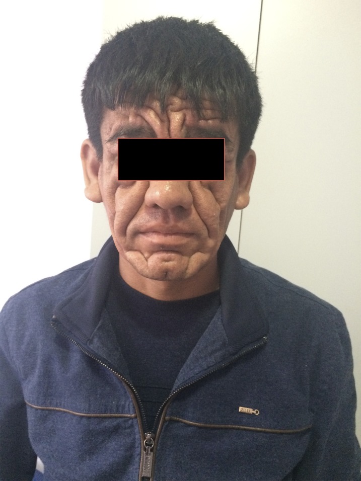
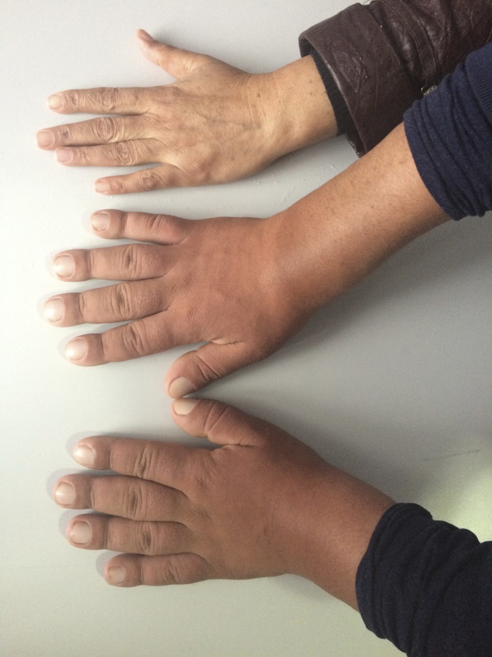
On examination, the patient was 1.7 m tall and weighed 75 kg. There was a rugose folding of his scalp, with the formation of tender, painful, rough skin folds in the parietal-occipital region, resembling cerebral gyri (i.e., cutis verticis gyrate). There was also a thickening and enlargement of the eyelids, due to cartilaginous hypertrophy, dystrophic changes of the conjunctiva, and atrophy of the Meibomian glands, with the formation of multiple cysts and granulomas (Fig. 1). The patient perspired excessively. There was thickening of the facial skin, with increased oiliness, increased rugosity, and seborrheic dermatitis. The skin over the hands was thick and apparently fixed to the underlying tissues, with finger clubbing (Fig. 2). On the abdomen were multiple scattered café-au-lait patches.
Figure 1.
Photograph of the patient with pachydermoperiostosis, published with the patient’s permission.
Figure 2.
Photograph of the patient’s hands and a normal subject’s left hand, for comparison.
A diagnosis of acromegaly was suspected. Results of routine baseline investigations were normal, but the serum insulin-like growth factor-1 level, 115μg/L, was below the normal range for his age (normal 95% confidence limits, 219 to 644 mg/L), and a random growth hormone level was 2.2μg/L, suggesting that a diagnosis of acromegaly was extremely unlikely [1]. However, the highly rugose nature of his facial and scalp skin, and the presence of finger clubbing suggested the alternative diagnosis of pachydermoperiostosis, also known as hypertrophic pulmonary osteoarthropathy (primary hypertrophic osteoarthropathy autosomal recessive 2, PHOAR2) [2]. Therefore, blood was collected for germline testing of the SLCO2A1 and HPGD genes by next-generation sequencing and dosage analysis (Leeds Genetics Laboratory, Leeds, UK). The patient was homozygous for a pathogenic SLCO2A1 mutation, c.764G>A (p.Gly255Glu), thus confirming the diagnosis as pachydermoperiostosis. On further questioning, the patient stated that both his maternal and paternal grandfathers had a similar appearance, with thick furrowing of the face and enlargement of their hands and feet, but milder than his, as did his mother’s brother. His parents were unrelated and he had two unaffected siblings. Family members were not available for further verification.
Pachydermoperiostosis has an autosomal recessive inheritance with variable, usually postpubertal, expressivity. The male-to-female ratio of the condition among patients is approximately 8:1. The onset of disease is gradual, with the first signs appearing during puberty, and the full syndrome is usually formed by approximately 20 to 30 years of age. Morphologically, the disease is characterized, above all, by the massive growth of the fibrillar structures of the dermis and subcutaneous tissue with ingrowths of fibrous connective tissue in the underlying tissues, which causes intimate cohesion of skin. The epidermis is not markedly abnormal, but the dermis becomes thickened with increasing collagen and elastin fibers, marked proliferation of fibroblasts, and small perivascular and perifollicular lymphohistiocytic infiltrates; the ostia of hair follicles become expanded and contain accumulations of horny masses. The number of mature sweat and sebaceous glands substantially increases and sometimes hyperplasia and/or hypertrophy of glandular cells can be seen. Some bones undergo periosteal ossification, with a diffuse layering of osteoid tissue over the cortex. Changes affecting joints include hyperplasia of superficial synovial cells and marked thickening of the walls of small blood vessels because of fibrosis. The pathologic processes also involve all types of vessels, with marked fibrosis in the walls of blood vessels and involvement of the vasculature of the internal organs. Some patients develop myelofibrosis (the full blood cell count in this patient was normal).
Biochemically, the two major genetic lesions both lead to an increase in prostaglandin E2, either by decreased degradation due to enzymatic loss (HPGD mutations) [3, 4] or a transporter defect (with SLCO2A1 mutations) [5]; this excess can be detected in the urine of affected patients. Previous case reports have been published, but few present confirmatory genetic data [6–14].
There are few data on the long-term effects of the disorder other than the chronic effects of the articular disease; however, the association with myelofibrosis is likely to reduce life expectancy, although this has not been quantified. There have been attempts at pharmacotherapy, with some evidence of clinical improvement in certain symptoms with nonsteroidal anti-inflammatory agents [15]. There is often evidence of consanguinity, as for other autosomal recessive diseases.
In summary, we present the images of a patient initially misdiagnosed with acromegaly and who was shown to have pachydermoperiostosis. Endocrinologists should be aware of this rare but important differential diagnosis.
Acknowledgments
Disclosure Summary: The authors have nothing to disclose.
References and Notes
- 1.Katznelson L, Laws ER Jr, Melmed S, Molitch ME, Murad MH, Utz A, Wass JA; Endocrine Society . Acromegaly: an Endocrine Society clinical practice guideline. J Clin Endocrinol Metab. 2014;99(11):3933–3951. [DOI] [PubMed] [Google Scholar]
- 2.Saigal R, Chaudhary A, Pathak P, Singh A, Gupta D, Tank ML. Pachydermoperiostosis. J Assoc Physicians India. 2016;64(3):88–89. [PubMed] [Google Scholar]
- 3.Uppal S, Diggle CP, Carr IM, Fishwick CW, Ahmed M, Ibrahim GH, Helliwell PS, Latos-Bieleńska A, Phillips SE, Markham AF, Bennett CP, Bonthron DT. Mutations in 15-hydroxyprostaglandin dehydrogenase cause primary hypertrophic osteoarthropathy. Nat Genet. 2008;40(6):789–793. [DOI] [PubMed] [Google Scholar]
- 4.Zhang Z, Xia W, He J, Zhang Z, Ke Y, Yue H, Wang C, Zhang H, Gu J, Hu W, Fu W, Hu Y, Li M, Liu Y. Exome sequencing identifies SLCO2A1 mutations as a cause of primary hypertrophic osteoarthropathy. Am J Hum Genet. 2012;90(1):125–132. [DOI] [PMC free article] [PubMed] [Google Scholar]
- 5.Diggle CP, Parry DA, Logan CV, Laissue P, Rivera C, Restrepo CM, Fonseca DJ, Morgan JE, Allanore Y, Fontenay M, Wipff J, Varret M, Gibault L, Dalantaeva N, Korbonits M, Zhou B, Yuan G, Harifi G, Cefle K, Palanduz S, Akoglu H, Zwijnenburg PJ, Lichtenbelt KD, Aubry-Rozier B, Superti-Furga A, Dallapiccola B, Accadia M, Brancati F, Sheridan EG, Taylor GR, Carr IM, Johnson CA, Markham AF, Bonthron DT. Prostaglandin transporter mutations cause pachydermoperiostosis with myelofibrosis. Hum Mutat. 2012;33(8):1175–1181. [DOI] [PubMed] [Google Scholar]
- 6.Rahaman SH, Kandasamy D, Jyotsna VP. Pachydermoperiostosis: incomplete form, mimicking acromegaly. Indian J Endocrinol Metab. 2016;20(5):730–731. [DOI] [PMC free article] [PubMed] [Google Scholar]
- 7.Sharma ML, Singh Y, Garg MK, Mishra S. Pachydermoperiostosis - mimic to acromegaly. Med J Armed Forces India. 2015;71(Suppl 1):S187–S189. [DOI] [PMC free article] [PubMed] [Google Scholar]
- 8.Shimizu C, Kubo M, Kijima H, Uematsu R, Sawamura Y, Ishizu A, Koike T. A rare case of acromegaly associated with pachydermoperiostosis. J Endocrinol Invest. 1999;22(5):386–389. [DOI] [PubMed] [Google Scholar]
- 9.Singh GR, Menon PS. Pachydermoperiostosis in a 13 year-old boy presenting as an acromegaly-like syndrome. J Pediatr Endocrinol Metab. 1995;8(1):51–54. [DOI] [PubMed] [Google Scholar]
- 10.Stoker DJ. Pachydermoperiostosis mimicking acromegaly. J R Soc Med. 1992;85(5):306. [PMC free article] [PubMed] [Google Scholar]
- 11.Mahy IR, Wiggins J. Pachydermoperiostosis mimicking acromegaly. J R Soc Med. 1992;85(1):46–47. [DOI] [PMC free article] [PubMed] [Google Scholar]
- 12.Donnelly S, Sugrue D. It looks like agromegaly [sic] but it is not. Ir J Med Sci. 1991;160(1):8–9. [DOI] [PubMed] [Google Scholar]
- 13.Harbison JB, Nice CM Jr. Familial pachydermoperiostosis presenting as an acromegaly-like syndrome. Am J Roentgenol Radium Ther Nucl Med. 1971;112(3):532–536. [DOI] [PubMed] [Google Scholar]
- 14. doi: 10.1136/bcr-2016-214624. Chakraborty PP, Bhattacharjee R, Mukhopadhyay S, Chowdhury S. Pseudoacromegaly in pachydermoperiostosis [published online ahead of print February 17, 2016]. BMJ Case Rep. doi:10.1136/bcr-2016-214624. [DOI] [PMC free article] [PubMed] [Google Scholar]
- 15. doi: 10.1111/bjd.14480. Li ZT, Wang D, Wang S. Successful treatment of pachydermoperiostosis with etoricoxib in a patient with a homozygous splice-site mutation in the SLCO2A1 gene [published online ahead of print February 15, 2016]. Br J Dermatol. doi:10.1111/bjd.14480. [DOI] [PubMed] [Google Scholar]


