Abstract
Hyperparathyroidism-jaw tumor syndrome (HPT-JT) is a rare autosomal dominant cause of familial hyperparathyroidism associated with benign, ossifying fibromas of the maxillofacial bones and increased risk of parathyroid carcinoma. The putative tumor suppressor gene CDC73 has been implicated in the syndrome, with a multitude of inactivating mutations identified; however, HPT-JT due to large-scale deletion of the chromosomal region containing the gene is exceedingly rare, and the clinical significance of this variant remains unclear. We report the case of a 32-year-old woman with a history of mandibular ossifying fibroma who presented with primary hyperparathyroidism and was found to harbor a large-scale, germline deletion on chromosome 1q31, including the CDC73 locus. HPT-JT is associated with loss of function of the putative tumor suppressor gene CDC73. Over 100 mutations and small insertions/deletions have been identified within the gene, the majority of which result in premature truncation of the parafibromin protein product. We report a case of HPT-JT associated with a large chromosomal deletion (4.1 Mb) encompassing the CDC73 gene locus. In the future, molecular testing in this autosomal dominant disorder should use techniques that allow for the detection of large-scale deletions in addition to the more commonly observed mutations and smaller-scale copy number alterations. Further investigation is needed to determine whether HPT-JT associated with a large-scale deletion carries increased risk of malignancy relative to the more common truncating mutations and what the implications are for genetic counseling.
Keywords: hyperparathyroidism-jaw syndrome, 1q31 deletion, CDC73
A case of hyperparathyroidism-jaw tumor syndrome associated not with a point mutation, but rather with a large chromosomal deletion encompassing the CDC73 gene locus.
Hyperparathyroidism-jaw tumor syndrome (HPT-JT) is a rare autosomal dominant cause of familial hyperparathyroidism associated with benign, ossifying fibromas of the mandible or maxillary facial bones and increased risk of parathyroid carcinoma. The putative tumor suppressor gene cell division cycle protein 73 homolog (CDC73) has been implicated in the syndrome, with a multitude of inactivating mutations identified, mostly comprised of point mutations and small insertions and deletions [1]. Historically, primary hyperparathyroidism associated with HPT-JT has been considered a more aggressive entity than its sporadic counterpart, with an increased risk of parathyroid carcinoma. The magnitude of this increased risk ranges from 10% to 15% [2, 3]. A greater understanding of the multitude of potential CDC73 variants is leading to more accurate estimates of risk that may be specific to certain classes of variant [4]. Here, we report the case of a 32-year-old woman with a history of ossifying mandibular fibroma and primary hyperparathyroidism who carries a previously unreported 4.1-Mb deletion on chromosome 1 encompassing the CDC73 locus.
1. Case Report
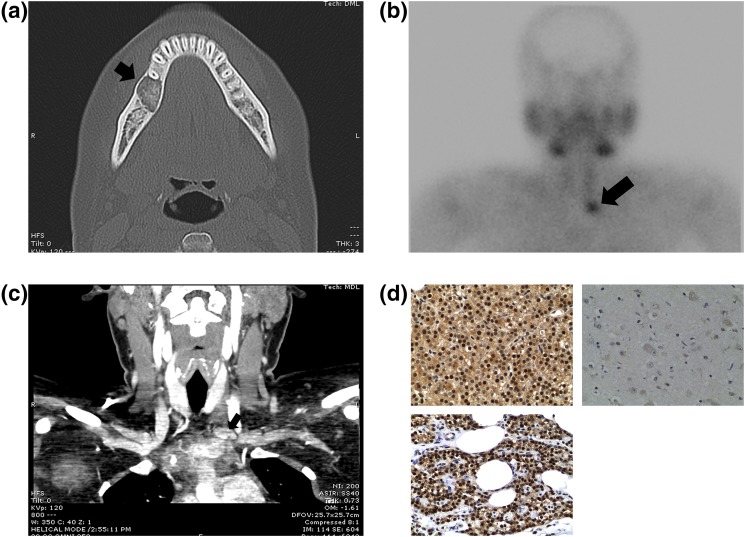
A 32-year-old woman presented with mildly elevated serum calcium (10.4 mg/dL; reference range, 8.4 to 10.2 mg/dL), a parathyroid hormone (PTH) level more than 3 times the upper limit of normal (162.1 pg/mL; reference range, 7.5 to 53.5 pg/mL), normal renal function (creatinine, 0.62 mg/dL; reference range, 0.52 to 1.04 mg/dL), normal 25-hydroxy vitamin D (34.6 ng/mL; reference range, 30.0 to 100.0 ng/mL), normal alkaline phosphatase (80 U/L; reference range, 38 to 126 U/L), and a mildly elevated 24-hour urine calcium (320 mg/24 h; reference range, 100 to 300 mg/24 h), consistent with a diagnosis of primary hyperparathyroidism. Her past history included resection of a 1.7-cm ossifying fibroma of the mandible 1 year prior [Fig. 1(a)] and a kidney stone several years prior. Thyroid, abdominal, and pelvic ultrasounds were performed, demonstrating normal-appearing thyroid gland, kidneys, uterus, and ovaries. Dual-energy X-ray absorptiometry testing demonstrated bone density within normal range, with T scores of –0.33 and –0.2 in the spine and femoral neck, respectively (z scores and forearm measures were unavailable). Nuclear medicine scanning utilizing Tc-99m sestamibi was suggestive of parathyroid adenoma inferior to the left thyroid lobe, at the level of the thoracic inlet [Fig. 1(b)], and four-dimensional parathyroid computed tomography (CT) demonstrated a corresponding 2.0-cm lesion [Fig. 1(c)].
Figure 1.
(a) Axial CT scan demonstrating a lucent, well-circumscribed 1.7-cm lesion with homogenous matrix of the right mandible consistent with biopsy-proven ossifying fibroma. (b) Planar image from Tc-99m sestamibi scan demonstrates a focus of delayed radiotracer washout inferior to the left thyroid lobe, at the level of the thoracic inlet, consistent with parathyroid adenoma. (c) Coronal image from four-dimensional, contrast-enhanced CT scan confirming a 2.0-cm lesion in the left inferior position. (d) Immunohistochemistry for parafibromin (×20 magnification): Tumor (top) demonstrates an admixture of strong nuclear positivity and scattered negative cells, imparting a mosaic pattern of protein expression. Normal parathyroid (bottom) demonstrates strong, diffuse nuclear expression of parafibromin. Brain tissue negative control (right). Paraffin-embedded, formalin-fixed parathyroid gland tissue was used to prepare 4-μm sections. Dehydration of the sections and a 40-minute epitope retrieval process in Leica Bond ER 1 (catalog no. AR9961) were performed on an automated Leica Bond III stainer. Sections were incubated with the primary mouse monoclonal anti-parafibromin antibody (Research Resource Identification Initiative AB_628102, clone 2H1, SC-33638; Santa Cruz Biotechnology, Santa Cruz, CA) at a dilution of 1:50 for 10 minutes on the automated stainer. This antibody is raised against a peptide corresponding to amino acids 87 to 100 of mouse parafibromin. Antibody detection was achieved using the Leica Bond Polymer Refine DAB Detection Kit (catalog no. DS9800, Leica Biosystems Newcastle, Ltd, Newcastle Upon Tyne, United Kingdom) with diaminobenzidine as the chromogen, and sections were counterstained in hematoxylin.
The combination of findings was suggestive of HPT-JT, and high-throughput sequencing was undertaken at the Yale Center for Genome Analysis. DNA was extracted and purified from a peripheral blood sample, array capture was performed with Roche/Nimblegen MedExome, and sequencing was performed with the Illumina HiSeq platform. A 4.1-Mb deletion was identified on chromosome 1q31.2-31.3 (chr1:192127840-196227528x1), encompassing the locus of the candidate CDC73 gene (chr1:193121958-193254815). Additional deleted genes in the region include multiple members of the Regulator of G-protein Signaling Gene family (RGS1, RGS2, RGS13, and RGS18) as well as TROVE Domain Family Member 2, Ubiquitin C-terminal Hydrolase L5, and portions of Potassium Sodium-Activated Channel Subfamily Member 2 (TROVE2, UCHL5, and KCNT2). The identified deletion is not documented in the curated International Standards for Cytogenomic Arrays database of pathogenic copy number variations (http://dbsearch.clinicalgenome.org). The deletion partially overlaps with known 1q deletions, specifically the intermediate category, which encompass the 1q25-32 region and include the CDC73 gene locus. The most common clinical manifestation of these intermediate 1q deletions is developmental delay; however, specific causative genes have not been identified [5]. No mutations were detected in the following genes: Multiple Endocrine Neoplasia 1, Ret Proto-Oncogene, Calcium Sensing Receptor, and Cyclin Dependent Kinase Inhibitors 1B, 2B, 2C, and 1A (MEN1, RET, CASR, CDKN1B, CDKN2B, CDKN2C, or CDKN1A). Family history is remarkable for two biologic adolescent sons who underwent genetic testing. One was a carrier of the same 4.1-Mb deletion of chromosome 1q31.2-31.3, including the entire CDC73 gene identified in the index case. The other child was not a carrier of this genotype, and no family members have been diagnosed with hyperparathyroidism and tumors of the jaw, kidney, or uterus.
The patient underwent en bloc parathyroidectomy of the preoperatively localized lesion. PTH levels were monitored at 5-minute intervals and fell from a baseline of 154.0 to 12.0 pg/mL 20 minutes post excision. Surgical pathology demonstrated a well-circumscribed parathyroid gland weighing 1.1 gm consisting of an enlarged hypercellular nodule of chief cells devoid of cytologic atypia, invasion, and significant mitotic activity. These findings are consistent with a parathyroid adenoma and rule out atypical adenoma or carcinoma. Immunohistochemistry did demonstrate staining for parafibromin [Fig. 1(d)]. A positive control, a normal parathyroid gland, was reviewed. The test slide was interpreted in a semiquantitative fashion. Nuclear expression of the immunomarker in >5% of parathyroid cells with a staining intensity approximately equal to that of the control was considered positive staining. Total serum calcium and PTH levels were normal at 9.2 mg/dL and 25.7 pg/mL during follow-up at 4 months.
2. Discussion
HPT-JT is a rare autosomal dominant syndrome with variable penetrance of parathyroid adenoma and carcinoma, benign jaw tumors, and occasional renal and uterine tumors. The syndrome has been linked to the cell division cycle protein 73 homolog gene (CDC73), with a multitude of reported variants predicted to result in either truncation or loss of parafibromin expression protein through nonsense-mediated mechanisms. Gross deletions represent only 1% of the mutations found in the CDC73 gene [1, 6]. Bellido et al. report a case of HPT-JT caused by a 24-nucleotide deletion involving the start codon of CDC73, presumably leading to complete loss of transcription, although parafibromin immunohistochemistry was not performed to confirm loss of protein expression [7]. In another HPT-JT kindred, including six patients over two generations, Cascón et al. identified a 547-kb germline deletion involving the CDC73 locus with confirmed loss of parafibromin staining [6].
As part of a cohort of 16 HPT-JT patients, Mehta et al. reported seven members from a single family with whole-gene deletion of CDC73 as detected by array-comparative genomic hybridization; however, the exact size and location of the deletion was not reported [8]. Six of the 16 patients (37.5%) in the cohort were found to have parathyroid carcinoma, which is significantly higher than the reported 15% incidence in the syndrome [2, 3]. Interestingly, of the seven patients harboring whole-gene deletions of CDC73, four were among those with malignant lesions, meaning that fully 57% of the patients with this deletion developed parathyroid carcinoma. The remaining two carcinoma patients carried small duplications in exon 7. In total, there were two uterine tumors, three renal lesions, and two jaw tumors reported, all among the parathyroid carcinoma patients. These findings raise the possibility that not all genomic variants confer an equal risk of parathyroid carcinoma or of the other HPT-JT–related lesions. Interestingly, biallelic loss of CDC73 expression has been reported in cases of sporadic parathyroid carcinoma [9]. Further study is needed to determine if HPT-JT patients with whole-gene deletion of at least one CDC73 allele may carry a higher risk of carcinoma than those with point mutations or small insertions and deletions.
The case presented here contributes to a relatively small body of literature devoted to HPT-JT, a syndrome with variable penetrance resulting in a spectrum of diseases spanning from the benign to the highly morbid. To our knowledge, this is the first case of HPT-JT arising in a context of large-scale germline chromosomal deletion of the region containing the CDC73 locus, with a presumed second-hit mutation in the remaining allele rendering it nonfunctional, yet still immunogenic on immunohistochemistry (fresh tumor material was unavailable to provide DNA for genetic testing of the presumed second-hit mutation). It is important to note that these large variants may not be detected by standard screening methods. High-resolution genomic assessment has become an integral component of the clinical evaluation of our patients, and the growing library of genomic variants in HPT-JT may explain the variable penetrance, severity, and constellation of phenotypes encountered in clinical practice. In the future, it will be critical to elucidate the specific clinical presentation associated with each genomic variant, as well as the impact of a germline heterozygous deletion, to best inform operative planning, surveillance regimens, and genetic counseling for both patients and their families.
Acknowledgments
Acknowledgments
Disclosure Summary: The authors have nothing to disclose.
Footnotes
- CDC73
- cell division cycle protein 73 homolog
- CT
- computed tomography
- HPT-JT
- hyperparathyroidism-jaw tumor syndrome
- PTH
- parathyroid hormone.
References and Notes
- 1.Newey PJ, Bowl MR, Cranston T, Thakker RV. Cell division cycle protein 73 homolog (CDC73) mutations in the hyperparathyroidism-jaw tumor syndrome (HPT-JT) and parathyroid tumors. Hum Mutat. 2010;31(3):295–307. [DOI] [PubMed] [Google Scholar]
- 2.Marx SJ. Hyperparathyroid and hypoparathyroid disorders. N Engl J Med. 2000;343(25):1863–1875. [DOI] [PubMed] [Google Scholar]
- 3.Simonds WF, James-Newton LA, Agarwal SK, Yang B, Skarulis MC, Hendy GN, Marx SJ. Familial isolated hyperparathyroidism: clinical and genetic characteristics of 36 kindreds. Medicine (Baltimore). 2002;81(1):1–26. [DOI] [PubMed] [Google Scholar]
- 4.Iacobone M, Masi G, Barzon L, Porzionato A, Macchi V, Ciarleglio FA, Palù G, De Caro R, Viel G, Favia G. Hyperparathyroidism-jaw tumor syndrome: a report of three large kindred. Langenbecks Arch Surg. 2009;394(5):817–825. [DOI] [PubMed] [Google Scholar]
- 5.Hu P, Wang Y, Meng LL, Qin L, Ma DY, Yi L, Xu ZF. 1q25.2-q31.3 deletion in a female with mental retardation, clinodactyly, minor facial anomalies but no growth retardation. Mol Cytogenet. 2013;6(1):30. [DOI] [PMC free article] [PubMed] [Google Scholar]
- 6.Cascón A, Huarte-Mendicoa CV, Javier Leandro-García L, Letón R, Suela J, Santana A, Costa MB, Comino-Méndez I, Landa I, Sánchez L, Rodríguez-Antona C, Cigudosa JC, Robledo M. Detection of the first gross CDC73 germline deletion in an HPT-JT syndrome family. Genes Chromosomes Cancer. 2011;50(11):922–929. [DOI] [PubMed] [Google Scholar]
- 7.Bellido V, Larrañaga I, Guimón M, Martinez-Conde R, Eguia A, Perez de Nanclares G, Castaño L, Gaztambide S. A novel mutation in a patient with hyperparathyroidism-jaw tumour syndrome. Endocr Pathol. 2016;27(2):142–146. [DOI] [PubMed] [Google Scholar]
- 8.Mehta A, Patel D, Rosenberg A, Boufraqech M, Ellis RJ, Nilubol N, Quezado MM, Marx SJ, Simonds WF, Kebebew E. Hyperparathyroidism-jaw tumor syndrome: results of operative management. Surgery 2014;156(6):1315–1325. [DOI] [PMC free article] [PubMed]
- 9.Shattuck TM, Välimäki S, Obara T, Gaz RD, Clark OH, Shoback D, Wierman ME, Tojo K, Robbins CM, Carpten JD, Farnebo LO, Larsson C, Arnold A. Somatic and germ-line mutations of the HRPT2 gene in sporadic parathyroid carcinoma. N Engl J Med. 2003;349(18):1722–1729. [DOI] [PubMed] [Google Scholar]


