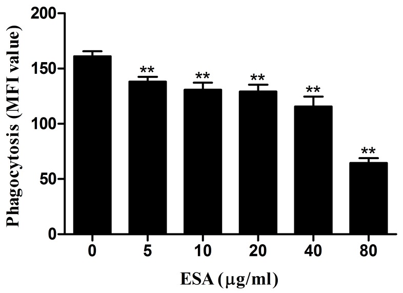
Figure 3. Effect of TgESAs on the phagocytosis of Ana-1 cells by flow cytometry.
Ana-1 cells were treated for 48 h with different concentrations (0, 5, 10, 20, 40, 80 μg/mL) of TgESAs. Group histograms showing the median fluorescence intensity (MFI) values. Values are mean ± standard deviation of three independent experiments. *P < 0.05 and **P < 0.01 compared with untreated group (0 μg/ml).

