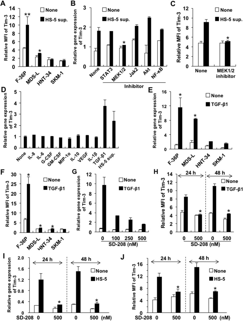
Figure 2. Upregulated Tim-3 expression in MDS cell lines.
(A) MDS cell lines were cultured with or without HS-5 sup. for 48 h. The cells were pretreated with signal transduction inhibitors of STAT3, MEK1/2, JAK2, Akt/PI3K, and NF-κB for 2 h and then cultured in complete medium containing HS-5 sup., after which Tim-3 mRNA expression (B) and protein were analyzed (C). The concentrations of each inhibitor were 500 nM of STAT3 inhibitor V, 20 μM of U0126 (MEK1/2 inhibitor), 25 μM of AG490 (JAK2 inhibitor), 25 μM of LY294002 (PI3K/AKT signaling inhibitor), and 5 nM of PDTC (NF-κB inhibitor). (D) F-36P cells were cultured with the following cytokines for 2 days: 5 ng/ml of IL-8, 5 ng/ml of IL-6, 100 pg/ml of G-CSF, 10 ng/ml of GM-CSF, 10 ng/ml of MIP-1α, 10 ng/ml of IL-10, 10 ng/ml of VEGF, 10 ng/ml of IL-1β, and 2.5 ng/ml of TGF-β1. (E, F) MDS cell lines were cultured with 2.5 ng/ml of TGF-β1 for 48 h. (G–J) F-36P cells were pretreated with SD208, a selective inhibitor of TGF-βRI kinase, at optimal concentrations for 2 h, followed by incubation with 2.5 ng/ml of TGF-β1 (G, H) or HS-5 sup. (I, J) for 24–48 h. After incubation with HS-5 sup. or TGF-β1, the cell surface (A, C, F, H, J) and mRNA (B, D, E, G, I) expression of Tim-3 was analyzed by FCM and real-time qPCR, respectively. Data represent mean ± SD. *P<0.05, **P<0.005 compared with the results without HS-5 sup. or cytokines (A, E, F) and with the results without inhibitors (B, C, H, I, J).

