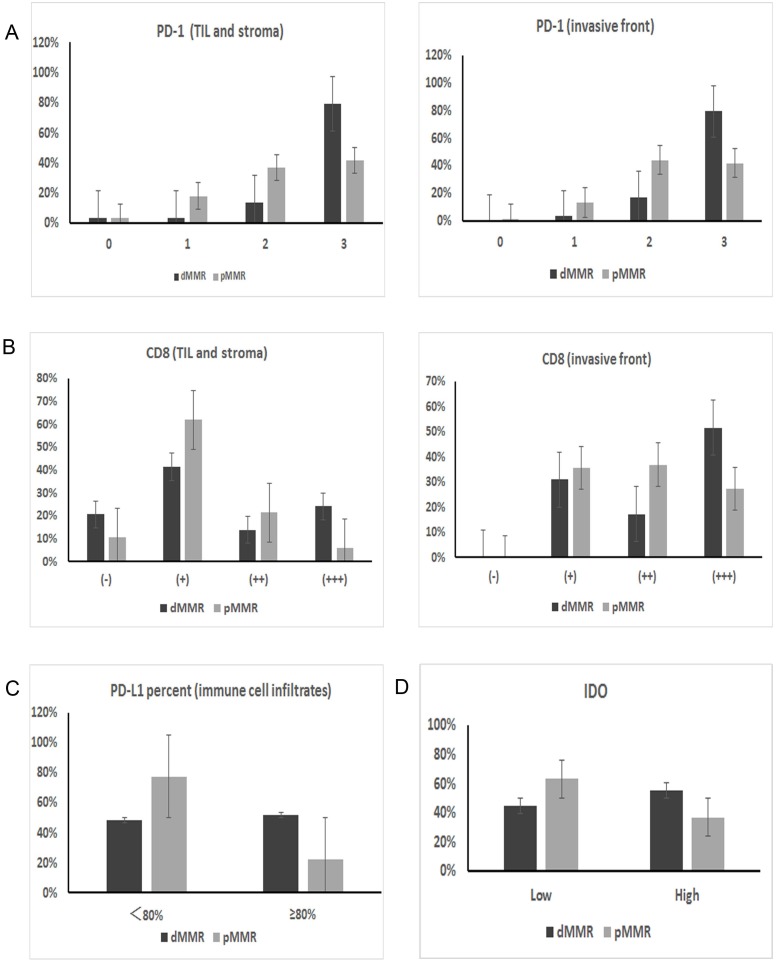
Figure 3. IHC expression differences in immune checkpoint proteins between dMMR and pMMR patients.
(A) PD-1 expression on TILs from the TIL and stromal compartment and the invasive front compartment was significantly higher in dMMR tumors than in pMMR tumors (p=0.003 for both compartments). (B) Similarly, the number of CD8+ T cells in the two compartments was also higher in dMMR tumors than in pMMR tumors (p=0.017 for TIL and stroma; p=0.038 for invasive front). (C) PD-L1 expression on immune cell infiltrates; the expression percentage differed significantly between the two groups when the cut-off value was set as 80% (p=0.003). (D) IDO expression in tumor cells also differed significantly between dMMR and pMMR tumors (p=0.026).

