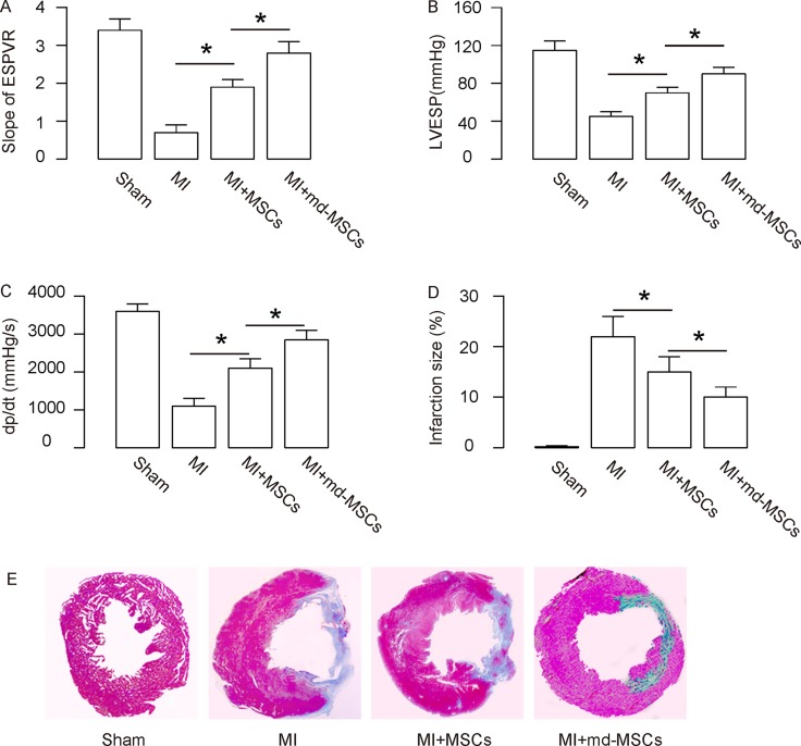
Figure 4. Better heart function is detected in MI-mice transplanted with md-MSCs.
The heart function of the mice from 4 groups was assessed using ventricular catheterization. (A) Endsystolic pressure-volume relationship (ESPVR). (B) Left ventricular end systolic pressure (LVESP). (C) Positive maximal pressure derivative (+dP/dt). (D–E) Masson’s trichrome staining to determine the collagen deposition in the heart tissue of each group, shown by quantification (D), and by gross images (E). N = 10. *p < 0.05.

