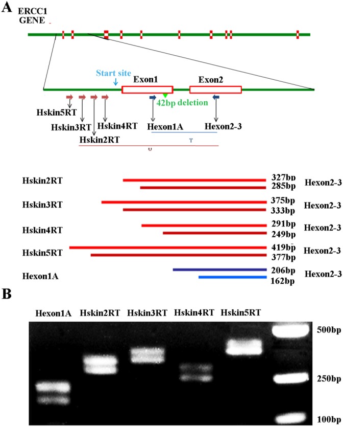
Figure 1. An upstream ERCC1 transcript in human cell lines.

(A) Schematic of human ERCC1 gene showing the exons, location of the normal transcription initiation site (green arrow), transcriptional start site (blue arrow) and presumed location of the upstream initiation site (red arrow). The positions of primers used to detect all ERCC1 transcripts (T) and predicted sizes of PCR products are indicated. The positions of the various primers used to detect the upstream ERCC1 transcripts (U) and predicted size of PCR products are also indicated. (B) RT-PCR of mRNA extracted from human ovarian cancer A2780 cells. All ERCC1 transcripts were detected with primer pair of Hexon1A and H-exon 2-3. ERCC1 transcripts initiating upstream were detected with primer pairs of hskin2RT, hskin3RT, hskin4RT, hskin5RT and H-exon2-3.
