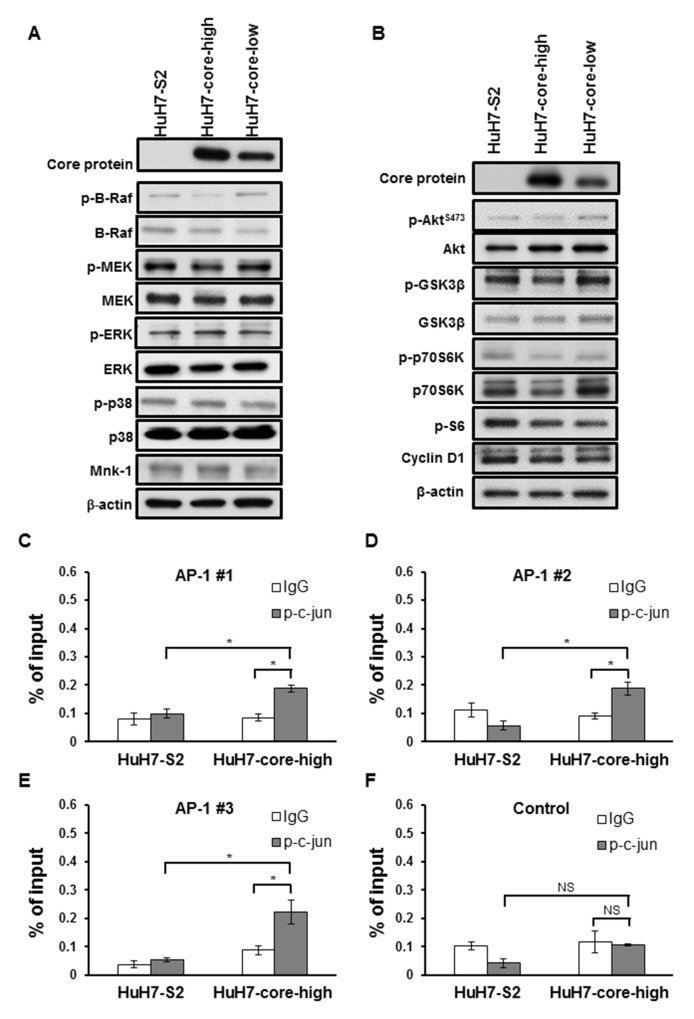
Figure 5.
(A-B) Western blots showing the expression of proteins associated with the MAPK (E) and the PI3K (F) pathways in HCC cells. (C-F) Chromatin immunoprecipitation assay using control IgG or the anti-p-c-jun antibody. We compared the DNA fragments harboring AP-1 binding sites (C-E) and those without (F) after enrichment with control IgG or anti-p-c-jun antibody. The total DNA input amount was used as the reference. All data are presented as mean ± SEM. *: p < 0.05; NS = not significant.

