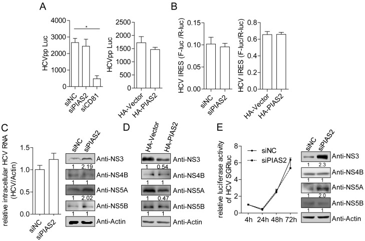
Figure 3.
PIAS2 restricts HCV protein expression in the subgenomic replicon. (A) Huh7 cells were transfected with siRNA or plasmids as indicated for 48 h and then transduced with HCVpp. The luciferase activity was measured 48 h post-transduction; (B) Huh7 cells were transfected with the siRNA or plasmids as indicated and then transfected with the pHCV-internal ribosome entry site (IRES) reporter plasmid. The dual-luciferase assay was performed 48 h later. The IRES translation efficiency was determined by the ratio of firefly luciferase (F-Luc) activity to Renilla luciferase (R-Luc) activity; (C) Con1 cells were transfected with the indicated siRNAs. The HCV RNA and protein expression levels were detected at 72 h post-transfection. The numbers below each blot show the relative density of the blots normalized to actin; (D) Con1 cells were transfected with vector or pHA-PIAS2 plasmids and HCV NS3, NS4B, NS5A and NS5B protein expression were detected by western blotting; (E) Huh7 cells were electroporated with the JFH1-SGR-luc RNA (10 µg) together with the siNC or siPIAS2 (20 nM). Luciferase activity was measured at the indicated time points and normalized to the value obtained at 4 h post-electroporation. The HCV protein expression levels were detected at 72 h, * p < 0.05.

