Abstract
Objective:
To compare the healing process of pressure ulcers treated with cryopreserved human amniotic membrane allograft and routine pressure ulcer care in our hospital.
Methods:
From January 2012 to December 2013, in a prospective randomized clinical trial (IRCT201612041335N2), 24 patients with second and third stage of pressure ulcers were enrolled in this study. All patients needed split-thickness skin grafts for pressure ulcer-wound coverage. Selected patients had symmetric ulcers on both upper and lower extremities. The patients were randomly divided into two groups: amnion and control. In the amnion group, the ulcer was covered with cryopreserved amniotic membrane and in the control group it was treated with local Dilantin powder application. The duration and success rate of complete healing was compared between the two groups.
Results:
The study group was composed of 24 pressure ulcers in 24 patients (19 males and 5 females) with a mean age of 44±12.70 years. The demographic characteristics, ulcer area, and underlying diseases were similar in both groups. The early sign of response, such as decrease in wound discharge, was detected 12-14 days after biological dressing. Complete pressure ulcer healing occurred only in the amnion group (p< 0.001). Partial healing was significantly higher in the amnion group (p< 0.03). Healing time in this group was faster than that the control group (20 days versus 54 days). No major complication was recorded with amniotic membrane dressing
Conclusion:
Cryopreserved amniotic membrane is an effective biologic dressing that promotes re-epithelialization in pressure ulcers
Key Words: Pressure ulcer, Dressing, Amniotic membrane, Grafting
Introduction
Pressure ulcer is a localized injury to the skin and/or underlying tissue and usually develops over a bony prominence, as a result of pressure, shear and/or friction. The sacrum and the heel are especially prone to development of pressure ulcers [1, 2]. Patients with impaired mobility such as stroke, unconsciousness, or spinal cord injury are most vulnerable to pressure ulcers. The overall prevalence of pressure ulcers was 27% in long term [1- 3]. Other factors such as old age, poor nutrition, poor sensation, urinary and fecal incontinence, and individuals with dementia or other cognitive disorders are susceptible to pressure ulcer [2-4]. Wounds usually heal in three phases including inflammatory phase, a proliferative phase with migration of fibroblasts and deposition of extracellular matrix, and a remodeling phase with cross-linking and reorganization of the collagen matrix [1, 5]. In pressure ulcers, the above processes are often locked into a state of chronic inflammation and no healing occurs [1, 5]. Pressure ulcers need frequent hospitalization. For example, in pressure ulcer with stage III or IV, complete healing may take as long as 6 months [1]. The cost of healing in pressure ulcer is high and also it affects the quality of life.
This event has a four-fold increased risk of death, especially among the elderly patients [3, 6, 7].Current practice for treatment of pressure ulcers includes cleansing with normal saline solution, debridement to remove the necrotic tissues and dressings to provide a moist wound environment [8, 9]. Treatment of the underlying cause, correcting the nutritional deficiencies and frequent position changing of patients to provide pressure relief are other important points that accelerate the process of healing [9, 10].
Traditional dressings include gauze moistened with saline or paraffin impregnated gauze. Other types of dressing such as hydrocolloid, polyurethane foam, hydropolymer, hydrocellular, and alginate are also used with different advantages and disadvantages [11-13]. Conventional dressings require frequent changes which can be painful and may even require anesthesia. Additionally, superimposed infection may also develop in this setting [1,11]. Human Amniotic Membranes (AM) is a natural biological scaffold with many documented clinical applications for more than one century [14,15].
The first documented use of amniotic membranes as a surgical dressing in skin transplantation was reported by Davis in 1910. They reported that this biological material had better results in wound healing when compared with xenograft or cadaveric coverings [16]. After that, AM has been frequently used in burned and ulcerated skin surfaces to facilitate the epithelial ell migration and promote healing [17].
Another popular application of the AM is in ophthalmology [15]. It has been used in conjunctival reconstruction [18] glaucoma [19], burn [20], pterygium [21] and bullous keratopathy [22]. AM has also been successfully applied in different clinical conditions such as vaginal reconstruction [23], abdominal surgery with an enterocutaneus fistula, gastroschisis and omphalocele in infants [24]. Several medical reports have described the amnion tissue as a biological dressing in treatment skin loss in Stevens-Johnsons diseases [25], replacement of normal nasal mucosa in Rendu-Osler-Weber diseases [26], periodontal surgery [27,28], and management of conjunctival defects [29,30].
Amnion has also been used in chronic varicose ulcers, decubitus ulcers and open infected wounds with good outcome [31,32]. Excellent results have been obtained in treatment of second and third degree burns and open infected wounds [32,33].
The AM contains many growth factors that participate in wound healing, including platelet-derived growth factor, basic fibroblast growth factor (bFGF), transforming growth factor beta 1 (TGF-β1), epidermal growth factor (EGF), and placental growth factor [34,35]. Other cytokines like interleukins (IL-1Ra, IL-4 and IL-10) and the Tissue Inhibitors of Metalloproteinase (TIMP-1, TIMP-2, TIMP-4, TIMP-b) are also present in the amniotic membrane [34,35]. TIMPs are involved in degradation of the extracellular matrix and may play a key role in the release of pressure ulcers. The purpose of this study was to compare the duration and success rate of complete healing in pressure ulcer with and without using amniotic membrane.
Material and Methods
Preparation of amniotic membrane
Amnion from elective cesarean sections was used with no history of premature rupture membranes, endometritis or meconeum ileus. The informed consent was obtained from pregnant women for donation and use of AM. The mothers' blood was tested for Hepatitis B (HBS Ag, anti HBe Ab) and C (anti HCV Ab), Rapid Plasma Reagin for Syphilis, and Human Immunodeficiency Virus 1 and 2 (anti HIV- Ab 1&2). In sterile condition, under lamellar flow hood, the placenta with adherent fetal membranes was washed with Phosphate Buffer Saline (PBS) containing 50 μg/ml penicillin, 50 μg/ml streptomycin, and 2.5 μg/ml amphotericin B. The amnion was easily separated from the chorion by blunt dissection and washed several times with PBS. Then, it was flattened onto a nitrocellulose membrane (Whatman, Schleicher and Schuell optitaran BA-S 85) with the epithelial surface up. The membrane with the filter was washed with PBS and 5 ×5 and 10 ×10 cm pieces were made. Each of them was subsequently immersed in 4%, 8% and 12% Dimethyl Sulphoxide (DMSO) phosphate buffered saline for 5 minutes and finally placed in a sterile vial containing 12% DMSO medium (Sigma).
DMSO is a standard cryopreservative substance. One placenta can provide four to five AM fragments 5 ×5 cm in diameter or 2 fragment of 10 ×10 cm. Vials were frozen at -80°C in our "Amnion Bank" [36,37]. The microbiology culture was used in order to ensure the sterility of the membrane. The membrane was easily defrosted before use by warming in room temperature, and rinsed two times in normal saline and after that it was used to cover the patient's ulcer. For evaluation of cell viability, Fluorescein Di acetate (FDA) (Sigma, Germany) was used to detect the viable cell. After thawing in room temperature and 2 times washing with normal saline, the membrane was incubated with FDA at room temperature for 5 minutes in the darkness. Staining solution was removed and washed with PBS. The sample was analyzed with fluorescent microscopy (Olympus, Japan). There were few scattered viable cells. When compared with a fresh sample before freezing, the number of viable cells was not different.
Patient Screening, Eligibility, and Randomization
This prospective, randomized, controlled trial (IRCT 201612041335N2) was conducted in hematology-oncology, neurology and trauma wards in Nemazee Hospital, which is affiliated to Shiraz University of Medical Sciences, from January 2012 to December 2013.
The study is registered at the Iranian Registry of Clinical Trials (IRCT 201612041335N2). All eligible patients who wished to participate and meet the inclusion criteria were enrolled in this study. The study design conformed to the ethical guidelines of the 1975 Declaration of Helsinki, and the Ethics committee of our university confirmed the study design. The informed consent was obtained from all participants.
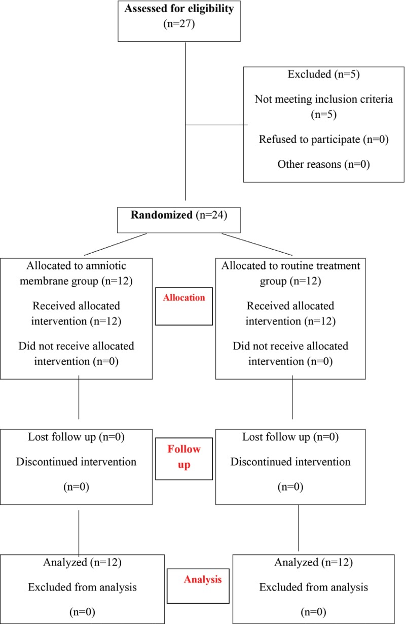
In order to facilitate the complete and un-bias reporting of trials findings, we followed the CONSORT (Consolidated Standards of Reporting Trials) statement which was updated in 2010. This is an evidence-based minimum set of recommendations for reporting of randomized clinical trials and consists of a checklist and flow diagram. The CONSORT flow diagram of the study is demonstrated in details (Figure 1).
Fig. 1.
CONSORT Flow diagram of trial participants
Simple randomization was carried out using a block randomization and no blinding was carried out. Also, staging of pressure ulcer was done by using the classification system made by National Pressure Ulcer Advisory Panel (NPUAP) and EPUAP [38].
The inclusion criteria were patients with stage II and III pressure ulcer; age ≥18 years; those able and willing to participate in study procedures with follow-up evaluations; and those with no clinical signs of infection.
The exclusion criteria included known or suspected malignancy of the current ulcer; stage I and IV pressure ulcer; pregnant or breast feeding women; previous treatment with biomedical or topical growth factor for wound healing; and participation in another clinical trial.
Study treatment
Subjects who continued to meet the study inclusion criteria were randomized to receive either the amniotic membrane allograft (amnion group) or routine regimen of wound care alone (control group). The pressure ulcers were cleansed with a sterile physiologic saline solution (rinsing, swabbing or irrigating), followed by debridement of the superficial dead skin with scalpel and encrusted exudates by washing with a mild povidone–iodine soap solution. Repeated changing position every 2-3 hours was done in all patients.
In the control group, the ulcer care was only debridement of the necrotic parts with scalpel, washing and cleaning with mild povidone –iodine soap solution, and local therapy with Dilantin powder [13, 39].
In the amnion group, after debridement and washing, the sterile AM was applied aseptically on the ulcer bed to cover the entire area. The cryopreserved AM was cut to size with a 15-blade scalpel, and placed over the ulcer site, ensuring that the membrane was consistently covering the entire wound surface. The membrane was applied in a way that no air bobble enclosures occured under the membrane.
It was left uncovered and undisturbed for a few minutes and then covered by moist gauze dressing until changing the next dressing. In the areas that were likely to come in contact with the bed, like the back, the ulcer area was protected with a gauze dressing and balloon ring bandage.
Application of AM occurred every 2-3 days during the study period until complete epithelialization occurred, the patient was withdrawn, or the study was completed.
The site of pressure ulcer for evaluation of infection and healing was assessed by the residents and the attending staff. Daily follow-up was done to detect the ulcer size and healing rate. The recovery was evaluated daily by the measurement of the wound surface area. Ulcer measurement was done with a graded centimeter ruler (length, width and depth) in all patients. The wound area was also calculated by multiplying the width (cm) ×length (cm). Any unusual signs and symptoms of clinical infection or allergic reactions were recorded. After wound measurement, the entire wound was digitally photographed.
A scar assessment of the ulcer sites was performed using the Modified Vancouver Scar Scale (MVSS).
The patients completed the study 8 weeks after the first treatment visit. In patients whose ulcer closed prior to this point were considered as having completed the study. In every visit, the percentage area reduction was calculated for the wound. Partial healing means reduction in the ulcer area by 50% or less. Wounds were defined healed if complete (100%) epithelialization occurred without drainage and need for dressing. Validation of healing was confirmed by an independent panel of physicians, including a vascular surgeon, a plastic surgeons, and a general surgeon.
Statistical analysis
Sample sizes of 12 in each group were selected to achieve at least 80% power and the level of significance was α=0.05 and β=20%. The study primary endpoint was comparison of proportion of wound healing at eight weeks between the two treatment groups.
All the analyses were performed using the SPSS statistical software, version 16 (SPSS Inc, Chicago, Ill, USA). The results are expressed as mean ± SD. The mean and standard deviation (SD) were calculated, using the aforementioned software. The χ2 test was used for comparing non-continuous variables (sex) and T-test was applied for comparing the continuous variables such as age and laboratory data. P0.05 was considered statistically significant.
Results
Twenty four patients, 19 male and 5 female, with a mean age of 44±12.70 years with pressure ulcer divided into the controls and amnion groups were studied. The size of pressure ulcers ranged from 2 x 1.6 cm to 9x6 cm, and the area of ulcers in the amnion group was 3.2 cm2 to 54cm2. In the control arm, the sizes of ulcers were from 1.5x2 cm to 7x8 cm and the areas of ulcers were 3 to 56 cm2. The demographics, clinical and para-clinical data of the patients are summarized in Tables 1 and 2.
Table 1.
Demographic features of enrolled patients
| Patients Characteristics | Amnion group (N=12) | Control group (N=12) | P -value |
|---|---|---|---|
| Age years , Mean±SDRange Median | 44±10.730-7546.5 | 43±16.1 27-6044.4 | 0.93 |
| Male/Female Ratio | 10/2 | 9/3 | 1.00 |
| Anemia due to underlying disease (Hemoglobin level) | 10 (9.8±0.83) | 12(9.8±2.30) | 0.97 |
| White blood cell count | 6391±2760 | 6716.7±2360 | 0.73 |
| Platelet count | 218250.00±100233 | 242333.33±117224 | 0.713 |
| Systemic infection | 6(50%) | 6(50%) | 0.68 |
| Fever during hospital course | 7(58%) | 8(66%) | 1.00 |
| Nutritional status Oral feeding/ NG tube feeding | 10(83%)/ 2(16%) | 10(83%)/ 2(16%) | 1.00 |
| Antibiotic consumption IV/PO/NOT | 6(50%)/5(42%)/1(8%) | 6(50%)/5(42%)/1(8%) | 1.00 |
| Underlying disease: | |||
| CancerCord injuryDiabetesMultiple sclerosis | 5(45%)1(8%)1(8%)1(8%) | 7(59%)2(17%)1(8%)1(8%) | 1.00 |
| Other complications a | 3(25%) | 3(25%) | 1.00 |
| Disease related mortality | 3(25%) | 5(42%) | 0.67 |
Other complications: renal failure, bleeding diathesis
Table 2.
Pressure Ulcer Characteristics
| Amnion group | Control group | P -value | |
|---|---|---|---|
| Number of Pressure Ulcer | 12 | 12 | 1.00 |
| Pressure Ulcer stage 2/3 | 11(92%)/1(8%) | 10(8%)/2(17%) | 1.00 |
| Pressure Ulcer size range (cm ) | 2×1.6 - 6×9 (5.3±2.3) | 1.5×2 - 7×8 (4.9±3.2) | |
| Pressure Ulcer surface areas (cm2) | 3.2-5439.3±20.5 | 3-5631.5±25.2 | |
| Pressure Ulcer Site | |||
| sacralOccipital areaGreater trochanterChest wallLateral malleolus | 5(41%)2(16%)3(25%)2(16%)1(8%) | 7(58%)1(8%)3(25%)11(8%)0 | 1.00 |
| Complete healing (100% wound closure) | 9(75%) | 0 | <0.001 |
| Partial healing a | 9(75%) | 3(25%) | 0.03 |
| Complete healing time (days) (Median) | 16-30 (20.5±10) | 0 (NSb) | <0.001 |
| Median duration of partial healing (days) | 20 | 54 | 0.03 |
| Decrease wound discharge time (days) | 2-3 | 10-12 | 0.03 |
| Granulation tissue formation time (days) | 12-14 (13±1.5) | 35-40 (38±4) | 0.03 |
| Number of used amniotic membrane (Median) | 4-8(6) | 0 | - |
| Sore infection | 0 | 1 | - |
| No response | 3(25%) | 9(75%) | 0.01 |
| Scar assessment by Modified Vancouver Scar Scale (MVSS) | 4 (Range: 3-5) | 8 (Range: 6-8) | 0.03 |
Partial healing means that ≤50% of sore area was healed;
NS: not seen after 8 weeks
The ulcer distribution was also similar in two arms, with the most prevalent area being in the sacrum. The underlying diseases were solid tumors, multiple traumas, multiple sclerosis, cord injury and diabetes. Six patients in both groups had symptoms of infection and received intravenous antibiotic.
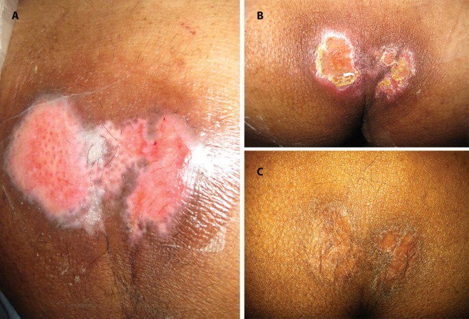
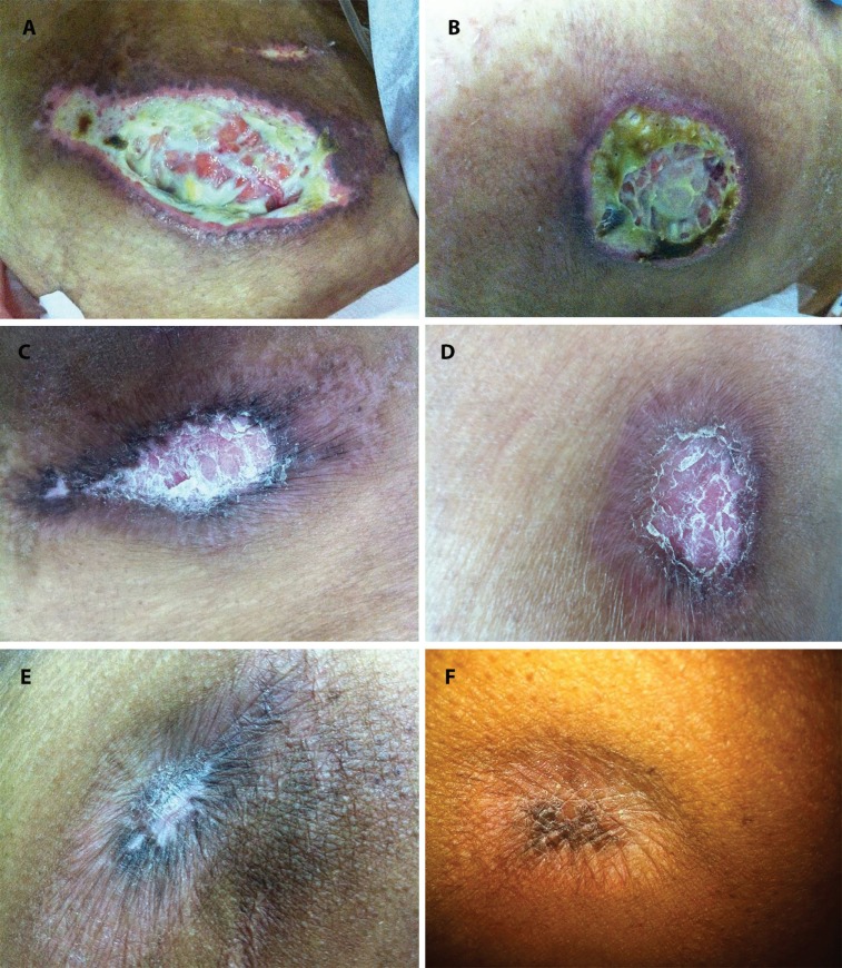
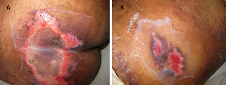
Complete ulcer healing only occurred in the amnion group during 16 to 30 days (p< 0.001) (Figures 2 and 3). In the control group, none of the patients had complete healing, and only partial healing was detected in three patients. Partial healing was significantly higher in the amnion group (p<0.03). In these cases, re-epithelialization occurred underneath the membrane (Figure 4). Healing time in the amnion group was faster than that in the control group (20 days versus 54 days); however, this difference was not statistically significant.
Fig. 2.
Patient with pressure ulcer on the buttocks. Clinical course of the ulcer from days 0 (A), 15 (B) and 30 (C) in the AM treated group. The size of lesion was decreased (more than 50%) from day 0 to day 15
Fig. 3.
The ulcer was located on the greater trochanter. Clinical course of the ulcer from days 0 (A), 7 (B), 14 (C), 28 (D), 32 (E) to 50 (F) in the AM treated group. Note the complete re-epithelialization in Fig 2F on day 50
Fig. 4.
Local application of allogenic amniotic membrane over the pressure ulcer located on the buttocks (A) and greater trochanter (B). The membrane was applied in a way that no air bobble enclosures occurred under the membrane
The early response to the use of biological dressing was decreased wound discharge during the first 2-3 days. The time required for decreasing the amount of wound discharge was less in the amnion group (2-3 days) in comparison to the control group (10-12 days). Only one patient in the control group showed signs of local infection, and based on the result of wound culture a systemic antibiotic was prescribed.
The number of amniotic membranes used in healed pressure ulcers was 4 to 8 membranes, which was changed every 2-3 days (Table 2). Patients with ulcer healing in the amnion group were evaluated according to the number of amniotic membranes used. Those with complete ulcer healing (9 patients) used 6.11±1.75 pieces of amniotic membrane (range = 4-8) compared with the three patients with partial wound healing that used 4.33 ± 2.5 (range =2-7) pieces (p=0.28). All cases were free from specific or dangerous complications such as erythema, bleeding and infection after using the biologic membrane. In regular intervals, scar assessment score was performed by a physician. The difference in MVSS score was found to be statistically significant, with the amnion group at a lower score than the control group (P<0.03).
Discussion
Nowadays, pressure ulcer prevention and care has improved significantly, but its occurrence is inevitable in both hospital and community settings. A cross-sectional study in Jordan showed that the majority of nurses had inadequate knowledge of pressure ulcer care [40]. The suboptimal clinical practices are the most important factor for creating pressure ulcer [40,41].
Several underlying pathologies and risk factors including severity of primary illness, co-morbidities, nutritional status, and degree of social and emotional support are considered for pressure ulcer development [42,43]. Other predicting factors for ulcer development are anemia, weight, sex, height, and the use of repositioning sheet [43].
In the treatment of pressure ulcers, some steps are mandatory. The first step is to manage the tissue load including pressure, shear, and friction, and the second one is to clean and dress the wound bed. Consumption of healthy diet and avoidance of smoking are also important [44]. The usefulness of amniotic membrane as a biological dressing was confirmed in the management of many clinical situations such as diabetic foot ulcers, and venous leg ulcers [45-50]. When this article was written, more than 300 clinical trials evaluating AM were registered on the NIH Clinical Trials website (https://clinicaltrials.gov).
Amnion was able to adhere immediately to the ulcer, reduce pain, decrease infection, and reduce fluid loss. Paracrine mechanisms of the intact or decellularized AM induces anti-inflammatory responses on both innate and adaptive immunity system, promote re-epithelialization, and also possess pro-angiogenic properties; all these might explain the therapeutic effects of this product. The intact AM is also used as an intact basement membrane that promotes re-epithelialization by enhancing cell migration, differentiation, as well as decreased cell apoptosis. Amniotic membrane is composed of collagen types IV, V and VII which facilitates the cellular growth [18,19]. Different cytokines such as angiopoietin-2, epidermal growth factor (EGF), basic fibroblast growth factor (bFGF), heparin binding epidermal growth factor (HB-EGF), HGF, TGFβ, platelet-derived growth factor BB (PDGF-BB), placental growth factor (PlGF), and VEGF have been detected in dehydrated human amnion/chorion membrane. These factors might enhance the epithelial cell proliferation [18,19,51-53].
No HLA matching is necessary for AM transplantation. As previously mentioned by Hori et al. , no expression of polymorphic HLA-A, -B(Class IA), and HLA -DR (class II) antigens was observed by amniotic epithelial cells, but expression of non-polymorphic, non-classical human leukocyte antigen (HLA-G) HLA-G was documented. HLA-G is involved in the induction of immune tolerance [54,55]. Therefore, AM is considered as a low immunogenic biological dressing.
The AM can be used both freshly or after processing and sterilization for transplantation. The quality of preserved AM is important. Different methods such as freeze drying, γ-sterilization, glycerol and DMSO cryopreservation have been used [56]. However, different preservation methods influence the viability of amnion epithelial cells, mesenchymal stromal cells, and growth factor content. The viability of cells was more preserved in DMSO than glycerol [55-57]. In comparison, the immunogenicity of cryopreserved AM tissue was less than the freshly prepared amnion [56].
Amnion epithelial cells were a major source of growth factors and removal of these cells eliminates many important growth factors [57,58]. Amnion epithelial cells have many characteristics similar to stem cells such as expression of Oct-4 and Nanog as pleuripotent stem cell-specific transcription factors [59]. Its stem cell potential as well as rich cytokine content and good extracellular matrix prepare an excellent niche that promotes the healing process. Therefore, amnion is a good candidate for dressing of pressure ulcer.
Besides the standard visual wound inspection (photography), there are several non-invasive methods to monitor the wound healing both in research and practice. Using computer-assisted three-dimensional quantification for evaluation of wound volume [60], flexible pressure monitoring system for monitoring of local pressure [61], high-resolution ultrasound, and optical coherence tomography (OCT) [62] are reported as non-invasive diagnostic tools in monitoring of cutaneous wound healing.
However, tissue biopsy and histopathology provides the best morphological assessment of wound healing. In this way, the wound healing process can be evaluated with 6 mm tissue punch biopsies on definite post-treatment days. Tissue samples are investigated histologically by hematoxylin/eosin (HE) staining for general epidermal and dermal architecture. Ancillary studies such as immunohistochemistry are suitable for evaluation of neovascularization (CD31, von Willebrand factor), or integrity evaluation of newly formed basement membrane (laminin).In our study, the pressure ulcers were covered with cryopreserved AM. The exudates and inflammation decreased and most of the patients were more comfortable and experienced less pain. We observed a higher and faster rate of wound healing with lower MVSS score than the routine dressing regimens. No significant complications such as pain, infection, necrosis, allergic reaction and bleeding were documented. Blood oozing from the ulcer was controlled by amniotic membrane covering. Other advantages of amniotic membrane are abundant supply, inexpensiveness, easy processing and storage, high tensile strength, and transparency that allows wound healing monitoring and reduction in fluid loss.
Limitations of our study include small sample size and lack of blinding (patient and physician). This study included different locations of pressure sores and the sample size was not sufficient to stratify them by location of ulcers. Another major weakness of this study stands in the lack of proper and scientific measurements for the cutaneous wound healing such as skin biopsy and histopathologic evaluation of healing process. Therefore, larger studies are underway to determine the efficacy of AM applications, in order to validate our results in this initial RCT. In future studies, considering histopathology is necessary. The cost effectiveness of amniotic membrane by considering product handling and storage must be evaluated. Improvement of cryopreservation techniques is another promising aspect for further clinical applications. In conclusion, cryopreserved amniotic membrane is an effective biologic dressing that promotes re-epithelialization in pressure ulcers.
Ethical Statement
An informed consent was obtained from pregnant women in order to collect the amnion and chorion. All patients read and signed an informed consent. The study design conformed to the ethical guidelines of the 1975 Declaration of Helsinki and the Ethics committee of Shiraz University of Medical Science approved the study design.
Conflict of Interest:
None declared.
References
- 1.Coleman S, Nixon J, Keen J, Wilson L, McGinnis E, Dealey C, et al. A new pressure ulcer conceptual framework. J Adv Nurs. 2014;70(10):2222–34. doi: 10.1111/jan.12405. [DOI] [PMC free article] [PubMed] [Google Scholar]
- 2.Capon A, Pavoni N, Mastromattei A, Di Lallo D. Pressure ulcer risk in long-term units: prevalence and associated factors. J Adv Nurs. 2007;58(3):263–72. doi: 10.1111/j.1365-2648.2007.04232.x. [DOI] [PubMed] [Google Scholar]
- 3.Gehin C, Brusseau E, Meffre R, Schmitt PM, Deprez JF, Dittmar A. Which techniques to improve the early detection and prevention of pressure ulcers? Conf Proc IEEE Eng Med Biol Soc. 2006;1:6057–60. doi: 10.1109/IEMBS.2006.259506. [DOI] [PubMed] [Google Scholar]
- 4.Berthe JV, Bustillo A, Melot C, de Fontaine S. Does a foamy-block mattress system prevent pressure sores ? A prospective randomised clinical trial in 1729 patients. Acta Chir Belg. 2007;107(2):155–61. [PubMed] [Google Scholar]
- 5.Eldad A, Stark M, Anais D, Golan J, Ben-Hur N. Amniotic membranes as a biological dressing. S Afr Med J. 1977;51(9):272–5. [PubMed] [Google Scholar]
- 6.Health Quality Ontario. Management of chronic pressure ulcers: an evidence-based analysis. Ont Health Technol Assess Ser. 2009;9(3):1–203. [PMC free article] [PubMed] [Google Scholar]
- 7.Yastrub DJ. Relationship between type of treatment and degree of wound healing among institutionalized geriatric patients with stage II pressure ulcers. Care Manag J. 2004;5(4):213–8. doi: 10.1891/cmaj.2004.5.4.213. [DOI] [PubMed] [Google Scholar]
- 8.Kramer JD, Kearney M. Patient, wound, and treatment characteristics associated with healing in pressure ulcers. Adv Skin Wound Care. 2000;13(1):17–24. [PubMed] [Google Scholar]
- 9.Bellingeri R, Attolini C, Fioretti O, Traspadini M, Costa M. Evaluation of the efficacy of a preparation for the cleansing of cutaneous injuries. Minerva Medica. 2004;95:1–9. [Google Scholar]
- 10.Falabella AF. Debridement and wound bed preparation. Dermatol Ther. 2006;19(6):317–25. doi: 10.1111/j.1529-8019.2006.00090.x. [DOI] [PubMed] [Google Scholar]
- 11.Kaya AZ, Turani N, Akyuz M. The effectiveness of a hydrogel dressing compared with standard management of pressure ulcers. J Wound Care. 2005;14(1):42–4. doi: 10.12968/jowc.2005.14.1.26726. [DOI] [PubMed] [Google Scholar]
- 12.Sayag J, Meaume S, Bohbot S. Healing properties of calcium alginate dressings. J Wound Care. 1996;5(8):357–62. [PubMed] [Google Scholar]
- 13.Hollisaz MT, Khedmat H, Yari F. A randomized clinical trial comparing hydrocolloid, phenytoin and simple dressings for the treatment of pressure ulcers [ISRCTN33429693] BMC Dermatology. 2004;4(1) doi: 10.1186/1471-5945-4-18. [DOI] [PMC free article] [PubMed] [Google Scholar]
- 14.Silini AR, Cargnoni A, Magatti M, Pianta S, Parolini O. The Long Path of Human Placenta, and Its Derivatives, in Regenerative Medicine. Front Bioeng Biotechnol. 2015;3:162. doi: 10.3389/fbioe.2015.00162. [DOI] [PMC free article] [PubMed] [Google Scholar]
- 15.Dua HS, Gomes JA, King AJ, Maharajan VS. The amniotic membrane in ophthalmology. Surv Ophthalmol. 2004;49(1):51–77. doi: 10.1016/j.survophthal.2003.10.004. [DOI] [PubMed] [Google Scholar]
- 16.Davis JS Skin transplantation. Johns Hopkins Hospital Reports. 1910;15:307–96. [Google Scholar]
- 17.Fetterolf DE, Snyder RJ. Scientific and clinical support for the use of dehydrated amniotic membrane in wound management. Wounds. 2012;24(10):299–307. [PubMed] [Google Scholar]
- 18.Gheorghe A, Pop M, Burcea M, Serban M. New clinical application of amniotic membrane transplant for ocular surface disease. J Med Life. 2016;9(2):177–9. [PMC free article] [PubMed] [Google Scholar]
- 19.Tawara A, Miyamoto N, Ishibashi S, Nagata T, Harada Y, Tou N, et al. Reconstruction of a nonfunctional trabeculectomy bleb using an amniotic membrane-wrapped silicone sponge to treat refractory glaucoma. Graefes Arch Clin Exp Ophthalmol. 2013;251(8):2013–8. doi: 10.1007/s00417-013-2348-x. [DOI] [PubMed] [Google Scholar]
- 20.Sharma N, Singh D, Maharana PK, Kriplani A, Velpandian T, Pandey RM, et al. Comparison of Amniotic Membrane Transplantation and Umbilical Cord Serum in Acute Ocular Chemical Burns: A Randomized Controlled Trial. Am J Ophthalmol. 2016;168:157–63. doi: 10.1016/j.ajo.2016.05.010. [DOI] [PubMed] [Google Scholar]
- 21.Katırcıoglu YA, Altiparmak U, Engur Goktas S, Cakir B, Singar E, Ornek F. Comparison of Two Techniques for the Treatment of Recurrent Pterygium: Amniotic Membrane vs Conjunctival Autograft Combined with Mitomycin C. Semin Ophthalmol. 2015;30(5-6):321–7. doi: 10.3109/08820538.2013.874468. [DOI] [PubMed] [Google Scholar]
- 22.Siu GD, Young AL, Cheng LL. Long-term symptomatic relief of bullous keratopathy with amniotic membrane transplant. Int Ophthalmol. 2015;35(6):777–83. doi: 10.1007/s10792-015-0038-x. [DOI] [PubMed] [Google Scholar]
- 23.Fotopoulou C, Sehouli J, Gehrmann N, Schoenborn I, Lichtenegger W. Functional and anatomic results of amnion vaginoplasty in young women with Mayer-Rokitansky-Küster-Hauser syndrome. Fertil Steril. 2010;94(1):317–23. doi: 10.1016/j.fertnstert.2009.01.154. [DOI] [PubMed] [Google Scholar]
- 24.Seashore JH, MacNaughton RJ, Talbert JL. Treatment of gastroschisis and omphalocele with biological dressings. J Pediatr Surg. 1975;10(1):9–17. doi: 10.1016/s0022-3468(75)80003-0. [DOI] [PubMed] [Google Scholar]
- 25.Sharma N, Thenarasun SA, Kaur M, Pushker N, Khanna N, Agarwal T, et al. Adjuvant Role of Amniotic Membrane Transplantation in Acute Ocular Stevens-Johnson Syndrome: A Randomized Control Trial. Ophthalmology. 2016;123(3):484–91. doi: 10.1016/j.ophtha.2015.10.027. [DOI] [PubMed] [Google Scholar]
- 26.Kesting MR1, Wolff KD, Nobis CP, Rohleder NH. Amniotic membrane in oral and maxillofacial surgery. Oral Maxillofac Surg. 2014;18(2):153–64. doi: 10.1007/s10006-012-0382-1. [DOI] [PubMed] [Google Scholar]
- 27.Zohar Y, Talmi YP, Finkelstein Y, Shvili Y, Sadov R, Laurian N. Use of human amniotic membrane in otolaryngologic practice. Laryngoscope. 1987;97(8 Pt 1):978–80. [PubMed] [Google Scholar]
- 28.Mohan R, Bajaj A, Gundappa M. Human amnion membrane: Potential applications in oral and periodontal field. Journal of International Society of Preventive & Community Dentistry. 2017;7(1):15. doi: 10.4103/jispcd.JISPCD_359_16. [DOI] [PMC free article] [PubMed] [Google Scholar]
- 29.Riau AK, Beuerman RW, Lim LS, Mehta JS. Preservation, sterilization and de-epithelialization of human amniotic membrane for use in ocular surface reconstruction. Biomaterials. 2010;31(2):216–25. doi: 10.1016/j.biomaterials.2009.09.034. [DOI] [PubMed] [Google Scholar]
- 30.Grewal DS, Mahmoud TH. Dehydrated Allogenic Human Amniotic Membrane Graft for Conjunctival Surface Reconstruction Following Removal of Exposed Scleral Buckle. Ophthalmic Surg Lasers Imaging Retina. 2016;47(10):948–951. doi: 10.3928/23258160-20161004-08. [DOI] [PubMed] [Google Scholar]
- 31.Gajiwala K, Gajiwala AL. Evaluation of lyophilised, gamma-irradiated amnion as a biological dressing. Cell Tissue Bank. 2004;5(2):73–80. doi: 10.1023/B:CATB.0000034076.16744.4b. [DOI] [PubMed] [Google Scholar]
- 32.Litwiniuk M, Bikowska B, Niderla-Bielińska J, Jóźwiak J, Kamiński A, Skopiński P, et al. Potential role of metalloproteinase inhibitors from radiation sterilized amnion dressings in the healing of venous leg ulcers. Mol Med Rep. 2012;6(4):723–8. doi: 10.3892/mmr.2012.983. [DOI] [PubMed] [Google Scholar]
- 33.Mohammadi AA, Johari HG, Eskandari S. Effect of amniotic membrane on graft take in extremity burns. Burns. 2013;39(6):1137–41. doi: 10.1016/j.burns.2013.01.017. [DOI] [PubMed] [Google Scholar]
- 34.Koob TJ, Lim JJ, Massee M, Zabek N, Denozière G. Properties of dehydrated human amnion/chorion composite grafts: Implications for wound repair and soft tissue regeneration. J Biomed Mater Res B Appl Biomater. 2014;102(6):1353–62. doi: 10.1002/jbm.b.33141. [DOI] [PubMed] [Google Scholar]
- 35.Koob TJ, Rennert R, Zabek N, Massee M, Lim JJ, Temenoff JS, et al. Biological properties of dehydrated human amnion/chorion composite graft: implications for chronic wound healing. Int Wound J. 2013;10(5):493–500. doi: 10.1111/iwj.12140. [DOI] [PMC free article] [PubMed] [Google Scholar]
- 36.Nejabat M, Masoumpour M, Eghtedari M, Azarpira N, Ashraf M, Astane A. Amniotic membrane transplantation for the treatment of pseudomonas keratitis in experimental rabbits. Iran Red Crescent Med J. 2009;11(2):149–54. [Google Scholar]
- 37.Khademi B, Bahranifard H, Azarpira N, Behboodi E. Clinical application of amniotic membrane as a biologic dressing in oral cavity and pharyngeal defects after tumor resection. Arch Iran Med. 2013;16(9):503–6. [PubMed] [Google Scholar]
- 38.Defloor T, Clark M, Witherow A, Colin D, Lind¬holm C, Schoonhoven , et al. EPUAP statement on prevalence and incidence moni¬toring of pressure ulcer occurrence. J Tissue Viability. 2005;15(3):20–7. doi: 10.1016/s0965-206x(05)53004-3. [DOI] [PubMed] [Google Scholar]
- 39.Anstead GM, Hart LM, Sunahara JF, Liter ME. Phenytoin in wound healing. Ann Pharmacother. 1996;30(7-8):768–75. doi: 10.1177/106002809603000712. [DOI] [PubMed] [Google Scholar]
- 40.Qaddumi J1, Khawaldeh A. Pressure ulcer prevention knowledge among Jordanian nurses: a cross- sectional study. BMC Nurs. 2014;13(1) doi: 10.1186/1472-6955-13-6. [DOI] [PMC free article] [PubMed] [Google Scholar]
- 41.Pinkney L1, Nixon J, Wilson L, Coleman S, McGinnis E, Stubbs N, et al. Why do patients develop severe pressure ulcers? A retrospective case study. BMJ Open. 2014;4(1):e004303. doi: 10.1136/bmjopen-2013-004303. [DOI] [PMC free article] [PubMed] [Google Scholar]
- 42.Jaul E. Assessment and management of pressure ulcers in the elderly: current strategies. Drugs Aging. 2010;27(4):311–25. doi: 10.2165/11318340-000000000-00000. [DOI] [PubMed] [Google Scholar]
- 43.Lee TT1, Lin KC, Mills ME, Kuo YH. Factors related to the prevention and management of pressure ulcers. Comput Inform Nurs. 2012;30(9):489–95. doi: 10.1097/NXN.0b013e3182573aec. [DOI] [PubMed] [Google Scholar]
- 44.Lachenbruch C1, Tzen YT, Brienza DM, Karg PE, Lachenbruch PA. The relative contributions of interface pressure, shear stress, and temperature on tissue ischemia: a cross-sectional pilot study. Ostomy Wound Manage. 2013;59(3):25–34. [PubMed] [Google Scholar]
- 45.Zelen CM, Serena TE, Snyder RJ. A prospective, randomised comparative study of weekly versus biweekly application of dehydrated human amnion/chorion membrane allograft in the management of diabetic foot ulcers. Int Wound J. 2014;11(2):122–8. doi: 10.1111/iwj.12242. [DOI] [PMC free article] [PubMed] [Google Scholar]
- 46.Fairbairn NG1, Randolph MA, Redmond RW. The clinical applications of human amnion in plastic surgery. J Plast Reconstr Aesthet Surg. 2014;67(5):662–75. doi: 10.1016/j.bjps.2014.01.031. [DOI] [PubMed] [Google Scholar]
- 47.DiDomenico LA, Orgill DP, Galiano RD, Serena TE, Carter MJ, Kaufman JP, et al. Aseptically Processed Placental Membrane Improves Healing of Diabetic Foot Ulcerations: Prospective, Randomized Clinical Trial. Plast Reconstr Surg Glob Open. 2016;4(10):e1095. doi: 10.1097/GOX.0000000000001095. [DOI] [PMC free article] [PubMed] [Google Scholar]
- 48.Zelen CM, Gould L, Serena TE, Carter MJ, Keller J, Li WW. A prospective, randomised, controlled, multi-centre comparative effectiveness study of healing using dehydrated human amnion/chorion membrane allograft, bioengineered skin substitute or standard of care for treatment of chronic lower extremity diabetic ulcers. Int Wound J. 2015;12(6):724–32. doi: 10.1111/iwj.12395. [DOI] [PMC free article] [PubMed] [Google Scholar]
- 49.Serena TE, Yaakov R, DiMarco D, Le L, Taffe E, Donaldson M, et al. Dehydrated human amnion/chorion membrane treatment of venous leg ulcers: correlation between 4-week and 24-week outcomes. J Wound Care. 2015;24(11):530–4. doi: 10.12968/jowc.2015.24.11.530. [DOI] [PubMed] [Google Scholar]
- 50.Serena TE, Carter MJ, Le LT, Sabo MJ, DiMarco DT. EpiFix VLU Study GroupA multicenter randomizedcontrolled clinical trial evaluating the use of dehydrated humanamnion/chorion membrane allografts and multilayer compression therapy vs multilayer compression therapy alone in the treatment of venous leg ulcers. Wound Repair Regen. 2014;22(6):688–93. doi: 10.1111/wrr.12227. [DOI] [PubMed] [Google Scholar]
- 51.Koob TJ, Lim JJ, Massee M, Zabek N, Rennert R, Gurtner G, et al. Angiogenic properties of dehydrated human amnion/chorion allografts: therapeutic potential for soft tissue repair and regeneration. Vasc Cell. 2014;6:10. doi: 10.1186/2045-824X-6-10. [DOI] [PMC free article] [PubMed] [Google Scholar]
- 52.Insausti CL, Alcaraz A, García-Vizcaíno EM, Mrowiec A, López-Martínez MC, Blanquer M, et al. Wound Repair Regen Amniotic membrane induces epithelialization in massive posttraumatic wounds. Wound Repair Regen. 2010;18(4):368–77. doi: 10.1111/j.1524-475X.2010.00604.x. [DOI] [PubMed] [Google Scholar]
- 53.Dua HS, Gomes JA, King AJ, Maharajan VS. The amniotic membrane in ophthalmology. Surv Ophthalmol. 2004;49(1):51–77. doi: 10.1016/j.survophthal.2003.10.004. [DOI] [PubMed] [Google Scholar]
- 54.Hori J, Wang M, Kamiya K, Takahashi H, Sakuragawa N. Immunological characteristics of amniotic epithelium. Cornea. 2006;25(10 Suppl 1):S53–8. doi: 10.1097/01.ico.0000247214.31757.5c. [DOI] [PubMed] [Google Scholar]
- 55.Strom SC, Gramignoli R. Human amnion epithelial cells expressing HLA-G as novel cell-based treatment for liver disease. Hum Immunol. 2016;77(9):734–9. doi: 10.1016/j.humimm.2016.07.002. [DOI] [PubMed] [Google Scholar]
- 56.Kubo M, Sonoda Y, Muramatsu R, Usui M. Immunogenicity of human amniotic membrane in experimental xenotransplantation. Invest Ophthalmol Vis Sci. 2001;42(7):1539–46. [PubMed] [Google Scholar]
- 57.Niknejad H, Peirovi H, Jorjani M, Ahmadiani A, Ghanavi J, Seifalian AM. Properties of the amniotic membrane for potential use in tissue engineering. Eur Cell Mater. 2008;15:88–99. doi: 10.22203/ecm.v015a07. [DOI] [PubMed] [Google Scholar]
- 58.Hennerbichler S, Reichl B, Pleiner D, Gabriel C, Eibl J, Redl H. The influence of various storage conditions on cell viability in amniotic membrane. Cell Tissue Bank. 2007;8(1):1–8. doi: 10.1007/s10561-006-9002-3. [DOI] [PubMed] [Google Scholar]
- 59.Miki T, Mitamura K, Ross MA, Stolz DB, Strom SC. Identification of stem cell marker-positive cells by immunofluorescence in term human amnion. J Reprod Immunol. 2007;75(2):91–6. doi: 10.1016/j.jri.2007.03.017. [DOI] [PubMed] [Google Scholar]
- 60.Körber A, Rietkötter J, Grabbe S, Dissemond J. Three-dimensional documentation of wound healing: first results of a new objective method for measurement. J Dtsch Dermatol Ges. 2006;4(10):848–54. doi: 10.1111/j.1610-0387.2006.06113.x. [DOI] [PubMed] [Google Scholar]
- 61.Kuhn C, Angehrn F. Use of high-resolution ultrasound to monitor the healing of leg ulcers : a prospective single-center study. Skin Res Technol. 2009;15(2):161–7. doi: 10.1111/j.1600-0846.2008.00342.x. [DOI] [PubMed] [Google Scholar]
- 62.Kuck M, Strese H, Alawi SA, Meinke MC, Fluhr JW, Burbach GJ, et al. Evaluation of optical coherence tomography as a non-invasive diagnostic tool in cutaneous wound healing. Skin Res Technol. 2014;20(1):1–7. doi: 10.1111/srt.12077. [DOI] [PubMed] [Google Scholar]


