Abstract
Rickettsiae are associated with a diverse range of invertebrate hosts. Of these, mosquitoes could emerge as one of the most important vectors because of their ability to transmit significant numbers of pathogens and parasites throughout the world. Recent studies have implicated Anopheles gambiae as a potential vector of Rickettsia felis. Herein we report that a metagenome sequencing study identified rickettsial sequence reads in culicine mosquitoes from the Republic of Korea. The detected rickettsiae were characterized by a genus-specific quantitative real-time PCR assay and sequencing of rrs, gltA, 17kDa, ompB, and sca4 genes. Three novel rickettsial genotypes were detected (Rickettsia sp. A12.2646, Rickettsia sp. A12.2638 and Rickettsia sp. A12.3271), from Mansonia uniformis, Culex pipiens, and Aedes esoensis, respectively. The results underscore the need to determine the Rickettsia species diversity associated with mosquitoes, their evolution, distribution and pathogenic potential.
Introduction
The genus Rickettsia contains a diverse collection of obligate intracellular bacteria that are primarily arthropod-associated [1]. In the last few decades, new species and strains of Rickettsia and geographic spread of known rickettsiae once thought to be geographically restricted have been discovered at an increasing rate from a varied host range [2–5].
In the Republic of Korea (ROK), several tick-borne spotted fever group rickettsiae (SFGR) have been detected by molecular methods including R. japonica detected in Haemaphysalis longicornis collected from Chungju, North Chungcheong province by flagging [6]; Rickettsia monacensis in Ixodes nipponensis collected from small mammals, South Jeolla, Gyeonggi and Gangwon provinces [7, 8]; and several unidentified Rickettsia spp. detected in Haemaphysalis and Ixodes spp. collected by tick drag from five provinces [9], and in northern Gyeonggi and Southwestern provinces [10, 11]. Rickettsia japonica was isolated from a patient and confirmed by PCR and sequencing [12]. Additionally, antibodies reactive to SFGR were detected in sera from febrile Korean patients [13, 14] and from US Army soldiers that were deployed to the ROK that conducted training at US/ROK operated training sites located near the demilitarized zone (DMZ) [15]. In addition to tick-borne rickettsiae, flea-borne rickettsiae, R. felis and R. typhi, were identified in fleas collected from live-captured small mammals from Gyeonggi province [16].
Historically, the vector-hosts for rickettsiae have been ticks, fleas, lice and mites [5], but recent studies have also shown the existence of rickettsiae in other invertebrates including leeches, ladybirds, sheep keds, and mosquitoes [1, 3, 17, 18]. A recent study also demonstrated that Anopheles gambiae may act as a potential vector for R. felis through induction of transient rickettsemia in mice [19]. This study and the observations of the presence of R. felis infections in humans, particularly in regions that are endemic for malaria, have sparked a lot of interest in mosquitoes as possible vectors for R. felis [4, 5, 20, 21].
Other evidence that suggests an association between rickettsiae and mosquitoes includes an early study that demonstrated the presence of an intracellular Rickettsia-like microorganism in the gonad cells of Culex pipiens in 1924 [22], which was later identified and named Wolbachia pipientis [23]. With the development of mosquito cell lines (Aedes albopictus Aa23, Ae. albopictus C6/36, and An. gambiae Sua5B), it has been determined that Wolbachia and Rickettsia species can utilize gonad cells for propagation [24]. These results have led several investigators to search for the presence of rickettsiae in mosquitoes. Several studies have now detected Rickettsia species by molecular techniques in mosquitoes from China [18, 25, 26] and Africa (Aedes spp. [21, 27], Anopheles spp. [21, 23], and Mansonia uniformis [21]). We report on the utilization of next-generation sequencing (NGS) technology using metagenome sequencing based approach to detect rickettsiae in field-collected mosquitoes near/in the DMZ of the ROK. Subsequently, additional gene sequencing was performed to further characterize the Rickettsia agents detected by NGS.
Materials and methods
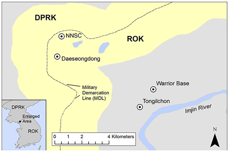
Mosquitoes were collected by Mosquito Magnet® (Pro model, Woodstream Corp. Lititz, Pennsylvania, USA) at military installations and training sites and villages near/in the DMZ [Collection sites: the Neutral Nations Supervisory Commission (NNSC) camp (37°57'16.39" N,26°40'50.03" E), Daeseongdong village (37°56'26.92" N, 126°40'37.42" E), Warrior Base Training Area (WBTA) (37°55'17.01" N, 126°44'30.22" E), and Tongilchon (beef farm ≈50 cattle) (37°54'32.18" N, 126°44'01.88" E)] in northern Gyeonggi Province, ROK from May-October 2012 (Fig 1). The NNSC camp is located inside the DMZ and is adjacent to the Military Demarcation Line (MDL) and Panmunjeom. Daeseongdong village is located inside the DMZ and adjacent to the MDL with approximately 200 residents. WBTA (a US Army training site) and Tongilchon village, with ≈200 residents, are located approximately 3 km from the southern entrance to the DMZ.
Fig 1. Map of the northern part of Gyeonggi province denoting collection sites of mosquitoes at NNSC (Neutral Nations Supervisory Commission camp adjacent to the Panmunjeom), Daeseongdong (located inside the Demilitarized Zone), Warrior Base (US Army training site) and Tongilchon (beef farm) located 2 km and 3 km, respectively from the MDL southern boundary of the Demilitarized Zone.
Mosquitoes were identified morphologically using standard keys [28, 29] and then placed in pools of up to 30 mosquitoes in 1.5 ml cryovials by species, date of collection, and collection site. The pooled specimens were homogenized in 0.6 ml of cell culture medium by bead-beating for 1 min using BioSpec Mini-BeadBeater-16 (Bio Spec Products Inc., Bartlesville, OK). After centrifugation at 6,000 × g for 10 min at 4°C, 130 μl clear supernatant was removed and treated with RNase free-DNase (QIAGEN Sciences, Germantown, MD) and subjected to nucleic acid extraction using QIAamp viral RNA kit (QIAGEN Sciences, Germantown, MD).
Purified nucleic acid samples were used in random reverse transcription and PCR amplification as described previously [30]. Nuclease-Free water (Invitrogen) (used as a blank control) and viral RNA of MS2 bacteriophage, a positive-sense single-stranded RNA virus (Roche Applied Science, Indianapolis, IN) were processed in parallel in separate reaction wells and analyzed to monitor the level of cross contamination. Random amplicons were sequenced using MiSeq sequencing system and reagents (Illumina, San Diego, CA). Sequence reads were subjected to metagenomics analyses using a bioinformatics pipeline [31], for sequence-based taxonomical identification of components in the specimens.
For mosquito specimens with rickettsial sequence reads identified in random metagenome sequencing, a total of 200 μl of the remaining cell culture medium homogenates were subjected to DNA extraction using QIAamp blood and tissue kits (QIAGEN), and the purified DNA was eluted in 100 μl of elution buffer. Two negative extraction controls (ECs) using molecular biology grade water (Invitrogen) were incorporated to ensure that the process was free from sample-to-sample contamination. DNA was diluted later using 10-fold serial dilutions (10–1000×) to reduce the effect of PCR inhibitors. The DNA preps were tested by a genus-specific quantitative real-time PCR (qPCR) assay (Rick17b) that amplifies and detects a 115-bp segment of the 17-kDa antigen gene [32].
PCR and nested PCR (nPCR) amplification and sequencing of rrs, gltA, 17 kDa antigen gene, ompB, ompA, and sca4 gene fragments were attempted on all Rick17b positive DNA preparations using primers and procedures previously described [33–36] and summarized in Table 1. To amplify the ompB gene, additional new primers targeting the conserved region of the ompB gene were used for a semi-nested PCR (sn-PCR) (Table 1). To amplify the gltA gene, an additional pair of primers was used: CS151F and CS1259R [25]. Two microliters of specimen DNA was used in a total reaction volume of 25 μl, containing 0.3 μM of each primer (Eurofins MWG Operon, Louisville, KY), 1× Platinum® PCR Supermix, High Fidelity (Invitrogen Corp., Carlsbad, CA) and RNase/DNase free water (GIBCO BRL Life Technologies, Inc., Gaithersburg, MD, USA). One microliter of PCR product was used in the nPCR or snPCR reactions performed in a Veriti Thermocycler (Applied Biosystems, Foster City, CA). For the new primers, the reaction mix was incubated at 94°C for 1 min followed by 40 cycles (35 cycles for nPCR and snPCR) of denaturation at 94°C for 30 seconds, annealing at 54°C (ompB) for 30 seconds and elongation at 68°C for 1 min, and final elongation at 72°C for 7 min. All PCR products were visualized using gel red (Biotium, Hayward, CA) on 1% (w/v) agarose gel (Sigma, St Louis, MO, USA). The size of the amplified product was determined by comparison with 1 kb plus molecular marker (Invitrogen). No positive controls were used in the PCR, nPCR, or snPCR procedures to decrease chances of contamination; however, negative controls (molecular biology grade water, GIBCO) were assessed with the samples and they were consistently negative in all runs.
Table 1. Oligonucleotide primers.
| Gene | Primer | Sequence (5–3) | Reference |
|---|---|---|---|
| 17kDa | R17K128F2∞ | GGGCGGTATGAAYAAACAAG | (32) |
| R17K238R∞ | CCTACACCTACTCCVACAAG | (32) | |
| R17K202TaqP∞ | FAM-CCGAATTGAGAACCAAGTAATGC-TAMRA | (32) | |
| rrs | 16SU17F* | AGAGTTTGATCCTGGCTCAG | (34) |
| 16SR34F#^ | CAGAACGAACGCTATCGGTA | (34) | |
| 16SU1592R* | AGGAGGTRATCCAGCCGCA | (34) | |
| 16SOR1198R#^ | TTCCTATAGTTCCCGGCATT | (34) | |
| 16sOR155F^ | TCAGTACGGAATAACWTTTAGAAATAA | This study | |
| 16SU547F^ | CAGCAGCCGCGGTAATAC | This study | |
| 16sU 833R^ | CTACCAGGGTATCTAATCCTGTT | This study | |
| 17kDa | R17k31F*#^ | GCTCTTGCAGCTTCTATGTTACA | 34 |
| R17k469R* | ACTTGCCATTGTCCGTCAGGTTG | 34 | |
| R17k2608R#^ | CATTGTCCGTCAGGTTGGCG | 34 | |
| ompB | 120-M59F* | CCGCAGGGTTGGTAACTGC | 35 |
| 120-1570R* | TCGCCGGTAATTRTAGCACT | 34 | |
| 120-2788F*^ | AAACAATAATCAAGGTACTGT | 36 | |
| 120-4346R*^ | CGAAGAAGTAACGCTGACTT | 36 | |
| RompB3521F^ | GATAATGCCAATGCAAATTTCAG | 36 | |
| RompB4224F^ | ACCAAGATTATAAGAAAGGTGATAA | 36 | |
| RompB3637R^ | GAAACGATTACTTCCGGTTACA | 36 | |
| RompB3008R^ | CGCCTGTAGTAACAGTTACAC | 36 | |
| RompB3503F*#^ | ACAACCATTAACGTACAAGATAA | This study | |
| RompB4293R* | GCACTACCTTGAGCAAAGAA | This study | |
| RompB4246R#^ | CACCTTTCTTATAATCTTGGTGTTTTAT | This study | |
| gltA | CS49F*# | ACCTATACTTAAAGCAAGTATYGGT | This study |
| CS1273R* | CATAACCAGTGTAAAGCTG | (36) | |
| CS1234R# | TCTAGGTCTGCTGATTTTTTGTTCA | (36) | |
| sca4 | RrD749F* | TGGTAGCATTAAAAGCTGATGG | (36) |
| RrD928F#^ | ATTTATACACTTGCGGTAACAC | (36) | |
| RrD2685R*#^ | TTCAGTAGAAGATTTAGTACCAAAT | (36) | |
| RrD_Sca4_F3^ | CAGGACCTCCTTTGCCTTCAT | This study | |
| RrD_Sca4_R2^ | TGCTTTATCTTGACCGTTCAA | This study | |
| ompA | 190-70F* | ATGGCGAATATTTCTCCAAAA | (33) |
| 190-701R* | GTTCCGTTAATGGCAAGCATCT | (33) | |
| 190-602n* | AGTGCAGCATTCGCTCCCCCT | (33) | |
| 190-3588F*# | AACAGTGAATGTAGGAGCAG | (33) | |
| 190-5238R*# | ACTATTAAAGGCTAGGCTATT | (33) |
∞primers used for qPCR;
*Primers used for PCR amplification;
#primers used for nested PCR;
^Primers used for sequencing
PCR products were purified using QIAquick PCR purification kit (QIAGEN). Sequencing reactions were performed utilizing both DNA strands with Big Dye Terminator v3.1 Ready Reaction Cycle Sequencing Kit (Life Technologies, Foster City, CA), according to the manufacturer’s instructions on an ABI 3500 genetic analyzer (Applied Biosystems, Foster City, CA). The rrs, gltA, 17kDa, ompB, and sca4 sequences were assembled using CodonCode Aligner version 5.0.1 (CodonCode Corporation, Centerville, MA) and exported to Molecular Evolutionary Genetics Analysis (MEGA) version 7 software (CEMI, Tempe, AZ) where they were compared with other historical strains available in GenBank. Phylogenetic analyses were performed using MEGA v7 [37] using Maximum Likelihood (ML) method using the Tamura-Nei model [38] for the individual genes. Multi-locus sequence typing (MLST) phylogeny was performed utilizing concatenated gene fragments of rrs (881-bp), gltA (993-bp), ompB (604-bp), and 17kDa gene (410-bp) of the novel Rickettsia genotypes from the mosquitoes in this study and validly published Rickettsia species from GenBank. The concatenated alignment tree was also estimated using the ML method and the Tamura-Nei model. Additionally, comparison of the gltA and sca4 sequences from this study and other mosquito molecular isolates was performed to determine their relatedness. Bootstrap analyses were performed with 1,000 replications.
GenBank accession numbers
The sequences of the new rickettsial molecular isolates A12.2646 from Mansonia uniformis, A12.2638 from Culex pipiens and A12.3271 from Aedes esoensis have been deposited in GenBank with accession numbers KY799063-KY799067, KY799068-KY799071 and KY799072-KY799074, MF590070 for rrs, 17 kDa antigen gene, ompB, gltA, and sca4, respectively.
All of the surveillance was done under a United States Forces Korea (USFK) regulation (USFK 2009, Reg. 40–2). We obtained verbal permission from the residents to set a mosquito magnet at two private sites at Daeseongdong and Tongilchon. All the other sites were military installations [Neutral Nations Supervisory Commission (NNSC) camp] or training sites (South Gate to the DMZ which is part of the Joint Security Area and Warrior Base).
Results
Metagenome sequencing were conducted for a total of 240 mosquito pools collected at four locations (NNSC camp, Daeseongdong village, beef farm at Tongilchon, and WBTA near/in the DMZ in the ROK. Pools included a total of 2,248 mosquitoes belonging to five genera and seven species, including: Aedes esoensis (n = 456), Armigeres subalbatus (n = 94), Culex bitaeniorhynchus (n = 1007), Culex pipiens (n = 627), Mansonia uniformis (n = 19), Ochlerotatus koreicus (n = 40), and Ochlerotatus nipponicus (n = 5).
Analyses of metagenome sequencing data identified rickettsial ribosomal RNA genes, suggesting the presence of rickettsiae in eight pools (Table 2). The eight positive pools and seven additional M. uniformis pools (negative for rickettsial RNA in NGS) were subjected to further molecular analyses. To confirm the presence of Rickettsia nucleic acid, a genus-specific qPCR (Rick17b) assay was used that showed that 7/8 (87.5%) rickettsial rRNA positive mosquito DNA pools were also positive for Rickettsia DNA, while, all seven pools of rickettsial rRNA negative M. uniformis were confirmed negative for rickettsial DNA. The seven Rickettsia-positive mosquito samples included: 3 pools of Cx. pipiens, 3 pools of Ae. esoensis and 1 pool of Mn. uniformis. One pool of Cx. bitaeniorhynchus that was positive for rickettsial RNA by NGS was negative by the Rick17b assay.
Table 2. Summary of qPCR and sequencing results for mosquito samples obtained from the Republic of Korea.
| Sequencing | ||||||||||||
|---|---|---|---|---|---|---|---|---|---|---|---|---|
| Sample Name | Location | Mosquito Species | Mosquitoes/ pool | Date collected | NGS | Rick17b qPCR | rrs | 17kDa | gltA | ompB | ompA | sca4 |
| A12.2552 | Daeseongdong | Cx. bitaeniorhynchus | 30 | 20-Aug-12 | Pos | Neg | - | - | - | - | - | - |
| A12.2638 | Daeseongdong | Cx. pipiens | 30 | 27-Aug-12 | Pos | Pos | + | + | + | + | - | - |
| A12.2639 | Daeseongdong | Cx. pipiens | 19 | 27-Aug-12 | Pos | Pos | + | + | + | - | - | - |
| A12.2646 | Daeseongdong | Mn. uniformis | 2 | 27-Aug-12 | Pos | Pos | + | + | + | + | - | + |
| A12.2784 | Warrior base | Cx. pipiens | 17 | 3-Sep-12 | Pos | Pos | + | + | + | + | - | - |
| A12.2856 | Daeseongdong | Ae. esoensis | 30 | 6-Sep-12 | Pos | Pos | + | + | + | + | - | - |
| A12.3106 | NNSC | Ae. esoensis | 20 | 17-Sep-12 | Pos | Pos | + | + | + | + | - | - |
| A12.3271 | NNSC | Ae. esoensis | 13 | 24-Sep-12 | Pos | Pos | + | + | + | + | - | - |
NGS- Next Generation Sequencing
Amplicons were generated for one or more of the rickettsial gene targets; rrs (7/7), gltA (7/7), 17kDa (7/7), ompB (6/7) and sca4 (1/7) (Table 1). Attempts to amplify the ompA fragments were unsuccessful for all samples. No culicine mosquitoes collected from Tongilchon assayed by NGS were positive for rickettsiae.
The sequences of the rrs from the seven Rick17b positive mosquito pools were compared to those available in the GenBank. The sequence from one mosquito pool of Cx. pipiens (A12.2638) shared 100% sequence identity with Rickettsia sp. WHANSA-97 from An. sinensis from China (KU586119) and 99.9% with Rickettsia bellii RML369-C. The sequences of the other 6 pools shared 99.3% sequence identity with R. bellii RML369-C. The rrs sequences from all the other samples exhibited ≤98.2 sequence homology with rickettsiae identified in mosquitoes Cx. quinquefasciatus, An. sinensis and Cx. tritaeniorhynchus from China [25] (Table 3).
Table 3. Percent sequence identity of the mosquito genotypes to Rickettsia species with validly published names and other mosquito rickettsial strains provided in the GenBank.
| Gene | Mosquito pool | Mosquito species | Sequence length | Percent identity with the closest Rickettsia sp. (number of matches/sequence length) | Accession number | Fournier et al. (41) Cutoff values |
|---|---|---|---|---|---|---|
| rrs | A12.2638 | Culex pipiens | 1091 | 99.9% (1087/1088) R. bellii | CP000087 | ≥99.8% |
| 99.9% (806/807) R. bellii WHANSA-97_An. sinensis | KU586119 | |||||
| 98.1% (1006/1026) R. monacensis_Cx. quinquefasciatus | KU586120 | |||||
| 97.5% (384/394) Candidatus R. sp. An. sinensis | KU586118 | |||||
| 95.5% (362/379) Candidatus R. sp. Cx. tritaeniorhynchus | KU586123 | |||||
| A12.2646 | Mansonia uniformis | 1096 | 99.3% (1083/1091) R. bellii | CP000087 | ||
| A12.2639 | Culex pipiens | 1090 | 99.3% (1082/1090) R. bellii | CP000087 | ||
| A12.2784 | Culex pipiens | 1090 | 99.3% (1082/1090) R. bellii | CP000087 | ||
| A12.2856 | Aedes esoensis | 1091 | 99.3% (1083/1091) R. bellii | CP000087 | ||
| A12.3106 | Aedes esoensis | 1090 | 99.3% (1082/1090) R. bellii | CP000087 | ||
| A12.3271 | Aedes esoensis | 1419 | 99.3% (1409/1419) R. bellii | CP000087 | ||
| 99.3% (1093/1101) R. bellii WHANSA-97_An. sinensis | KU586119 | |||||
| 98.2% (1291/1314) R. monacensis_Cx. quinquefasciatus | KU586120 | |||||
| 97.5% (668/685) Candidatus R. sp. An sinensis | KU586118 | |||||
| 96.4% (646/670) Candidatus R. sp. Cx. tritaeniorhynchus | KU586123 | |||||
| 17kDa | A12.2638 | Culex pipiens | 410 | 89.3% (367/411) R. helvetica | LN794217 | NA |
| A12.2646 | Mansonia uniformis | 98.0% (402/410) R. monacensis | LN794217 | |||
| 100.0% (394/394) Rickettsia sp. Tx140 | EF689738 | |||||
| A12.2639 | Culex pipiens | 89.3% (367/411) R. helvetica | LN794217 | |||
| A12.2784 | Culex pipiens | 89.3% (367/411) R. helvetica | LN794217 | |||
| A12.2856 | Aedes esoensis | 89.3% (367/411) R. helvetica | LN794217 | |||
| A12.3106 | Aedes esoensis | 89.3% (367/411) R. helvetica | LN794217 | |||
| A12.3271 | Aedes esoensis | 89.3% (367/411) R. helvetica | LN794217 | |||
| ompB | A12.2638 | Culex pipiens | 640 | 85.0% (192/226) R. hoogstraalii | EF629536 | ≥99.2% |
| A12.2646 | Mansonia uniformis | 690 | 94.1% (649/690) R. hoogstraalii | EF629536 | ||
| 91.1% (626/687) Rickettsia sp. Anopheles gambiae | JN620080 | |||||
| 90.7% (514/567) Rickettsia sp. Culex pipiens | KU761260 | |||||
| A12.2639 | Culex pipiens | - | - | |||
| A12.2784 | Culex pipiens | 646 | 85.3% (198/232) R. hoogstraalii | EF629536 | ||
| A12.2856 | Aedes esoensis | 646 | 85.3% (198/232) R. hoogstraalii | EF629536 | ||
| A12.3106 | Aedes esoensis | 646 | 85.3% (198/232) R. hoogstraalii | EF629536 | ||
| A12.3271 | Aedes esoensis | 649 | 85.5% (201/235) R. hoogstraalii | EF629536 | ||
| gltA | A12.2638 | Culex pipiens | 1004 | 97.8% (982/1004) R. bellii | CP000087 | ≥99.9% |
| 100% 1004/1004) R. bellii WHANSA-97_An. sinensis | KU586331 | |||||
| A12.2646 | Mansonia uniformis | 995 | 96.5% (960/995) R. asembonensis | |||
| 96.7% (695/719) Rickettsia sp. Anopheles melas | JQ354961 | |||||
| 96.5% (920/953) Rickettsia sp. Anopheles gambiae | JN620082 | |||||
| 96.1% (366/381) Rickettsia sp. Culex pipiens | KU761259 | |||||
| 94.7% (942/995) R. monacensis_Cx. quinquefasciatus | KU586332 | |||||
| A12.2639 | Culex pipiens | 1000* | 85.4% (854/1000) Rickettsia felis | CP000053 | ||
| A12.2784 | Culex pipiens | 84.9% (849/1000) R. bellii | CP000087 | |||
| A12.2856 | Aedes esoensis | 96.5% (920/953) Rickettsia sp. Anopheles gambiae | JN620082 | |||
| A12.3106 | Aedes esoensis | |||||
| A12.3271 | Aedes esoensis | |||||
| sca4 | A12.2638 | Culex pipiens | - | - | ≥99.3% | |
| A12.2646 | Mansonia uniformis | 1681 | 92.3% (1559/1689) R. felis | CP000053 | ||
| 98.5% (326/331) Rickettsia sp. Culex pipiens | KU761261 | |||||
| 81.8% (516/631) Rickettsia sp. Anopheles gambiae | JN620081 | |||||
| A12.2639 | Culex pipiens | - | - | |||
| A12.2784 | Culex pipiens | - | - | |||
| A12.2856 | Aedes esoensis | - | - | |||
| A12.3106 | Aedes esoensis | - | - | |||
| A12.3271 | Aedes esoensis | - | - |
NA: not applicable—Not amplified
*-gltA sequences from the five pools were identical represented (A12.3271 pool)
Seven 410-bp sequences of the 17kDa antigen gene were generated from all seven positive mosquito pools (Table 3). The sequence from pool A12.2646 was only 98.0% identical to the closest Rickettsia with a validly published name, R. monacensis IrR/Munich. A 394-bp fragment of this gene sequence was 100% identical to an uncultured Rickettsia sp. Tx140 (EF689738) from an Ixodes scapularis tick from Texas, USA. The other six sequences (A12.2638, A12.2639, A12.2784, A12.2856, A12.3106 and A12.3271) were 100% identical to each other and were most similar to, yet very disparate from R. helvetica (89.3%) and R. monacensis (88.3%).
Attempts to amplify the ompB gene using previously described primers [34, 35], were unsuccessful. However, successful amplification of the pool A12.2646 using previously described primers [36] was accomplished. While using the new ompB primers, snPCR produced amplicons of the appropriate size from 6 mosquito pools (Table 3). A 690-bp ompB sequence from the Mn. uniformis pool, A12.2646 was identical to the sequence produced by 120-2788F/120-4346 primer pair, and was distantly related to R. hoogstraalii (94.1%). An additional 687-bp and 567-bp sequence of the ompB gene shared only 91.1% and 90.7% nucleotide similarity with the uncultured Rickettsia sp. from An. gambiae and Rickettsia endosymbiont of Cx. pipiens, respectively. The ompB sequences from five other mosquito pools (A12.2638, A12.2784, A12.2856, A12.3106 and A12.3271) were quite different from any other known Rickettsia sp. Only 34% of the ompB sequence aligned to R. hoogstraalii at ≤85.5% sequence homology.
First, attempts to amplify the gltA gene fragment using primer CS49F and previously described primers [36] were unsuccessful. Alternatively, primers previously described [25] were used and a 995-1004-bp gltA gene sequences were amplified from all the seven mosquito pools. Pool A12.2638 gltA sequence was 100% similar to Rickettsia sp. WHANSA-97 from An. sinensis but exhibited only 97.8% similarity with R. bellii RML369-C. Pool A12.2646 gltA sequence was most similar to that of R. asembonensis (96.5%) isolated from cat fleas. The sequences from mosquito samples A12.2639, A12.2784, A12.2856, A12.3106 and A12.3271 were identical and shared only 84.9% sequence similarity with R. bellii-the closest Rickettsia species. However, the gltA sequences shared ≤96.7% nucleotide identity in the respective aligned regions with uncultured Rickettsia spp. from An. melas, An. gambiae, and Rickettsia endosymbiont of Cx. pipiens (Table 3). The gltA sequences indicate that the rickettsiae from the pooled mosquitoes are genetically unique.
A 1,681-bp sequence from the sca4 gene was produced in 1/7 (14.3%) mosquito pools (A12.2646). Based on this gene, the sequence exhibited the highest similarities to R. felis URRWXCal2 (92.3%), and shared 98.5% and 81.8% nucleotide identity with the Rickettsia endosymbiont of Cx. pipiens and uncultured Rickettsia sp. from An. gambiae, respectively (Table 3).
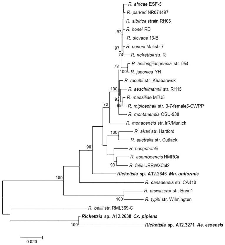
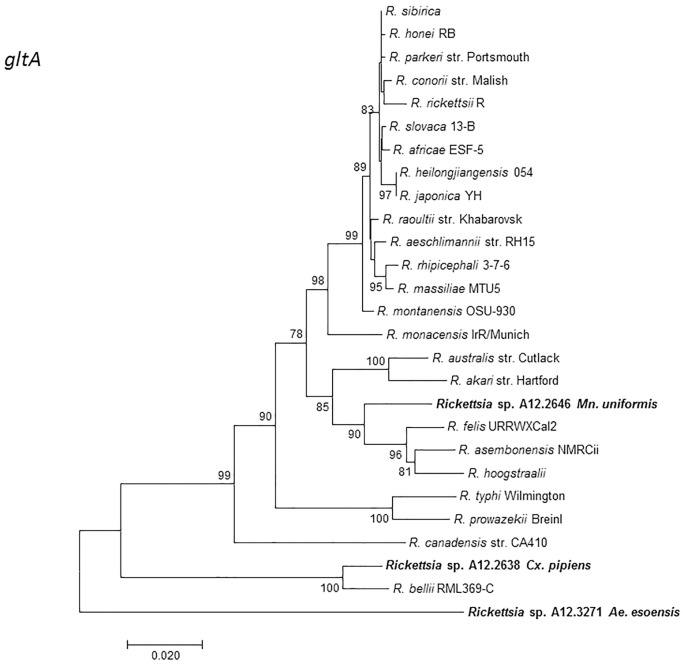
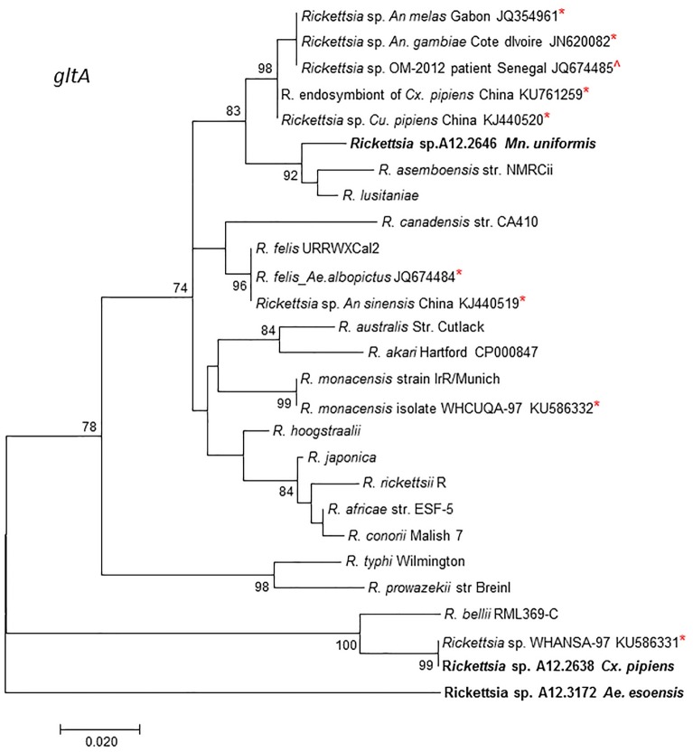
MLST analysis using concatenated fragments of rrs, gltA, 17kDa, and ompB genes showed that the Rickettsia sp. A12.2646 from Mn. uniformis did not cluster with any Rickettsia species but branched separately between the SFGR group and R. canadensis. Rickettsia sp. A12.2638 from Cx. pipiens and Rickettsia sp. A12.3271 from Ae. esoensis branched separately in a clade basal to R. bellii (Fig 2). Phylogenetic analysis based on the gltA gene sequences showed that Rickettsia sp. A12.2638 from Cx. pipiens clustered together with R. bellii RML369-C while Rickettsia sp. A12.3271 from Ae. esoensis was placed in a separate branch basal to the R. bellii. Rickettsia sp. A12.2646 from Mn. uniformis branched separately from the other members of SFGR (Fig 3). Incorporating other mosquito rickettsial genotypes detected in Africa and China, Rickettsia sp. A12.2638 from Cx. pipiens clustered together with R. bellii RML369-C and Rickettsia sp. WHANSA-97 detected in An. sinensis from China, while Rickettsia sp. A12.2646 from Mn. uniformis was placed in a clade together with other members of SFGR, but in a different branch away from other Rickettsia species from mosquitoes (Fig 4). Similar divergence was observed with the sca4 gene sequence from Rickettsia sp. A12.2646 (S1 Fig). The sequences from mosquito samples A12.2639, A12.2784, A12.2856, A12.3106 and A12.3271 were identical for rrs, ompB and 17kDa, and for the purpose of illustration, they are represented by A12.3271 from Ae. esoensis. The ompB sequence from A12.2639 was not amplifiable.
Fig 2. MLST dendrogram consisting of concatenated gene fragments of: rrs (881), gltA (993), ompB (604) and 17kDa gene (410) showing the phylogenetic position of Rickettsia sp. A12.2646 from Mansonia uniformis, Rickettsia sp. A12.2638 from Cx. pipiens and A12.3271 from Ae. esoensis among Rickettsia species in the GenBank.
The evolutionary history was inferred by the maximum likelihood method.
Fig 3. A dendrogram showing the phylogenetic position of Rickettsia sp. A12.2646 from Mn. uniformis, Rickettsia. sp. A12.2638 from Cx. pipiens and A12.3271 from Ae. esoensis from the Republic of Korea in relation to other Rickettsia species with validly published names for the gltA gene (993-bp).
The evolutionary history was inferred by the maximum likelihood method.
Fig 4. A dendrogram based on the gltA gene (345-bp) showing the phylogenetic position of Rickettsia sp. A12.2646 from Mn. uniformis, Rickettsia sp. A12.2638 from Cx. pipiens and A12.3271 from Ae. esoensis from Republic of Korea in relation to other rickettsial genotypes from mosquitoes and other historical strains provided in GenBank.
The evolutionary history was inferred using maximum likelihood method. *Represent other Rickettsia sp. from mosquitoes and ^from a patient’s blood.
Discussion
Recent reports suggesting that mosquitoes may play a role in the epidemiology of R. felis rickettsiosis [19] led us to conduct this study to determine whether R. felis and/or other rickettsiae could be detected in mosquitoes collected near/in the DMZ using NGS metagenome sequence-based pathogen discovery studies [30, 39]. Utilizing NGS, qPCR and MLST, three novel Rickettsia agents were detected in three different mosquito species: Cx. pipiens, Mn. uniformis and Ae. esoensis.
NGS has been used to unearth novel microbes including rickettsiae in ticks in western Europe [40, 41]. Our study used NGS for the detection of rickettsiae in mosquitoes, which resulted in the discovery of three potentially new Rickettsia genotypes in mosquito hosts. Though the random unbiased manner of metagenome NGS led to the production of a limited amount of species specific sequences that were insufficient to discriminate rickettsiae to species level with confidence, it is a great means of screening for rickettsiae as shown in this report. To overcome this limitation, mosquito pools positive for rickettsiae by NGS were subsequently evaluated using a Rickettsia-genus specific qPCR assay and the species determined by MLST. This application demonstrated a good concordance between NGS and the genus-specific qPCR assay, with 7/8 NGS positive samples being confirmed positive by Rick17b qPCR assay. Rickettsia DNA was not detected by the Rick17b assay in the mosquito DNA preparation from Cx. bitaeniorhynchus that was positive by NGS. The inability to amplify rickettsial DNA from Cx. bitaeniorhynchus preparation using the Rickettsia genus-specific qPCR (Rick17b) may be due to its presence in low copy numbers that were below the detection limit of the Rick17b assay or because the gene may be too divergent for amplification using the Rick17b primers.
Previous studies have documented the presence of rickettsiae in mosquitoes using species-specific qPCR assays and/or by PCR and sequencing of one or more of the following gene fragments; rrs, gltA, ompA, ompB, sca4, and groEL. A high rate of infection was reported in a recent study, where 21% of 53 Cx. pipiens mosquitoes assessed were infected with a novel Rickettsia [18]. Additionally, extensive diversity of Rickettsiales in multiple mosquito species was reported by Guo et al. [25]. In this study, there were three novel Rickettsia genotypes in Mn. uniformis, Cx. pipiens, and Ae. esoensis identified, which corroborate previous reports of the occurrence of Rickettsia felis [21, 26, 27] and other novel genotypes [18, 23, 25] in numerous mosquito species. Although there is no proof that vertical transmission of rickettsiae in these blood feeders occur, Guo et al. [25] demonstrated rickettsiae in each life stage (eggs, larvae, pupae, and adults) of laboratory-cultivated Armigeres subalbatus and Cx. tritaeniorhynchus mosquitoes. This observation suggests transovarial and transstadial transmission of rickettsiae in mosquitoes, and is corroborated by a previous study that detected R. felis in a male An. arabiensis mosquito [21]. However, in a separate laboratory study, when mosquitoes were fed by membrane feeding on blood infected with R. felis, the mosquitoes became infected but the bacteria were not transmitted to the F1 progeny [19]. This is the first report of detection of rickettsiae in Ae. esoensis and in mosquitoes from ROK. It was not determined if the rickettsiae are endosymbionts with transovarial transmission or if the mosquitoes became infected with rickettsiae through an alternative method. This report adds to the list of mosquito species that harbor Rickettsia species and shows that they may be more prevalent in mosquitoes than previously thought.
To identify the mosquito-borne rickettsiae we utilized three conserved gene fragments (rrs, 17 kDa antigen gene, and gltA) and three variable gene fragments (ompA, ompB, and sca4). Successful amplification and sequencing was achieved for all seven mosquito preparations for the rrs and 17kDa genes, but was variably successful using other gene fragments (gltA, ompB, and sca4) or unsuccessful (ompA). The much conserved rrs gene fragment sequences from Rickettsia-positive mosquitos (Cx. pipiens, Mn. uniformis and Ae. esoensis) were closest to that of Rickettsia (99.3–99.9%) in GenBank and confirmed the presence of rickettsiae in mosquitoes. Attempts to amplify various fragments of the variable ompB gene were somewhat successful. Similarly, a previous study encountered [23] comparable problems with mosquito preparations that were positive for Rickettsia spp. by qPCR, but could not be amplified by various primers targeting the ompB gene. Amplification was achieved herein using the primer pair 120–2788 and 120–4346, for only one mosquito pool (A12.2646 from Mn. uniformis) and in 6/7 mosquito pools using a new set of primers. The ompB sequence from five mosquito DNA preps was very divergent with >65% of the ompB sequence being unable to identify any match in the GenBank. This indicates that the rickettsiae from these pooled mosquito samples are genetically unique. We were most likely unable to differentiate the sequences from these five mosquito pools because the amplified region was in a highly conserved region from which our primers were located (4293–3503) as shown in other studies [42].
BLASTN searches for the rrs, gltA, ompB, and sca4 genes among mosquito-borne rickettsiae identified numerous sequences from mosquito species from China and Africa. Even though these rickettsiae came from similar hosts, none of the uncultured Rickettsia spp. identified in these mosquitoes including Candidatus Rickettsia sp. An. sinensis, Candidatus Rickettsia sp. Culex tritaeniorhyncus [25], the two uncultured Rickettsia spp. detected in An. gambiae and An. melas mosquitoes from Gabon [23] and a R. endosymbiont found in Cx. pipiens from China [18] shared perfect identity with the ones detected in the present study. However, the highly conserved rrs and gltA sequences from A12.2638 shared high nucleotide similarities (99.9% and 100%, respectively) with a Rickettsia sp. WHANSA-97 detected in An. sinensis from China. While it is likely that these two are the same, or very closely related, BLASTN searches for the gltA gene sequences revealed 97.8% or less homology with any of the R. bellii with validly published names. Additionally, the ompB sequence from A12.2638 could not identify any similarities with R. bellii. Due to the significant differences in the gltA and ompB sequences, we do not believe that this is R. bellii, but maybe related.
A previous study detected R. felis in Mn. uniformis from Senegal [21]. This is in contrast to the finding from this study where a different genotype was identified in a pool (A12.2646) of two Mn. uniformis mosquitoes. This suggests that one species of mosquito is capable of hosting different rickettsial species. The rickettsiae identified from A12.2646 clustered with R. felis-like organisms and other uncultured Rickettsia sp. from An. gambiae and An. melas [18, 23], and shared only ≤ 96.7% gltA nucleotide sequence homologies with the latter. Interestingly, the gltA sequences from the uncultured Rickettsia spp. detected in An. gambiae and An. melas mosquitoes from Gabon were closely related (99.7%) to a Rickettsia sp. detected in blood from a febrile patient from Senegal [JQ674485] [23]. This close relationship might indicate that some of these uncultured Rickettsia species found in mosquitoes may be potential human pathogens. Further work to establish the transmission dynamics of mosquito-borne rickettsiae and their potential to infect mammalian hosts is needed.
To strengthen the characterization of the mosquito-borne rickettsiae within the genus we concatenated rrs, gltA, 17 kDa gene, and ompB gene fragments. Unfortunately, we did not include the other genes because there was only one sequence obtained using sca4, and none from ompA. Based on the phylogenetic relationship inferred by concatenation of the rrs, gltA, 17kDa, and the ompB gene fragments, Rickettsia sp. A12.2646, Rickettsia sp. A12.2638, and Rickettsia sp. A12.3271 branched separately to form distinct lineages. The novel mosquito species, Rickettsia sp. A12.2638 and Rickettsia sp. A12.3271 were placed in a branch basal to the R. bellii clade, which supports the presence of additional rickettsial lineages [1], other than the widely accepted groups of the SFGR, TG, transitional group Rickettsia (TGR), and an ancestral group Rickettsia (AGR).
The nucleotide sequence similarities of the rickettsiae from mosquitoes collected from ROK were lower than the cut-off values proposed for Rickettsia species definition [43] and suggest them as tentative new Rickettsia species. Further studies are needed to shed light on the evolutionary relationship of these mosquito-borne rickettsiae to previously described rickettsiae, and their seasonal, geographic and species diversity, prevalence, biology, and pathogenic potential.
Supporting information
The evolutionary history was inferred using maximum likelihood method. *Represent other Rickettsia sp. from mosquitoes.
(TIF)
Acknowledgments
We thank Dr. Penny Masuoka of Uniformed Services University of the Health Sciences, Bethesda, Maryland for her assistance with mapping.
Disclaimers
The views expressed in this article are those of the authors and do not reflect the official policy of the Department of Army, Department of the Navy, Department of Defense, or the U.S. Government. Trade names are used for identification purposes only and do not imply endorsement.
Data Availability
All relevant data are within the paper and its Supporting Information files.
Funding Statement
This research was funded by the Armed Forces Health Surveillance Branch, Global Emerging Infections Surveillance and Response System (AFHSB-GEIS), Work Unit Numbers: 188M.0931.001-A0074, P0015-17-ME and 17-WR-1.2.1. The grant(s) were awarded to ALR, TAK and RGJ. The funders had no role in the study design, data collection and analysis, decision to publish, or preparation of the manuscript.
References
- 1.Weinert LA, Werren JH, Aebi A, Stone GN, Jiggins FM. Evolution and diversity of Rickettsia bacteria. BMC biol. 2009;7:6 doi: 10.1186/1741-7007-7-6 [DOI] [PMC free article] [PubMed] [Google Scholar]
- 2.Hechemy KE, Avsic-Zupanc T, Childs JE, Raoult DA. Rickettsiology: present and future directions: preface. Ann NY Acad Sci 990: 17–20. [DOI] [PubMed] [Google Scholar]
- 3.Kikuchi Y, Sameshima S, Kitade O, Kojima J, Fukatsu T. Novel clade of Rickettsia spp. from leeches. Appl Environ Microbiol. 2002;68:999–1004. doi: 10.1128/AEM.68.2.999-1004.2002 [DOI] [PMC free article] [PubMed] [Google Scholar]
- 4.Parola P, Musso D, Raoult D. Rickettsia felis: the next mosquito-borne outbreak? The Lancet Infect Dis. 2016;16:1112–3. doi: 10.1016/S1473-3099(16)30331-0 [DOI] [PubMed] [Google Scholar]
- 5.Parola P, Paddock CD, Socolovschi C, Labruna MB, Mediannikov O, Kernif T, et al. Update on tick-borne rickettsioses around the world: a geographic approach. Clin Microbiol Rev. 2013;26:657–702. doi: 10.1128/CMR.00032-13 [DOI] [PMC free article] [PubMed] [Google Scholar]
- 6.Lee JH, Park HS, Jung KD, Jang WJ, Koh SE, Kang SS, et al. Identification of the spotted fever group rickettsiae detected from Haemaphysalis longicornis in Korea. Microbiol Immunol. 2003;47:301–4. [DOI] [PubMed] [Google Scholar]
- 7.Lee KM, Choi YJ, Shin SH, Choi MK, Song HJ, Kim HC, et al. Spotted fever group rickettsia closely related to Rickettsia monacensis isolated from ticks in South Jeolla province, Korea. Microbiol Immunol. 2013;57:487–95. doi: 10.1111/1348-0421.12062 [DOI] [PubMed] [Google Scholar]
- 8.Shin SH, Seo HJ, Choi YJ, Choi MK, Kim HC, Klein TA, et al. Detection of Rickettsia monacensis from Ixodes nipponensis collected from rodents in Gyeonggi and Gangwon Provinces, Republic of Korea. Exp Appl Acarol. 2013;61(3):337–47. doi: 10.1007/s10493-013-9699-1 [DOI] [PubMed] [Google Scholar]
- 9.Kang SW, Doan HT, Choe SE, Noh JH, Yoo MS, Reddy KE, et al. Molecular investigation of tick-borne pathogens in ticks from grazing cattle in Korea. Parasitol Inter. 2013;62(3):276–82. [DOI] [PubMed] [Google Scholar]
- 10.Chae JS, Yu DH, Shringi S, Klein TA, Kim HC, Chong ST, et al. Microbial pathogens in ticks, rodents and a shrew in northern Gyeonggi-do near the DMZ, Korea. J Vet Sci 2008;9:285–93. doi: 10.4142/jvs.2008.9.3.285 [DOI] [PMC free article] [PubMed] [Google Scholar]
- 11.Noh Y, Lee YS, Kim HC, Chong ST, Klein TA, Jiang J, et al. Molecular detection of Rickettsia species in ticks collected from the southwestern provinces of the Republic of Korea. Parasit Vectors. 2017;10(1):20 doi: 10.1186/s13071-016-1955-x [DOI] [PMC free article] [PubMed] [Google Scholar]
- 12.Chung MH, Lee SH, Kim MJ, Lee JH, Kim ES, Kim MK, et al. Japanese spotted fever, South Korea. Emer Infect Dis. 2006;12(7):1122–4. [DOI] [PMC free article] [PubMed] [Google Scholar]
- 13.Jang WJ, Choi YJ, Kim JH, Jung KD, Ryu JS, Lee SH, et al. Seroepidemiology of spotted fever group and typhus group rickettsioses in humans, South Korea. Microbiol Immunol. 2005;49:17–24. [DOI] [PubMed] [Google Scholar]
- 14.Jang WJ, Kim JH, Choi YJ, Jung KD, Kim YG, Lee SH, et al. First serologic evidence of human spotted fever group rickettsiosis in Korea. J Clin Microbiol. 2004;42:2310–3. doi: 10.1128/JCM.42.5.2310-2313.2004 [DOI] [PMC free article] [PubMed] [Google Scholar]
- 15.Jiang J, Myers TE, Rozmajzl PJ, Graf PC, Chretien JP, Gaydos JC, et al. Seroconversions to rickettsiae in US Military personnel in South Korea. Emer Infect Dis. 2015;21:1073–4. [DOI] [PMC free article] [PubMed] [Google Scholar]
- 16.Ko S, Kim HC, Yang YC, Chong ST, Richards AL, Sames WJ, et al. Detection of Rickettsia felis and Rickettsia typhi and seasonal prevalence of fleas collected from small mammals at Gyeonggi Province in the Republic of Korea. Vector Borne Zoonotic Dis. 2011;11:1243–51. doi: 10.1089/vbz.2010.0261 [DOI] [PubMed] [Google Scholar]
- 17.Liu D, Wang YZ, Zhang H, Liu ZQ, Wureli HZ, Wang SW, et al. First report of Rickettsia raoultii and R. slovaca in Melophagus ovinus, the sheep ked. Parasit Vectors. 2016;9:600 doi: 10.1186/s13071-016-1885-7 [DOI] [PMC free article] [PubMed] [Google Scholar]
- 18.Zhang J, John Kelly P, Lu G, Cruz-Martinez L, Wang C. Rickettsia in mosquitoes, Yangzhou, China. Emerg Microbes Infect. 2016;5:e108 doi: 10.1038/emi.2016.107 [DOI] [PMC free article] [PubMed] [Google Scholar]
- 19.Dieme C, Bechah Y, Socolovschi C, Audoly G, Berenger JM, Faye O, et al. Transmission potential of Rickettsia felis infection by Anopheles gambiae mosquitoes. Proc Natl Acad Sci USA. 2015;112(26):8088–93. doi: 10.1073/pnas.1413835112 [DOI] [PMC free article] [PubMed] [Google Scholar]
- 20.Maina AN, Knobel DL, Jiang J, Halliday J, Feikin DR, Cleaveland S, et al. Rickettsia felis infection in febrile patients, western Kenya, 2007–2010. Emerg Infect Dis. 2012;18:328–31. doi: 10.3201/eid1802.111372 [DOI] [PMC free article] [PubMed] [Google Scholar]
- 21.Mediannikov O, Socolovschi C, Edouard S, Fenollar F, Mouffok N, Bassene H, et al. Common epidemiology of Rickettsia felis infection and malaria, Africa. Emerg Infect Dis. 2013;19:1775–83. doi: 10.3201/eid1911.130361 [DOI] [PMC free article] [PubMed] [Google Scholar]
- 22.Hertig M, Wolbach SB. Studies on Rickettsia-Like Micro-Organisms in Insects. J Med Res. 1924;44:329–74.7. [PMC free article] [PubMed] [Google Scholar]
- 23.Socolovschi C, Pages F, Ndiath MO, Ratmanov P, Raoult D. Rickettsia species in African Anopheles mosquitoes. PLoS One. 2012;7:e48254 doi: 10.1371/journal.pone.0048254 [DOI] [PMC free article] [PubMed] [Google Scholar]
- 24.Sakamoto JM, Azad AF. Propagation of arthropod-borne Rickettsia spp. in two mosquito cell lines. Appl Environ Microbiol. 2007;73:6637–43. doi: 10.1128/AEM.00923-07 [DOI] [PMC free article] [PubMed] [Google Scholar]
- 25.Guo WP, Tian JH, Lin XD, Ni XB, Chen XP, Liao Y, et al. Extensive genetic diversity of Rickettsiales bacteria in multiple mosquito species. Sci Rep. 2016;6:38770 doi: 10.1038/srep38770 [DOI] [PMC free article] [PubMed] [Google Scholar]
- 26.Zhang J, Lu G, Kelly P, Zhang Z, Wei L, Yu D, et al. First report of Rickettsia felis in China. BMC Infect Dis. 2014;14:682 doi: 10.1186/s12879-014-0682-1 [DOI] [PMC free article] [PubMed] [Google Scholar]
- 27.Socolovschi C, Pages F, Raoult D. Rickettsia felis in Aedes albopictus mosquitoes, Libreville, Gabon. Emerg Infect Dis. 2012;18:1687–9. doi: 10.3201/eid1810.120178 [DOI] [PMC free article] [PubMed] [Google Scholar]
- 28.Lee KW. A revision of the illustrated taxonomic keys to genera and species of female mosquitoes in Korea (Diptera, Culicidae): 5th Medical Detachment, 168th Medical Battalion, 18th Medical Command, US Army; 1998.
- 29.Tanaka K M K S E. A revision of the adult and larval mosquitoes of Japan (including the Ryukyu Archipelago and the Ogasawara Islands) and Korea (Diptera: Culicidae). Contr Am Entomol Inst. 1979;16:43–85. [Google Scholar]
- 30.Hang J, Klein TA, Kim HC, Yang Y, Jima DD, Richardson JH, et al. Genome sequences of five arboviruses in field-captured mosquitoes in a unique rural environment of South Korea. Gene Announc. 2016;4. [DOI] [PMC free article] [PubMed] [Google Scholar]
- 31.Kilianski A, Carcel P, Yao S, Roth P, Schulte J, Donarum GB, et al. Pathosphere.org: pathogen detection and characterization through a web-based, open source informatics platform. BMC Bioinfo. 2015;16:416. [DOI] [PMC free article] [PubMed] [Google Scholar]
- 32.Jiang J, Stromdahl EY, Richards AL. Detection of Rickettsia parkeri and Candidatus Rickettsia andeanae in Amblyomma maculatum Gulf Coast ticks collected from humans in the United States. Vector Borne Zoonotic Dis. 2012;12:175–82. doi: 10.1089/vbz.2011.0614 [DOI] [PubMed] [Google Scholar]
- 33.Fournier PE, Roux V, Raoult D. Phylogenetic analysis of spotted fever group rickettsiae by study of the outer surface protein rOmpA. Inter J Syst Bacteriol. 1998;48:839–49. [DOI] [PubMed] [Google Scholar]
- 34.Jiang J, Maina AN, Knobel DL, Cleaveland S, Laudisoit A, Wamburu K, et al. Molecular detection of Rickettsia felis and Candidatus Rickettsia asemboensis in fleas from human habitats, Asembo, Kenya. Vector Borne Zoonotic Dis. 2013;13:550–8. doi: 10.1089/vbz.2012.1123 [DOI] [PMC free article] [PubMed] [Google Scholar]
- 35.Roux V, Raoult D. Phylogenetic analysis of members of the genus Rickettsia using the gene encoding the outer-membrane protein rOmpB (ompB). Inter J Syst Evol Microbiol. 2000;50:1449–55. [DOI] [PubMed] [Google Scholar]
- 36.Jiang J, Blair PJ, Felices V, Moron C, Cespedes M, Anaya E, et al. Phylogenetic analysis of a novel molecular isolate of spotted fever group rickettsiae from northern Peru: Candidatus Rickettsia andeanae. Ann NY Acad Sci. 2005;1063:337–42. doi: 10.1196/annals.1355.054 [DOI] [PubMed] [Google Scholar]
- 37.Kumar S, Stecher G, Tamura K. MEGA7: Molecular Evolutionary Genetics Analysis Version 7.0 for Bigger Datasets. Mol Biol Evol. 2016;33:1870–4. doi: 10.1093/molbev/msw054 [DOI] [PMC free article] [PubMed] [Google Scholar]
- 38.Tamura K, Nei M. Estimation of the number of nucleotide substitutions in the control region of mitochondrial DNA in humans and chimpanzees. Mol Biol Evol. 1993;10:512–26. [DOI] [PubMed] [Google Scholar]
- 39.Korkusol A, Takhampunya R, Hang J, Jarman RG, Tippayachai B, Kim HC, et al. A novel flavivirus detected in two Aedes spp. collected near the demilitarized zone of the Republic of Korea. The Journal of general virology. 2017;98(5):1122–31. doi: 10.1099/jgv.0.000746 [DOI] [PubMed] [Google Scholar]
- 40.Vayssier-Taussat M, Moutailler S, Michelet L, Devillers E, Bonnet S, Cheval J, et al. Next generation sequencing uncovers unexpected bacterial pathogens in ticks in western Europe. PLoS One. 2013;8:e81439 doi: 10.1371/journal.pone.0081439 [DOI] [PMC free article] [PubMed] [Google Scholar]
- 41.Xiang L, Pozniak B, Cheng TY. Bacteriological analysis of saliva from partially or fully engorged female adult Rhipicephalus microplus by next-generation sequencing. Antonie van Leeuwenhoek. 2017;110:105–13. doi: 10.1007/s10482-016-0780-8 [DOI] [PubMed] [Google Scholar]
- 42.Ma JS. Rickettsioses in South Korea, data analysis. Emerg Infect Dis. 2006;12:531–2. [DOI] [PMC free article] [PubMed] [Google Scholar]
- 43.Fournier PE, Dumler JS, Greub G, Zhang J, Wu Y, Raoult D. Gene sequence-based criteria for identification of new rickettsia isolates and description of Rickettsia heilongjiangensis sp. nov. J Clin Microbiol. 2003;41:5456–65. doi: 10.1128/JCM.41.12.5456-5465.2003 [DOI] [PMC free article] [PubMed] [Google Scholar]
Associated Data
This section collects any data citations, data availability statements, or supplementary materials included in this article.
Supplementary Materials
The evolutionary history was inferred using maximum likelihood method. *Represent other Rickettsia sp. from mosquitoes.
(TIF)
Data Availability Statement
All relevant data are within the paper and its Supporting Information files.


