Figure 3.
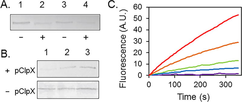
Degradation of AcrB-His6-ssrA. (A) Anti-AcrB Western blot analysis of degradation of detergent solubilized AcrB-His6-ssrA in vitro. Lanes 1 and 2 contain 0.2 µg of AcrB-His6-ssrA, and lanes 3 and 4 contain 0.1 µg of AcrB-His6-ssrA. Lanes 2 and 4 also contain ClpX and ClpP. (B) Anti-AcrB Western blot analysis of whole cell lysate prepared from DL41ΔacrBΔclpX transformed with pQE-AcrB-His6-ssrA and pBAD-ClpX (pClpX) (top) or pQE-AcrB-His6-ssrA alone (bottom). AcrB-His6-ssrA was expressed at the basal level without induction, and then the cell culture was divided equally into three samples: arabinose was added into the first sample to induce the expression of ClpX (lane 1). Sample 1 and sample 2 (lane 2) were incubated for an additional 2 h at 28 °C, while sample 3 (lane 3) was left on ice. (C) EtBr accumulation assay of DL41ΔacrBΔclpX transformed with the indicated plasmids and cultured with or without ClpX induction. pQE70 (red, negative control), pQE-AcrB-His6-ssrA/pClpX with arabinose (orange), pQE-AcrB-His6-ssrA/pClpX without induction (green), pQE-AcrB-His6-ssrA (blue), and pQE-AcrB-His6 (purple, positive control).

