Abstract
Spontaneous vaginal evisceration of the small bowel is a rare event. It is precipitated in the postmenopausal woman commonly by hysterectomy and in the premenopausal woman by vaginal trauma. We report a case of a 75-year-old woman presenting with a protruding mass in her vagina and associated abdominal pain. A combined laparoscopic and transvaginal method of repair is described and the advantage of using both techniques highlighted.
Keywords: Vaginal evisceration, Laparoscopy, Evisceration of small bowel
Vaginal evisceration of intestinal contents is very rare. McGregor reported the first case in 1907.1 Since then, there have been fewer than 100 reported cases in the literature. The typical patient is a postmenopausal women with a history of gynaecological surgery (most commonly hysterectomy) often with pelvic floor dysfunction. In premenopausal women, vaginal evisceration is associated with vaginal trauma most commonly following sexual intercourse, obstetric instrumentation or foreign body insertion.
The first case of laparoscopic-assisted repair of small bowel evisceration per vagina was published in 1996.2 As far as the authors are aware, only a further three cases have been described using laparoscopy as an adjunct to repair.3,4
The combined laparoscopic and transvaginal approach is advantageous: laparoscopy allows direct visualisation and assessment of the small bowel with the ability to reduce the herniated bowel contents. The transvaginal route allows optimum closure of the vaginal stump.
We report a second case using a combined laparoscopic and transvaginal approach without the use of omental mesh repair with subsequent early patient discharge.
Case history
A 75-year-old woman presented with lower abdominal pain and prolapsed small bowel protruding from her vagina, brought on by straining at defecation. She had a vaginal hysterectomy for prolapse 4 years earlier and suffered from stress incontinence. Her obstetric history was one normal vaginal delivery age 29 years with no documented tears. She was not currently sexually active and had no recent history of abdominal or vaginal trauma.
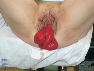
On examination, 40 cm of oedematous, but viable, small bowel (ileum) eviscerated through the vagina (Fig. 1). Her vital signs and blood investigations were normal.
Figure 1.

Spontaneous evisceration of small bowel.
The patient was taken to theatre and positioned in lithotomy and Trendelenburg position. Manual reduction of bowel was attempted unsuccessfully due to a tight vaginal neck. Laparoscopy allowed gentle dilatation of the vaginal neck and easy reduction of the small bowel into the peritoneal cavity, at which point its viability was further assessed. The vagina appeared atrophic. The remaining abdominal viscera were unremarkable.
The vagina was then everted laparoscopically allowing for direct inspection and manual assessment of the quality of tissue. The edge of the vagina was trimmed and the defect repaired via the vaginal route using continuous absorbable sutures.
Histology of the vaginal tissue showed some superficial ulceration; however, there was no evidence of dysplasia or malignancy.
The patient was given three doses of intravenous antibiotics and discharged early on day 2 postoperatively.
Discussion
Small bowel evisceration through the vagina is an uncommon surgical emergency with potential morbidity and mortality. Although ileum is most commonly involved, omentum, proximal bowel and fallopian tubes have all been described.
In many ways, the presentation of this postmenopausal case is fairly typical with a sudden onset of a protruding mass in the vagina and associated abdominal pain following an episode of increased intra-abdominal pressure. Vaginal bleeding or discharge may also occur, although not in this case. Risk factors included her previous gynaecological surgery on a background of pelvic floor deficiency. Several other factors may contribute to weakness of the vaginal stump following surgery including postoperative infection, haematoma, and advanced age often in the presence of hypo-oestrogenism. Constipation, coughing, trauma, radiation therapy and enterocoele, in turn, make evisceration more likely.3
Early recognition of evisceration is essential to avoid complications associated with prolonged incarceration of the bowel and strangulation. The ensuing combination of hypovolaemic and septic shock as well as the requirement for bowel resection adds considerable morbidity to an already vulnerable patient group.
The patient is ideally managed with prompt resuscitation and transfer to the operating theatre. Immediate administration of intravenous antibiotics is recommended due to extraperitoneal exposure of the intestine. The definitive management is by surgical repair. The approach can be transvaginal or transabdominal, although a combination of the two is usually the optimum method for ensuring adequate inspection of the bowel and repair of the vaginal defect. Some studies have used the additional placement of an omental patch or mesh to reinforce the repair.4
The advent of laparoscopy has revolutionised emergency surgical and gynaecological practice. The inherent morbidity of laparotomy is avoided; hence, postoperative recovery, pain and in-hospital stay are all reduced. In this particular case, laparoscopy has the added benefit of allowing an easy atraumatic reduction of the small bowel after dilatation of the hernial ring and also close inspection of the abdominal and pelvic viscera. Laparoscopy was also used to evert the vagina and enables optimal transvaginal repair and assessment of the quality of the vaginal cuff under direct vision. The direct visual and manual assessment of the defect and surrounding tissue is advantageous in terms of selecting the best method of repair. Sutures can be placed directly per vagina which reduces operative time and reduces the cost requirement of laparoscopic suture devices. The secure nature of the open repair may alleviate the need for placement of an omental patch and avoids the undesirable risks of placing a synthetic mesh in the setting of emergency surgery.
Recurrence rates, irrespective of the method of repair may be in the range of 6–33%.5
Conclusions
These cases, although rare, are best managed under the joint care of surgical and gynaecological specialties and the authors would advocate laparoscopy as the first-line procedure in their management. It allows for the immediate assessment of the viability of the herniated bowel as well as the potential to repair the underlying defect. The morbidity and mortality of emergency laparotomy are hence avoided.
References
- 1.McGregor A. Rupture of the vaginal wall with protrusion of small intestines in a woman 63 years of age: replacement, suture, recovery. J Obstet Gynecol Br Emp 1907; : 252. [Google Scholar]
- 2.Nezhat CH, Nezhat F, Seidman DS, Nezhat C. Vaginal vault evisceration after total laparoscopic hysterectomy. Obstet Gynecol 1996; : 868–70. [PubMed] [Google Scholar]
- 3.Lledo JB, Roig MP, Serra AS, Astaburuaga CR, Gimenez FD. Laparoscopic repair of vaginal evisceration. Surg Laparosc Endosc Percutan Tech 2002; : 446–8. [DOI] [PubMed] [Google Scholar]
- 4.Yaakovian MD, Hamad GG, Guido RS. Laparoscopic management of vaginal evisceration: case report and review of the literature. J Minim Invasive Gynecol 2008; : 119–21. [DOI] [PubMed] [Google Scholar]
- 5.Ramirez PT, Klemer DP. Vaginal evisceration after hysterectomy: a literature review. Obstet Gynecol Surv 2002; : 462–7. [DOI] [PubMed] [Google Scholar]


