

Abstract
Pancreatitis is a serious and common presentation to general surgery departments. There are many complications associated with pancreatitis and we present an undescribed presentation of a pancreatic pseudocyst and include its management strategy.
Keywords: Pancreatic pseudocyst, Abdominal swelling, Pancreatitis
Pancreatic pseudocysts are a well-recognised complication of acute and chronic pancreatitis. They are typically found within the abdominal cavity but, occasionally, such cysts can fistulate into the pleural cavity; a complication that we have documented previously.5 We present an undocumented complication of a pancreatic fistula into the lumbar region and its clinical and surgical management.
Case history
A 44-year-old man with a history of chronic pancreatitis secondary to alcohol was admitted with ischaemia of his toes. An incidental finding of left-sided pleural effusion was made during echocardiography to exclude cardiogenic sources of thrombus and 5 l of brown fluid with an amylase content of 14000 IU was drained over two occasions. A computer tomography (CT) scan showed a 5-cm fluid collection at the tail of the pancreas tracking to the left hemidiaphragm and communicating with the left pleural cavity. There was no re-accumulation and he was discharged on conservative management. The ischaemia of the toes resolved spontaneously and was attributed to peripheral vascular disease secondary to smoking. Five months later, he re-presented with a 30 × 15 × 10 cm swelling of the left flank (Figs 1 and 2). He denied any alcohol intake over the preceding 5 months. Computer tomography scan (Fig. 3) confirmed complete resolution of the left pleural effusion, anterior displacement of the left kidney and a large retroperitoneal fluid collection originating from the tail of the pancreas in free communication with the left flank. The pancreatic tail was slightly bulky suggestive of on-going inflammation. Needle aspiration of the swelling showed a dark viscous fluid with an amylase level in excess of 15,000 IU. Percutaneous drainage fell out after 24 h with rapid refilling of the flank swelling. At operation, the left crus of the diaphragm appeared to be fibrosed and there was a large fistula into the lumbar space. The tail of the pancreas was dissected and removed along with the adherent spleen. The dilated pancreatic duct was connected to a Roux loop. He made an uneventful recovery and is asymptomatic at 3 months’ postoperatively. A fistula of this kind has not been documented previously.
Figure 1.

Image (45º) demonstrating protrusion.
Figure 2.
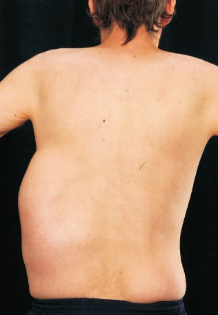
Posterior image demonstrating medial extension.
Figure 3.
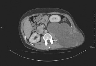
Computed tomography scan showing large pancreatic pseudocyst displacing left kidney anteriorly and extending to subcutaneous tissues.
Discussion
A pancreatic pseudocyst is defined as a localised collection of pancreatic juice enclosed by a wall of fibrous or granulation tissue arising as a result of pancreatitis or pancreatic trauma.1,2 Pancreatic pseudocysts are associated with 10–20% of cases of pancreatitis.1 They are more commonly found in men than women and solitary in 85% of cases.1 Alcohol-related pancreatitis is more commonly associated with pseudocyst formation accounting for between 59–65% of pseudocysts.1,2 What is unusual about this case is the location. Pancreatic pseudocysts are typically located intra-abdominally not within the subcutaneous tissues. Computed tomography (CT) is the preferred method of imaging for diagnosis,1,3 with serial ultrasonography for interval monitoring.3 There are mixed views on optimal management. Most surgeons offer conservative treatment initially as spontaneous resolution is observed in 50% of cases.1 However, 67% of patients with a pseudocyst measuring more than 6 cm ultimately required surgical intervention compared to 40% of patients with smaller pseudocysts.1 This is based on current imaging technologies with higher sensitivities detecting more pseudocysts.1
The management options for intra-abdominal pancreatic pseudocysts include surgical, percutaneous (for a mean of 48 days4) and endoscopic drainage. Pseudocysts persisting for longer than 6 weeks have been associated with a 3-fold increase in complication rates,3 which increases with time.4 The complications of pseudocysts include infection, haemorrhage, obstruction, rupture and erosion into neighbouring structures including major blood vessels.1–3 Pancreatic fistulae and ascites result when there is leakage or rupture of the pseudocyst into the abdominal cavity.2,3 In this case, the main complications are covered by rupture and erosion.
Our case represents repeated pleural effusions presumably resulting from rupture and fistulation of the pancreatic duct initially into the left pleural cavity. Such presentations account for less than 3% of cases and are even rarer to be an asymptomatic event1 as encountered here. In our case, two separate drainage procedures appear to have satisfactorily resolved the pancreaticopleural fistula but its subsequent presentation into the lumbar region has not been described previously.
Resection is the optimal treatment for persisting leakage from the tail of the pancreas such as in this case.3 We encountered a unique combination of normal alcoholic pancreatic pseudocyst with spontaneously resolving pancreaticopleural fistula but subsequent asymptomatic rupture posterior–superiorly into the abdominal cavity eroding through latissimus dorsi to form a sufficiently large subcutaneous collection to displace the left kidney anteriorly to almost touch the anterior abdominal wall. Surprisingly, this caused no inflammation or destruction of the skin overlying the lumbar region.
Conclusions
Pancreatic pseudocyst is a relatively common occurrence and a well-recognised complication of pancreatitis. This case not only highlights the variety of presentations of this condition, but also demonstrates the importance of knowing the anatomical structures involved in fluid accumulations. The possibility of creating pancreaticocutaneous fistulae from malpositioned pleural aspiration is raised by this case although it turned out not to be the case. Conservative management and percutaneous drain insertion was demonstrated to be of limited value in this case due to the posterior communication with the peritoneal cavity and on-going distal pancreatic drainage.
References
- 1.Lillemore KD, Yeo CJ. Management of complications of pancreatitis. Curr Problems Surg 1998; : 1–98. [PubMed] [Google Scholar]
- 2.Yeo CJ. Pancreatic pseudocysts, ascites and fistulas. Curr Opin Gen Surg 1994; 173–8. [PubMed] [Google Scholar]
- 3.Bradley EL. Complications of chronic pancreatitis. Surg Clin North Am 1989; : 481–97. [DOI] [PubMed] [Google Scholar]
- 4.Adams DB, Harvey TS, Anderson MC. Percutaneous catheter drainage of pancreatic pseudocysts. Am Surg 1991; : 29–33. [PubMed] [Google Scholar]
- 5.Burgess NA, Moore HE, Williams JO, Lewis MH. A review of pancreatico-pleural fistula in pancreatitis and its management. HPB Surg 1992; : 79–86. [DOI] [PMC free article] [PubMed] [Google Scholar]