Abstract
Appendicitis is one of the most common surgical emergencies, and is usually diagnosed clinically on point tenderness in the right iliac fossa. The diagnosis can be difficult if there is abnormal anatomy. This case looks into the presentation of appendicitis within an incisional hernia secondary to radical nephrectomy. Appendicitis may present within hernias, and there should be a low threshold for computed tomography assessment of hernias when there is clinical doubt about the symptoms associated with the hernia.
Keywords: Appendicitis, Incisional hernia, Radical nephrectomy
The appendix can be found in abnormal positions due to the variable location of the caecum due to different degrees of rotation during development or variations in caecal attachments or secondary to previous surgery. Whilst the appearance of the appendix in hernias is not uncommon, with the appendix been found in an inguinal hernia sac during elective hernia repair on 1% of occasions,1 the presence of a gangrenous appendix within an inguinal hernial sac is rare and even rarer to find in femoral, umbilical, obturator or incisional herniae.2,3 Here, we report the first case of appendicitis within an incisional hernia secondary to radical nephrectomy.
Case history
A 57-year-old Caucasian man presented to the emergency department with sudden onset of pain within his right-sided incisional hernia associated with nausea and loss of appetite. Incisional hernia was secondary to a right radical nephrectomy for renal cell cancer performed 9 months earlier. Examination was consistent with a reducible incisional hernia with an otherwise soft abdomen and no palpable masses. The patient was admitted under urology given previous renal cell cancer.
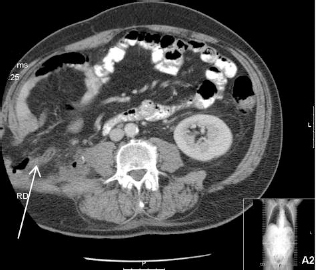
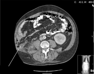
Despite analgesia and 24-h observation, the pain had not improved and the patient became pyrexial with a raised C-reactive protein level. A computed tomography (CT) scan was arranged which showed a large incisional hernia containing the caecum and within it an inflamed appendix (Figs 1 and 2).
Figure 1.

CT image of incisional hernia containing the caecum and hernia sac and surrounding stranding of fat.
Figure 2.

CT image showing incisional hernia and inflamed appendix.
The patient was taken to theatre where the abdomen was entered using the previous nephrectomy incision. The CT findings were confirmed of a healthy caecum present in the hernia with an inflamed appendix (Figs 1 and 2); appendicectomy was performed. After a thorough washout of the area, a prolene mesh was inserted for closure of the defect.
Postoperatively, the patient had an uneventful recovery with resolution of symptoms and pyrexia. Six-week followup revealed a well-healed operation site, with no recurrence of the hernia and no postoperative complications.
Discussion
In this case, the defect in the abdominal cavity was large enough to allow for the displacement of the caecum and appendix into the hernial sac. At the time of operation, the whole caecum was found to be in the hernial sac and it is likely that the caecum was highly mobile due to the mobilisation of the caecum during the previous nephrectomy.
The insidious onset of symptoms is not uncommon with appendicitis within a hernia due to the inflammation being isolated within the hernial sac preventing the spread of inflammation to the peritoneal cavity,4 thus making preoperative diagnosis difficult. However, because of our patient’s previous history of malignancy and non-classical symptoms, we were able to have a pre-operative diagnosis from CT.
There is some controversy as to the use of prolene meshes in the repair of infected abdominal wall hernias, with documented complications of re-infection and cystic swellings.5 It was not possible to close the abdominal wall defect without a mesh due to the large size of the defect. There is some literature supporting the use of biological meshes in these instances; however, a large study6 (1452 patients) revealed no statistical difference between the use of synthetic or biological meshes in the prevention of late onset deep mesh infection. It was, therefore, deemed appropriate to close the defect using a standard prolene mesh.
Conclusions
This case highlights a number of considerations. First, appendicitis may present in hernias and often it presents late as systemic manifestations are often not present earlier because of the sequestration of the infective process within the hernial sac. Second, a low threshold to request CT for assessment of hernias where clinical doubt about the significance of the symptoms associated with the hernia remains. Third, in cancer patients, one must still be aware of the possibility of acute surgical presentations.
Acknowledgement
The authors would like to thank Dr Dabbagh who performed and reviewed the CT images shown in this manuscript.
References
- 1.Thomas WE, Vowles KD, Williamson RC. Appendicitis in external herniae. Ann R Coll Surg Engl 1982; : 121–2. [PMC free article] [PubMed] [Google Scholar]
- 2.Doig CM. Appendicitis in umbilical hernial sac. BMJ 1970; : 113–4. [DOI] [PMC free article] [PubMed] [Google Scholar]
- 3.Arcampong EQ. Strangulated obturator hernia with acute gangrenous appendicitis. BMJ 1969; : 230. [DOI] [PMC free article] [PubMed] [Google Scholar]
- 4.Fukukura Y, Chang SD. Acute appendicitis within a femoral hernia: multidetector CT findings. Abdom Imaging 2005; : 620–2. [DOI] [PubMed] [Google Scholar]
- 5.Fawcett AN, Atherton WG, Balsitis M. A complication of the use of prolene mesh in the repair of abdominal wall hernias. Hernia 1998; : 173–4. [Google Scholar]
- 6.Selikoukos S, Tzovaras G, Liakou P, Mantzos F, Hatzitheofilou C. Late onset deep mesh infection after inguinal hernia repair. Hernia 2007; : 15–7. [DOI] [PubMed] [Google Scholar]


