



Abstract
For over a century, otolaryngologists have recognised the condition of aural exostoses, but their significance and aetiology remains obscure, although they tend to be associated with frequent swimming and cold water immersion of the auditory canal. The fact that this condition is usually bilateral is predictable since both ears are immersed in water. However, why do exostoses only grow in swimmers and why do they grow in the deep bony meatus at two or three constant sites? Furthermore, from an evolutionary point of view, what is or was the purpose and function of these rather incongruous protrusions?
In recent decades, paleoanthropological evidence has challenged ideas about early hominid evolution. In 1992 the senior author suggested that aural exostoses were evolved in early hominid Man for protection of the delicate tympanic membrane during swimming and diving by narrowing the ear canal in a similar fashion to other semiaquatic species. We now provide evidence for this theory and propose an aetiological explanation for the formation of exostoses.
Keywords: Aquatic theory of evolution, Exostoses, External ear canal
In recent decades, paleoanthropological evidence has modified our ideas about early hominid evolution, inspired by Sir Alister Hardy’s visionary article1 and Elaine Morgan’s ‘aquatic ape’ theory.2 In the context of this suggested littoral influence during evolution, the senior author (PHRE) explored various enigmatic anatomical features relating to the upper aerodigestive tract, unique in Man, that were not seen in other higher primates or other terrestrial mammals and suggested that these were much more logically explained by a historical period of aquatic adaptation during early hominid evolution.3 These findings were presented in a symposium on the aquatic ape theory organised by Sir David Attenborough at the annual meeting of the British Society for the Advancement of Science in September 1992.
One of these anatomical features concerned exostoses, which the senior author proposed were evolved for the purpose of protection of the delicate eardrum during swimming and diving, to narrow the ear canal in a similar fashion to other semiaquatic species. He suggested that if exostoses were identified in early hominid fossil skulls, this would provide good evidence to support the aquatic theory that early hominid Man spent significant periods of time in the water. In contrast to modern Man, early hominids would have been swimming and diving in search of food, hunting for their survival.3 We now believe that this evidence of Man’s aquatic past has been established in the form of fossil exostoses of the external ear canal.4
External ear canal bony swellings
‘Remnants of the past that don’t make sense in present terms – the useless, the odd, the peculiar, the incongruous – are the signs of history.’
Stephen Jay Gould (1941–2002)
Bony swellings of the external ear canal occur with varying frequency and are of two distinct types: osteomas and external auditory canal exotoses. Osteomas are almost always unilateral. They usually arise in the lateral part of the bony canal and have a pedunculated or lobular appearance. They are composed of dense ivory bone and are considered to be true pathological benign tumours of bone. There may be a hereditary factor in their aetiology since Roche has found an incidence of 27.9% in Australian Aborigines.5 Hrdlička has also described a particularly high incidence in Peruvian and American Indian populations.6
On the other hand, exostoses are broad-based hyperplastic bony swellings that are usually bilateral and are generally found on the posterior wall at the medial end of the bony external canal wall (70%)7,8 although they can be found situated on the anterior or superior aspect of the canal wall. While most cases are asymptomatic, they can predispose to otitis externa and conductive hearing loss with severe canal occlusion (Fig 1). The introduction of high resolution computed tomography (CT) has greatly enhanced the ability to demonstrate anatomical bony details (Fig 2).
Figure 1.

Normal external ear canal (left) and exostoses of ear canal (right)
Figure 2.
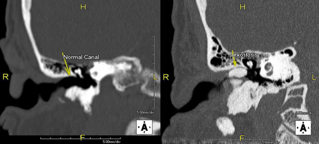
Computed tomography of temporal bones showing a normal ear canal (left) and exostoses (right)
Even recently, the aetiology of exostoses/osteomas has been considered an unresolved issue.9 In the past, it was thought to be predominantly genetic6 but it is now generally accepted that cold water exposure is the main aetiological factor in the formation of exostoses in the external ear canal.10–15 The exact way in which water stimulates bony hyperplasia at these specific sites remains uncertain but we believe that a review of the anatomical and developmental embryology may shed light on why water exposure results in hyperplasia in constant specific sites.4
Developmental embryology of the human external ear canal
The adult external auditory canal is divided into two portions: the cartilaginous portion in its outer third and the bony portion in its inner two thirds (Fig 3). It measures 24mm on average in length. Its development in utero is a complex process but could help explain the reasons behind the development of external ear canal exostoses.
Figure 3.

The human ear showing a normal ear canal
The canal develops from the dorsal part of the first branchial groove during the fourth and fifth week of gestation.16 From the skin surface, it extends inwards as a funnel shaped tube, which deepens, with proliferation of its ectoderm, forming an epithelial plug.17 The ectoderm at the deep end of the epithelial plug is briefly in contact with the endoderm lining of the pharyngeal pouch (derivatives: Eustachean tube and middle ear). A mesodermal membrane then grows between the pharyngeal pouch (middle ear) and branchial groove (external ear canal), and eventually separates the two spaces, forming the tympanic membrane.
From 18 weeks’ gestation, the latter stages of development include opening up and clearing of the deep canal, and widening of the whole canal. The bony part of the ear canal begins to ossify during the fourth and fifth month to form the tympanic ring, which supports the tympanic membrane. Three other centres of ossification just lateral to the eardrum (the tympanic, squamous and mastoid epiphyseal growth plates) grow outwards in a tubular fashion, forming the bony part of the external ear canal (Fig 4).17
Figure 4.

The three epiphyseal growth plates, just lateral to the tympanic ring
Formation of exostoses in the external ear canal
In the bony portion of the normal external auditory canal, there are no adnexal structures, and the distance between the epidermal (skin) surface and underlying bone is consequently small.18 Pilch believes that this close relationship could explain why external ear canal exostoses develop in this particular area.19 He suggests that cold water enters the deep canal and exerts a cooling effect that stimulates the nearby periosteum to produce new bone. In light of this, animal studies using guinea pigs have shown that repeated exposure to cold water in the canal causes fibrous proliferation of the subcutaneous tissue in the deep meatus and stimulates the periosteum to produce a layered formation of periosteal bone.20 Furthermore, Harrison also used guinea pigs in similar experiments and described comparable histological evidence of new bone formation in the deep meatus.21
The location of exostoses formation in the external auditory canal in one to three specific sites has led us to propose that these areas correspond exactly to the three centres of ossification (the tympanic, squamous and mastoid epiphyseal plates).4 With repeated cold water exposure to the ear canal, there is redness and hyperaemia at these ossification sites, and a resulting cellular inflammatory process subsequently initiates a repair response. Similar to fracture of a long bone, repair must result from the actions of osteoblasts and osteocytes, which work together to create new bone – exostoses in this case (Figs 5 and 6).
Figure 5.

Stimulation of the osteoblastic activity of the epiphyseal plates
Figure 6.

Formation of exostosis at the epiphyseal plate
Within external ear exostoses, the histology varies from compact bony tissue with some irregular Haversian canals to spongy centred protuberances.22 This inconsistent histology probably reflects the uncontrolled growth due to poorly coordinated osteoblast–osteoclast activity. Additionally, Sadé23 and Cruess24 also emphasised the importance of granulation tissue first stimulating the bone resorption. Cytokines (such as IL-1, IL-6 and TGF-β) are released by lymphocytes, macrophages and epithelial cells, and are involved in bone resorption.25 However, several growth factors are released to counteract this bone resorption and lead to net bone formation.26 Velasco-Vasquez et al deduce that it is possible that an imbalance between the local effects of the osteogenic growth factors and bone resorption cytokines during repeated episodes of hyperaemia acquired through exposure to cold water may eventually promote the formation of external auditory canal exostoses.27 Long-term exposure of the ear canal to water can therefore result in osteogenic activity.28,29
Evolutionary adaptations to the hearing mechanism in aquatic and semiaquatic mammals
We believe that the formation of external auditory canal exostoses in Homo species may represent an adaptive response to an aquatic environment. Land-based mammals, which depend on air mediated sound transmission at the tympanic membrane, usually have a widely patent external ear canal (Fig 1) for maximum reception of auditory stimuli. Marine mammals, on the other hand, have adapted to sound transmission in an aquatic medium and have narrow or vestigial canals. A wide meatus is no longer needed and may also be a dangerous liability predisposing to rupture of the tympanic membrane because of rapid increase in external pressure when swimming and diving. The evolutionary adaptations to the external ear canal depend on whether the species lives permanently in an aquatic or marine environment, or has a semiaquatic habitat.
Whales and other Cetacea have evolved very specialised mechanisms for sound transmission and reception. Since these great mammals left the land and took to the seas millions of years ago, their survival has been contingent on the evolution of ear structures that are highly specialised for underwater hearing. As their hearing no longer depended on air mediated transmission, the external ear canal became completely occluded with a keratin plug and they developed a highly sophisticated ‘sonar’ mechanism. They rely primarily on sound for sensory perception as well as for communication. Sound therefore has a profound influence on navigation, feeding, socialisation, breeding and other whale behaviour.
Extant whales (cetaceans) fall into two groups: toothed whales (odontocetes) and baleen whales (mysticetes). Toothed whales are fast swimmers and hunters, and include porpoises, dolphins, killer whales and sperm whales. Baleen whales are filter feeders that live on zooplanktons, and include the blue whale, the humpback whale, the gray whale and the bowhead whale.
Among the many differences between toothed and baleen whales is their specialisation for underwater hearing. The toothed whale’s biological sonar (echolocation) involves vocalisation from the nose and the reception of the echo in the ear. The whale recycles the air in its complex nasal passage and produces a high pitched sound that is beamed out to the environment through the melon, an oil body in its bulging forehead (Fig 7). The sound beamed from the melon into the environment is bounced back to the whale, providing information that helps the animal to accurately decipher its environment through highly specialised modification of the hearing mechanism.30
Figure 7.

The toothed whale forehead melon for echolocation
By contrast, the baleen whale can vocalise and hear very low pitched or infrasonic sound, which can travel great distances and scatter to large areas in water. With infrasonic sound, baleen whales can communicate with each other over geographic areas as large as an ocean basin.
Semiaquatic mammals, on the other hand, still need air transmitted sound reception on land but also require protection of the tympanic apparatus when underwater, for which they have evolved various mechanisms to temporarily narrow the ear canal. The hippopotamus can stay underwater for about 15 minutes, and closure of its auditory canal is achieved by contraction and angulating it backwards (Fig 8). The platypus achieves this with a skin fold and the desman by a glandular swelling.
Figure 8.

The hippopotamus closes its ear canal by angulation when diving.
The hooded seal shows an interesting model of evolutionary adaptation in that it has broad-based exostoses in the floor of the external ear canal lateral to the tympanic membrane. Along with cavernous tissue in the middle ear, these exostoses allow the seal to dive to depths of greater than 1,000m at pressures of 100atm without damage to the middle ear or tympanic membrane.31 Clearly, the presence of external auditory canal exostoses confers a selective advantage for the seal when in the marine environment. And so it might in hominids.
External ear exostoses in modern populations
From early years, the relationship between water exposure and external ear canal exostoses in modern populations has been described. Two of the most noted otolaryngologists, Belgraver32 and Van Gilse,10 found similar populations describing this cold water relationship. Belgraver, for example, found that members of a swimming club had an incidence of 42.8% while patients in his general clinic had an incidence recorded as low as 2.02%.32 In 1893 Field made the first association between external auditory canal exostoses and seawater.33 Shortly thereafter, in 1904, Körner similarly suggested salt water as an important causative factor when he first noted the greater incidence of external ear canal exostoses in coastal populations compared with inland populations in Germany.34
The natural history of external auditory canal exostoses has been observed by Wong et al in a population of 307 surfers.35 They found that the length of time that individuals spent surfing was proportional to the presence and severity of the exostoses. In surfers with external ear canal exostoses, 61.1% had surfed for ten years or less but in surfers who had surfed for more than ten years, 82.4% had severe exostoses. Mann further noticed in his study that the severity of external auditory canal exostoses was also influenced by the frequency of water exposure and he found an incidence of 64% in individuals swimming three times a week.36
Interestingly, it appears that the temperature of the water and wind chill interaction is also important.15 Specifically, Kennedy noted that external ear canal exostoses were more common in communities between the latitudes 308° and 458° both north and south of the equator, where the water temperature is below 19°C.12 More recently, Verhaegen has described that those who swim almost daily in water colder than 18–20°C presented with a greater incidence of external auditory canal exostoses.37 Kroon et al demonstrated in their surfer population that there was a higher prevalence of exostoses among the subjects who surfed more frequently in the colder waters.38
External ear exostoses in archaeological populations
Studies of exostoses in archaeological populations (Fig 9) may reflect the littoral activities of early hominids as they utilised the marine environment in search of food or for other activities. Homo fossils have typically been found in the vicinity of rivers, estuaries and lakes. Barr attributed the high incidence of exostoses (18%) in the preserved crania of native mound builders of central Tennesse to their frequent exposure to the river.39 Recently, Moore et al have observed that exostoses can develop in the presence of salt water as well as in inland populations who swim in river water.40 Verhaegen and Munro suggested that Pleistocene Homo populations similarly displayed littoral habits and that this is evidenced by the findings of pachyosteosclerosis.41
Figure 9.

Computed tomography of fossil skulls
Pérez et al have shown CT evidence of exostoses in the middle Pleistocene era (Fig 10).42 Further studies have provided direct evidence of consumption of aquatic foods during the early Pleistocene era.43,44
Figure 10.

Computed tomography of cranial remains from the Sima de los Huesos Middle Pleistocene site, Sierre de Atapuerca, Spain, showing stenosis of left auditory canal (a), left auditory meatus of the cranium (b) and right auditory meatus of the cranium (c). Scale bar = 1cm. Reprinted with permission from Elsevier.42
We propose that a period of marine exploitation for littoral habits or otherwise could result eventually in significant adaptive changes to the anatomy of the external ear canal. Exostoses have been found in several Neanderthal and some Homo erectus skulls (specifically, Homo erectus skull X from Zhoukoudian), suggesting they engaged in some type of aquatic activity.12
In 1990 a temporal bone from Lake Ndutu in the Olduvai Gorge was described with the diagnosis of external auditory exostosis.45 Additionally, Verhaegen presented convincing arguments that at least some Neanderthals were habitual divers.46 He noted bilateral exostoses in the skull of the middle aged Shanidar I and extensive exostoses in male skulls discovered at La Chapelle-aux-Saints. These fossil findings suggest that Neanderthals dived regularly and probably every day in cold rivers, along which they lived.
External ear exostoses in the context of hominid evolution
The ‘savannah’ theory
The evolution of early Man from our primate ancestral ape family (which included our closest relatives, the chimpanzee and the gorilla) became evident about 6–7 million years ago when there was a split from the common ancestral ape. The traditionally held ‘savannah’ theory of evolution proposed by Dart in 1924 postulates that gradual evolution of ancestral bipedal Man from the arboreal ape occurred because of climatic and behavioural changes. Loss of their forest habitat during the late Miocene drought period (7–8mybp) with corresponding extension of grassy plains or savannahs resulted in movement of prehominid apes from the tree habitat on to the plains for survival.47 This theory suggests that this branch of the family stood upright in order to see further over the grassy plains, and developed walking and running skills to escape from predators as well as to be able to stalk and catch game.
However, if this was so advantageous, why did other members of the primate family not adopt a similar upright gait and why is Homo the only terrestrial bipedal mammal? On the contrary, our cousins, the apes and the chimpanzees, have remained unchanged for over ten million years. In addition to our upright stance, Homo has further unique characteristics that are only seen in aquatic and semiaquatic mammals.
The ‘aquatic ape’ theory
In April 1960 an article appeared in the New Scientist by the distinguished marine biologist Sir Alister Hardy suggesting an alternative theory of evolution that seemed to explain many of the dramatic changes that occurred during Man’s development from the arboreal ape.1 He proposed that as a result of the changing environment during the Miocene drought with reduction of the forest habitat and vegetation, certain anthropod apes, ancestral to Man, were driven by competition from life in the trees to assume a new habitat on the shores of the sea, estuaries and inland waters, where they became adept at hunting for abundant supplies of food, shellfish and sea urchins in the shallow coastal waters.
The coincidental inundation of the Afar region in East Africa and formation of the Great Rift Valley, where most of these early hominid fossils have been found, would have provided an ideal environment for these changes. Initially wading on all fours, these aquatic apes gradually assumed a more upright posture, the buoyancy of the water enabling them to support their spine, allowing rotation of the pelvis into a more erect and streamlined position. They were able to extend their territory into deeper water, developing the ability to swim and dive. This aquatic ape theory of evolution was further explored in the writings of Elaine Morgan.2
Owing to the additional buoyancy, stability on two legs would have been much easier to acquire in the water environment than on the savannah, as exemplified by the only other primate to have ventured into the sea. The proboscis monkey lives in the mangrove swamps in the coastal waters of Borneo and although it retains a mainly terrestrial quadruped gait similar to other apes living in trees, it can also adopt a bipedal mode when wading in the shallows in order to keep its head above water. In certain instances, it has been noted to use this mode of movement for walking on dry land.
The compelling argument proposed by Hardy that ancestral Man spent a period of aquatic adaptation is not unique in evolutionary history. On the contrary, this process is well recognised, and several species of birds, reptiles and mammals are known to have abandoned their terrestrial existence to become adapted and modified to an aquatic life.
An early example is a member of the dinosaur family (ichthyosaur), which took to the water, evolving flippers instead of legs before becoming extinct. Among mammals, members of the cetacean family (whales, dolphins, porpoises) successfully adapted to an aquatic existence, as did some of the hoofed mammals related to the elephant (sea cows). Others include aquatic birds (penguins), aquatic carnivores (sea lions, seals, otters), aquatic rodents (beavers, water voles), aquatic reptiles (crocodiles, sea snakes) and aquatic insectivores (water shrews, desmans). There is therefore no reason to suppose that a similar transformation did not occur among the primate order.3,4
Over recent years, new research and evidence has demonstrated convincingly that at some stage during the last few million years, our human ancestors were exposed to a period of semiaquatic evolution that led to the acquisition of unique and primordial human characteristics not seen in chimpanzees, gorillas or other primates or terrestrial mammals. However, many of these aquatic adaptations can be seen in different combinations in waterside and aquatic mammals. These include our streamlined shape and upright stance, fur loss and tendency to obesity.
Other unique human characteristics such as enlarged brains, tool use and extreme dexterity as well as our ability to speak and voluntary breath control are unique and not seen in any other terrestrial mammal. These aquatic adaptations were important functions that allowed the genus Homo to become such a dominant species, permitting their evolution into a large brained, bipedal hominid that colonised the Old World as far as the island of Flores in Indonesia, Tafelbaai in South Africa, and Boxgrove and Pakefield in England, trekking along coasts and rivers. A unique thermoregulatory system, sweat and tear production, aquisition of subcutaneous fat, large paranasal sinuses and aural exostoses are among other characteristics seen in Man but not in other terrestrial mammals.4
Perhaps the most compelling argument supporting the littoral theory is that early hominid Man could not have evolved such a large brain in relation to body mass without two essential lipoproteins, arachidonic acid and docosahexaeionic acid, which are found predominantly in fish and seafood.48 Without these brain building proteins, living on the savannah, early Man could not have developed a big brain or evolved into such a dominant species. This evidence for the waterside habitat seems to have finally lain to rest the discredited savannah theory.
Medical considerations of the ‘aquatic ape’ theory
The consequences of this period of aquatic adaptation in early hominid evolution have not only resulted in unique anatomical and physiological characteristics not seen in other higher primates or terrestrial mammals but have also led to various pathological conditions that are only seen in Man, directly as a result of our unique bipedal gait. Some of the changes have been greatly advantageous, leading to enlargement of the brain and the acquisition of speech. Others might be associated with ‘unexpected’ or even pathological conditions, not seen in other terrestrial mammals, as diverse as neonatal swimming behaviour, obesity, hypertension, alopoecia and acne, hyperhidrosis, lumbar osteoarthritis, varices, inguinal hernias and vaginal prolapses. Other uniquely human conditions such as hyperventilation, sudden infant death syndrome, Cheyne–Stokes breathing and asthma, vasomotor rhinopathy and sinusitis as well as possibly thyroid disorders and cystic fibrosis may also be directly related to our different evolutionary pathway.
Conclusions
‘Nothing of him that doth fade, but doth suffer a sea-change into something rich and strange.’
The Tempest, William Shakespeare (1564–1616)
Ear exostoses may appear a peculiar characteristic unique only to Homo and none of the other primates. However, the embryological development of the external ear canal offers a logical explanation for the sites of development of exostoses and their pathogenesis.
This ability to develop bony growths may appear redundant now but it is highly likely that the presence of these growths constituted a selective survival advantage for early hominids during evolution. Furthermore, a comparative study of the external ear canals of other semiaquatic mammals reveals similar mechanisms or structures to partially or temporarily close off the ear canal to protect the tympanic membrane. Their development in Man as a physiological modification rather than a pathological entity may have provided an evolutionary advantage for early hominids in a marine or semiaquatic environment. It could therefore be considered crucial evidence of Man’s aquatic past and should provide enough weight to stimulate further discussion on the validity of the aquatic theory of evolution.
References
- 1.Hardy A. Was man more aquatic in the past? New Scientist 1960; : 642–645. [Google Scholar]
- 2.Morgan E. The Aquatic Ape Hypothesis. London: Souvenir Press: 1997. [Google Scholar]
- 3.Rhys Evans PH. The paranasal sinuses and other enigmas: an aquatic evolutionary theory. J Laryngol Otol 1992; : 214–225.> [DOI] [PubMed] [Google Scholar]
- 4.Rhys-Evans PH, Cameron M. Surfer’s ear (aural exostoses) provides hard evidence of Man’s aquatic past. Hum Evol 2014; : 75–90. [Google Scholar]
- 5.Roche AF. Aural exostoses in Australian Aboriginal skulls. Ann Otol Rhinol Laryngol 1964;: 82–91. [DOI] [PubMed] [Google Scholar]
- 6.Hrdlička A. Ear exostoses. Smithsonian Miscellaneous Collections 1935; : 1–100. [Google Scholar]
- 7.Gregg JB, Bass WM. Exostoses in the external auditory canals. Ann Otol Rhinol Laryngol 1970; : 834–839. [DOI] [PubMed] [Google Scholar]
- 8.Gregg JB, Gregg PS. Dry Bones: Dakota Territory Reflected. Vermillion, SD: University of South Dakota Press; 1988. [Google Scholar]
- 9.Aufderheide AC, Rodríguez-Martín C. The Cambridge Encyclopedia of Human Paleopathology. Cambridge: Cambridge University Press; 1998. pp254–256. [Google Scholar]
- 10.Van Gilse PH. Des observations ultérieures sur la genèse des exostoses du conduit externe par l’irritation d’eau froide. Acta Otolaryngol 1938; : 343–352. [Google Scholar]
- 11.Ascenzi A, Balistreri P. Aural exostoses in a Roman skull excavated at the ‘Baths of the Swimmer’ in the ancient town of Ostia. J Hum Evol 1975; : 579–584. [Google Scholar]
- 12.Kennedy GE. The relationship between auditory exostoses and cold water: a latitudinal analysis. Am J Phys Anthropol 1986; : 401–415. [DOI] [PubMed] [Google Scholar]
- 13.Deleyiannis FW, Cockcroft BD, Pinczower EF. Exostoses of the external auditory canal in Oregon surfers. Am J Otolaryngol 1996; : 303–307. [DOI] [PubMed] [Google Scholar]
- 14.Hutchinson DL, Denise CB, Daniel HJ, Kalmus GW. A reevaluation of the cold water etiology of external auditory exostoses. Am J Phys Anthropol 1997; : 417–422. [DOI] [PubMed] [Google Scholar]
- 15.Okumura MM, Boyadjian CH, Eggers S. Auditory exostoses as an aquatic activity marker: a comparison of coastal and inland skeletal remains from tropical and subtropical regions of Brazil. Am J Phys Anthropol 2007; : 558–567. [DOI] [PubMed] [Google Scholar]
- 16.Ballachanda B. The Human Ear Canal. San Diego: Singular Publishing Group; 1994. [Google Scholar]
- 17.Kerr AG. Scott-Brown’s Otolaryngology. 6th edn Boca Raton, FL: CRC Press; 1997. [Google Scholar]
- 18.Michaels L. The Ear : Sternberg SS. Histology for Pathologists. 2nd edn Philadelphia: Lippincott-Raven; 1997. pp337–366. [Google Scholar]
- 19.Pilch BZ. Head and Neck Surgical Pathology. Philadelphia: Lippincott Williams & Wilkins; 2000. [Google Scholar]
- 20.Fowler EP, Osmun PM. New bone growth due to cold water in the ears. Arch Otolaryngol 1942; : 455–466. [Google Scholar]
- 21.Harrison DF. The relationship of osteomata of the external auditory meatus to swimming. Ann R Coll Surg Engl 1962; : 187–201. [PMC free article] [PubMed] [Google Scholar]
- 22.van den Broek AJ. On exostoses in the human skull. Acta Neerl Morph 1943; : 95–118. [Google Scholar]
- 23.Sadé J. Secretory Otitis Media and Its Sequelae. New York: Churchill Livingstone; 1979. [Google Scholar]
- 24.Cruess RL. Physiology of Bone Formation and Resorption. : Cruess RL. The Musculoskeletal System. New York: Churchill Livingstone; 1982. pp219–252. [Google Scholar]
- 25.Graves DT. The potential role of chemokines and inflammatory cytokines in periodontal disease progression. Clin Infect Dis 1999; : 482–490. [DOI] [PubMed] [Google Scholar]
- 26.Mohammed S, Pack AR, Kardos TB. The effect of transforming growth factor beta one (TGF-beta 1) on wound healing, with or without barrier membranes, in a Class II furcation defect in sheep. J Periodontal Res 1998; : 335–344. [DOI] [PubMed] [Google Scholar]
- 27.Velasco-Vazquez J, Betancor-Rodriguez A, Arnay-De-La-Rosa M, Gonzalez-Reimers E. Auricular exostoses in the prehistoric population of Gran Canaria. Am J Phys Anthropol 2000; : 49–55. [DOI] [PubMed] [Google Scholar]
- 28.Lerner UH. Regulation of bone metabolism by the kallicrein–kinin system, the coagulation cascade, and the acute-phase reactants. Oral Surg Oral Med Oral Pathol 1994; : 481–493. [DOI] [PubMed] [Google Scholar]
- 29.Mundy GR, Boyce B, Hughes D et al. The effects of cytokines and growth factors on osteoblastic cells. Bone 1995; (2 Suppl): 71S–75S. [DOI] [PubMed] [Google Scholar]
- 30.Luo ZX, Ruf I, Martin T. The petrosal and inner ear of the Late Jurassic cladotherian mammal Dryolestes leiriensis and implications for ear evolution in therian mammals. Zool J Linn Soc 2012; : 433–463. [Google Scholar]
- 31.Stenfors LE, Sadé J, Hellström S, Anniko M. How can the hooded seal dive to a depth of 1000 m without rupturing its tympanic membrane? Acta Otolaryngol 2001; : 689–695. [DOI] [PubMed] [Google Scholar]
- 32.Belgraver P. Over exostoses van de uitwendige gehoorgang [dissertation]. Leiden, Netherlands: Leiden University; 1938. [Google Scholar]
- 33.Field GP. A Manual of Diseases of the Ear. London: Baillière, Tindall & Cox; 1893. [Google Scholar]
- 34.Körner O. Über den angeblich zyklischen Verlauf der akuten Paukenhöhlenentzündung. Z Ohrenheilkd 1904; : 369–372. [Google Scholar]
- 35.Wong BJ, Cervantes W, Doyle KJ et al. Prevalence of external auditory canal exostoses in surfers. Arch Otolaryngol Head Neck Surg 1999; : 969–972. [DOI] [PubMed] [Google Scholar]
- 36.Mann GE. The torus auditivus: a reappraisal. Palaeopathol Newsl 1986; : 5–9. [PubMed] [Google Scholar]
- 37.Verhaegen M. Aquatic versus savanna: comparative and paleo-environmental evidence. Nutr Health 1993; : 165–191. [DOI] [PubMed] [Google Scholar]
- 38.Kroon DF, Lawson ML, Derkay CS et al. Surfer’s ear: external auditory exostoses are more prevalent in cold water surfers. Otolaryngol Head Neck Surg 2002; : 499–504. [DOI] [PubMed] [Google Scholar]
- 39.Barr T. Manual of Diseases of the Ear: Including Those of the Nose and Throat in Relation to the Ear, for the Use of Students and Practitioners of Medicine. Glasgow: James MacLehose; 1901. [Google Scholar]
- 40.Moore RD, Schuman TA, Scott TA et al. Exostoses of the external auditory canal in white-water kayakers. Laryngoscope 2010; : 582–590. [DOI] [PubMed] [Google Scholar]
- 41.Verhaegen M, Munro S. Pachyosteosclerosis suggests archaic Homo frequently collected sessile littoral foods. Homo 2011; : 237–247. [DOI] [PubMed] [Google Scholar]
- 42.Pérez PJ, Gracía A, Martínez I, Arsuaga JL. Paleopathological evidence of the cranial remains from the Sima de Los Huesos Middle Pleistocene site (Sierra de Atapuerca, Spain). Description and preliminary inferences. J Hum Evol 1997; : 409–421. [DOI] [PubMed] [Google Scholar]
- 43.Joordens JC, Wesselingh FP, de Vos J et al. Relevance of aquatic environments for hominins: a case study from Trinil (Java, Indonesia). J Hum Evol 2009; : 656–671. [DOI] [PubMed] [Google Scholar]
- 44.Braun DR, Harris JW, Levin NE et al. Early hominin diet included diverse terrestrial and aquatic animals 1.95 Ma in East Turkana, Kenya. Proc Natl Acad Sci USA 2010; : 10,002–10,007. [DOI] [PMC free article] [PubMed] [Google Scholar]
- 45.Rightmire GP. The Evolution of Homo Erectus. Cambridge: Cambridge University Press; 1990. [Google Scholar]
- 46.Verhaegen M. Aquatic Features in Fossil Hominids. : Roede M, Wind J, Patrick J, Reynolds V. Aquatic Ape: Fact or Fiction. London: Souvenir Press; 1991. p83. [Google Scholar]
- 47.Dart RA. Australopithecus africanus: the man-ape of South Africa. Nature 1925; : 195–199. [Google Scholar]
- 48.Crawford MA, Broadhurst CL, Guest M et al. A quantum theory for the irreplaceable role of docosahexaenoic acid in neural cell signalling throughout evolution. Prostaglandins Leukot Essent Fatty Acids 2013; : 5–13. [DOI] [PubMed] [Google Scholar]