Abstract
Intralesional corticosteroid (IL-CS) injections have been used to treat a variety of dermatological and nondermatological diseases. Although an important therapeutic tool in dermatology, a number of local side effects, including skin atrophy, have been reported following IL-CS injections. We recently noticed that a subset of patients with steroid-induced atrophy presented with ivory-colored areas under trichoscopy. We performed a retrospective analysis of trichoscopic images and medical records from patients presenting ivory-colored areas associated with atrophic scalp lesions. In this paper, we associate this feature with the presence of steroid deposits in the dermis and report additional trichoscopic features of steroid-induced atrophy on the scalp, such as prominent blood vessels and visualization of hair bulbs.
Keywords: Alopecia, Corticosteroid, Dermoscopy, Intralesional injection, Skin atrophy, Steroids, Trichoscopy
Introduction
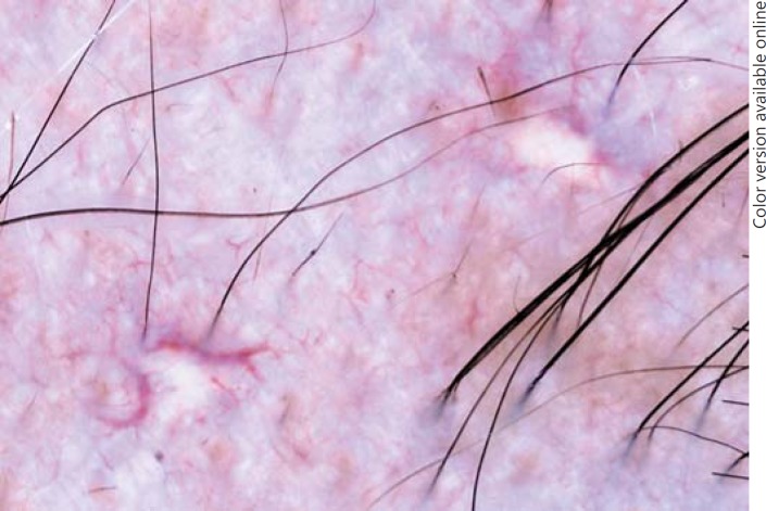
Intralesional corticosteroid (IL-CS) injections have been used to treat a variety of dermatological and nondermatological diseases [1]. IL-CS injections are, for example, considered as a first-line therapy in patchy alopecia areata [2]. Although an important therapeutic tool in dermatology, a number of local side effects, including skin atrophy, have been reported following IL-CS injections [1]. We recently noticed that a subset of patients with steroid-induced atrophy presented with ivory-colored areas under trichoscopy (Fig. 1). In this paper, we associate ivory-colored areas with the presence of steroid deposits in the dermis and report additional trichoscopic features of steroid-induced atrophy. To the best of our knowledge, there are no other studies reporting such data.
Fig. 1.
Ivory-colored areas seen under trichoscopy in an alopecia areata patient presenting corticosteroid-induced skin atrophy. Original magnification: ×20.
Materials and Methods
We performed a retrospective analysis of trichoscopic images and medical records from patients presenting ivory-colored areas associated with atrophic scalp lesions. All patients had been seen for different types of hair loss, including both scarring and nonscarring alopecias, at 4 referral centers in Brazil. Diagnosis was established clinically by dermatologists with experience in hair disorders and, in doubtful cases, was confirmed through pathology. Clinical information is summarized in Table 1. Trichoscopy and photographic documentation were performed using either FotoFinder Dermoscope®, FotoFinder Handyscope® (Teachscreen Software, Bad Birnbach, Germany), or DermLite® Foto (3Gen, San Juan Capistrano, CA, USA) attached to a Nikon® J1 camera.
Table 1.
Clinical information from patients with scalp lesions of steroid-induced atrophy presenting ivory-colored areas
| Patient | Gender | Age, years | Lesions, n | Condition | Corticosteroid and concentration, mg/mL |
|---|---|---|---|---|---|
| 1 | f | 30 | 7 | AA | TH 10 |
| 2 | f | 26 | 1 | HS | BD 5 + BDS 2 |
| 3 | f | 68 | 2 | DLE | TH 10 |
| 4 | f | 41 | 2 | AA | TA 10 |
| 5 | m | 28 | 2 | AA | TH 20 |
| 6 | f | 74 | 1 | LPP | TH 05 |
| 7 | f | 68 | 9 | FFA | unknown |
| 8 | f | 59 | 1 | AA | unknown |
AA, alopecia areata; HS, hypertrophic scar; DLE, discoid lupus erythematosus; LPP, lichen planopilaris; FFA, frontal fibrosing alopecia; TH, triamcinolone hexacetonide; BD, betamethasone dipropionate; BDS, betamethasone disodium phosphate; TA, triamcinolone acetonide.
Results
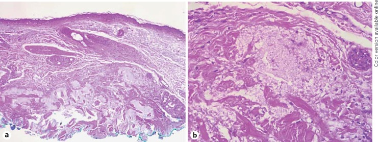
Twenty-five lesions from 8 patients were retrieved. All patients had been submitted to IL-CS injections as part of their treatment. In 3 patients, biopsies from ivory-colored areas were available and revealed circumscribed, fine pale foamy material in the dermis, suggestive of steroid deposits (Fig. 2) [3].
Fig. 2.
Biopsies from ivory-colored areas revealed circumscribed, fine pale foamy material present in the dermis (a), which can be better visualized in b. HE staining. Original magnification: a ×100 and b ×200.
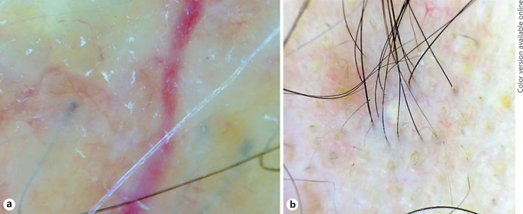
Another remarkable trichoscopic feature was the presence of prominent blood vessels in 23 (92%) lesions (Fig. 3), which were classified as thin (16 lesions; 64%) and thick arborizing (15 lesions; 60%) and which in 3 lesions (12%) were so numerous as to form a vascular network. Visualization of hair bulbs through the atrophic skin was evident in 5 lesions (20%) (Fig. 4a). Interestingly, in 5 (out of 12) lesions of patients with alopecia areata, regrowing hairs were organized as a collar that surrounded the steroid deposits (Fig. 4b).
Fig. 3.
Vascular changes in steroid-induced atrophy. Trichoscopy showed thin arborizing vessels (a), thick arborizing vessels (b), and blood vessels forming a vascular network (c). For classification purposes, vessels thinner than the average hair were considered as “thin” and vessels thicker than the average hair as “thick.” Original magnification: ×20.
Fig. 4.
Additional trichoscopic features included visualization of hair bulbs (a) and collars of regrowing hairs around the ivory-colored areas (b). Original magnification: ×20.
During our research, another 8 patients with 13 similar lesions in areas other than the scalp were also identified. Such patients had also been submitted to IL-CS injections for different reasons (clinical information can be found in Table 2).
Table 2.
Clinical information from patients with body lesions of steroid-induced atrophy presenting ivory-colored areas
| Patient | Gender | Age, years | Lesions, n | Body location | Condition | Corticosteroid and concentration, mg/mL |
|---|---|---|---|---|---|---|
| 1 | m | 31 | 2 | abdomen | HS | TH 20 |
| 2 | f | 32 | 2 | face/chest | acne cysts | TH 20 |
| 3 | f | 70 | 2 | arms | granuloma annulare | TH 20 |
| 4 | f | 34 | 1 | inframammary | HS | TH 10 |
| 5 | f | 41 | 1 | abdomen | HS | TH 20 |
| 6 | f | 35 | 2 | abdomen/arm | HS | TH 20 |
| 7 | f | 32 | 2 | breasts | HS | TH 20 |
| 8 | m | 32 | 1 | chest | HS | TH 20 |
HS, hypertrophic scarring; TH, triamcinolone hexacetonide.
Discussion
The presence of steroid deposits has already been reported after triamcinolone injections in different body areas, such as the vocal cords and eyes [4, 5]. In our case series, the reason for such deposits can only be speculated on. However, it is remarkable that in 25 lesions (out of 28 in which the steroid used is known) the corticosteroid injected was triamcinolone hexacetonide, which is known to be much less soluble than other steroids, including triamcinolone acetonide [6, 7]. Some authors have even considered triamcinolone hexacetonide to be unsuitable for intralesional injection due to its inordinately long half-life [1]. Furthermore, in the 25 lesions in which triamcinolone hexacetonide was used, concentrations were equal to or higher than 10 mg/mL in 24 lesions (96%), which could serve as an additional risk factor for steroid deposition and tissue atrophy. Another interesting point to consider is that triamcinolone hexacetonide should not be mixed with diluents or local anesthetics containing preservatives, such as parabens or phenols, since they may cause precipitation of the steroid [1]. Even though this information was not available, we cannot exclude that the type of diluent used may have played a role in the formation of cutaneous deposits in some cases.
Steroid-induced atrophy may be self-limited [8]. It is unknown whether the presence of steroid deposits in the dermis may hinder resolution of cutaneous atrophy. According to personal observations (B.D.-E. and L.S.A.), surgical removal of the deposits leads to partial regression of dermal atrophy, but prospective studies are necessary to confirm this information.
One limitation of the study was the limited access to patient data, since most of the patients were originally treated by other physicians. Information lacking includes the total number of injections received by each patient and the duration of atrophy.
In conclusion, we describe ivory-colored areas as a novel trichoscopic feature representing steroid deposits in the dermis and suggest that their presence may indicate the etiology of skin atrophy in cases in which a previous history of IL-CS injections is not clear. We further report additional features of steroid-induced atrophy on the scalp, such as prominent vessels and visualization of hair bulbs. Finally, we suggest the use of triamcinolone acetonide rather than of its less soluble salt hexacetonide for intralesional injections and at lower concentrations, typically at 2.5 and 5 mg/mL for scalp lesions.
Statement of Ethics
All patients have given their consent for their details to be described in this article.
Disclosure Statement
The authors have no conflicts of interest to disclose.
Funding Sources
There were no funding sources for this work.
References
- 1.Firooz A, Tehranchi-Nia Z, Ahmed AR. Benefits and risks of intralesional corticosteroid injection in the treatment of dermatological diseases. Clin Exp Dermatol. 1995;20:363–370. doi: 10.1111/j.1365-2230.1995.tb01351.x. [DOI] [PubMed] [Google Scholar]
- 2.Alkhalifah A, Alsantali A, Wang E, McElwee KJ, Shapiro J. Alopecia areata update: part II. Treatment. J Am Acad Dermatol. 2010;62:191–202. doi: 10.1016/j.jaad.2009.10.031. [DOI] [PubMed] [Google Scholar]
- 3.Kaur S, Thami GP, Mohan H. Intralesional steroid induced histological changes in the skin. Indian J Dermatol Venereol Leprol. 2003;69:232–234. [PubMed] [Google Scholar]
- 4.Wang CT, Lai MS, Hsiao TY. Comprehensive outcome researches of intralesional steroid injection on benign vocal fold lesions. J Voice. 2015;29:578–587. doi: 10.1016/j.jvoice.2014.11.002. [DOI] [PubMed] [Google Scholar]
- 5.Okka M, Bozkurt B, Kerimoglu H, et al. Control of steroid-induced glaucoma with surgical excision of sub-Tenon triamcinolone acetonide deposits: a clinical and biochemical approach. Can J Ophthalmol. 2010;45:621–626. doi: 10.3129/i10-055. [DOI] [PubMed] [Google Scholar]
- 6.Garg N, Perry L, Deodhar A. Intra-articular and soft tissue injections, a systematic review of relative efficacy of various corticosteroids. Clin Rheumatol. 2014;33:1695–1706. doi: 10.1007/s10067-014-2572-8. [DOI] [PubMed] [Google Scholar]
- 7.Porter D, Burton JL. A comparison of intra-lesional triamcinolone hexacetonide and triamcinolone acetonide in alopecia areata. Br J Dermatol. 1971;85:272–273. doi: 10.1111/j.1365-2133.1971.tb07230.x. [DOI] [PubMed] [Google Scholar]
- 8.Chang KH, Rojhirunsakool S, Goldberg LJ. Treatment of severe alopecia areata with intralesional steroid injections. J Drugs Dermatol. 2009;8:909–912. [PubMed] [Google Scholar]


