Abstract
Alterations in the social and cognitive domain are considered important indicators for increased disability in many stress-related disorders. Similar impairments have been observed in rodents chronically exposed to stress, mimicking potential endophenotypes of stress-related psychopathologies such as major depression disorder (MDD), anxiety, conduct disorder, and posttraumatic stress disorder (PTSD). Data from numerous studies suggest that deficient plasticity mechanisms in hippocampus (HC) and prefrontal cortex (PFC) might underlie these social and cognitive deficits. Specifically, stress-induced deficiencies in neural plasticity have been associated with a hypodopaminergic state and reduced neural plasticity persistence. Here we assessed the effects of unpredictable chronic mild stress (UCMS) on exploratory, social and cognitive behavior of females of two inbred mouse strains (C57BL/6J and DBA/2J) that differ in their dopaminergic profile. Exposure to chronic stress resulted in impaired circadian rhythmicity, sociability and social cognition in both inbred strains, but differentially affected activity patterns and contextual discrimination performance. These stress-induced behavioral impairments were accompanied by reduced expression levels of brain derived neurotrophic factor (BDNF) in the prefrontal cortex. The strain-specific cognitive impairment was coexistent with enhanced plasma corticosterone levels and reduced expression of genes related to dopamine signaling in hippocampus. These results underline the importance of assessing different strains with multiple test batteries to elucidate the neural and genetic basis of social and cognitive impairments related to chronic stress.
Introduction
Chronic stressful life events are a major risk factor in the development and maintenance of many psychopathologies [1]. Stress-exposure often perturbs one`s physiological and psychological functioning leading to behavioral dysfunctions in the affective, social and cognitive domain [2,3]. Among other well-defined symptoms, social and cognitive disturbances are considered to be a major contributor to the burden of disease in patients suffering from stress-related disorders such as major depression disorder (MDD), anxiety, Cushing`s syndrome, and posttraumatic stress disorder (PTSD) [4]. Impairments in attention [5], processing speed [6], executive functioning [7], learning and memory [8,9] have been widely reported in these patients. However, the neuropathological mechanisms underlying these behavioural dysfunctions are still not fully understood. Different animal models have been established and used to investigate what mediates these stress-induced anomalies [10–12].
In accordance with human studies, animals subjected to chronic stress mimic many cognitive impairments in different learning and memory protocols such as Morris water maze [13], passive avoidance [14] and radial arm maze [15]. Potentially underlying these behavioural anomalies, chronic stress has been shown to have detrimental effects on HC and PFC structure and function [16–19]. In these particular brain structures, expression of genes involved in neuronal plasticity [20] and dopamine-dependent memory persistence have been shown to be dysregulated in patients and animal models of PTSD, Cushing`s syndrome and other stress-related psychopathologies [17,21–25]. Brain derived neurotrophic factor (BDNF) is involved in the development, growth and differentiation of novel neurons as well as in the survival of existing neurons [26]. Long-term exposure to stress leads to decreased BDNF expression in HC [27] and PFC [28], which has been proposed to be related to cognitive impairments observed in chronically stressed mice [29]. Accordingly, post-mortem studies revealed a significant reduction of BDNF in PFC and HC of patients suffering from stress-related mood disorders [30].
Among other neural populations, ventral tegmental area (VTA) dopamine (DA) neurons have been shown to regulate the expression of proteins, such as BDNF, necessary for lasting neuronal plasticity in HC [31,32] and PFC [33]. DA release in NAc, PFC and HC is considered to be essential in motivational vigour [34], social behaviour [35] and long-term memory persistence [36,37]. In humans for example, genetic predisposition for increased DA availability is a putative resilience factor for negative emotionality and depression [38], whereas microstructural abnormalities in midbrain and subcortical regions (including VTA) have been observed in depressed patients [39]. In animal models, DAergic neurotransmission has been shown to be directly involved in mediating stress responses [40,41], depression-like behaviour [42], as well as in determining the balance between susceptibility versus resilience to stress-induced behavioural abnormalities [43]. However, the extent to which DAergic neurotransmission is involved in stress-induced cognitive decline remains unclear.
In this study we investigated the effects of unpredictable chronic mild stress (UCMS) on a battery of exploration, social and cognitive tests in two inbred mouse strains. UCMS is a well-established rodent model for stress-related psychopathologies, displaying changes at the molecular, anatomical, and behavioral level comparable to clinical observations [10,44–49]. The inbred strains C57BL/6J and DBA/2J are frequently used and have been shown to differ in their reaction to stressful manipulations [50], social and cognitive behavior [51,52], HC and PFC synaptic plasticity [53,54], as well as in their DAergic profile [55]. The goal of these experiments was to investigate the impact of genetic background on stress-induced social and cognitive impairments and how this correlates with the expression levels in the HC and PFC of genes implicated in dopaminergic neurotransmission and neuronal plasticity.
Materials and methods
Animals
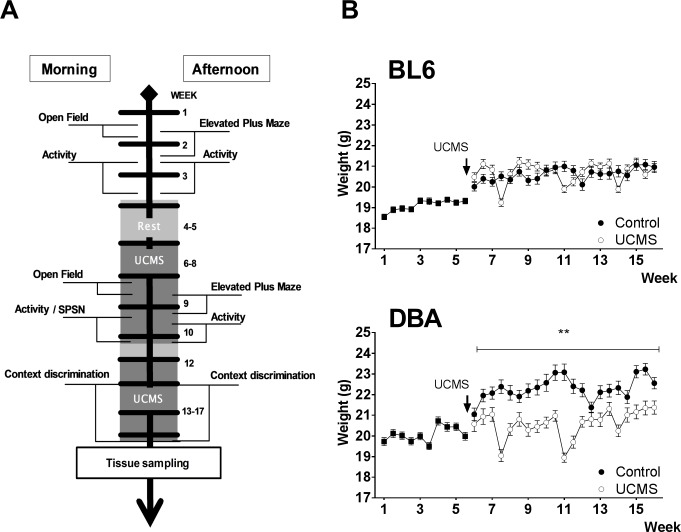
Subjects were 30 female mice from the DBA/2J inbred strain (DBA) and 30 female mice from the C57BL/6J inbred strain (BL6) (Elevage Janvier, Le Genest Saint Isle, France). Upon arrival mice were 8 weeks old. Mice were group housed (7/8 per cage) in standard animal cages throughout the entire experiment and kept under temperature and humidity controlled conditions (12h/12h light-dark cycle with lights on at 8:00 a.m., 22°C). Food and water were available ad libitum. Following baseline testing (week 1–3), mice were randomly assigned to one of two groups: controls and exposure to UCMS. Animals in the control group were provided cage enrichment (carboard rolls, nesting material), whereas UCMS-exposed mice were not. Body weight was closely monitored (Fig 1B). All behavioral testing was performed during the light phase of the activity cycle, with the exception of the 23h-activity test. For the first series of behavioral tests (open field, activity, elevated plus maze and sociability/preference for social novelty test), 60 animals (15/group) were tested. In contextual discrimination, 48 animals (12 /group) were tested. One mouse died during discrimination training and was excluded from analysis.
Fig 1. Schematic representation of experimental design.
(A) and effects of UCMS on body weight (B), measured over the complete behavioral testing period. Data are presented as mean +/- SEM. ** p < 0.01.
All experimental procedures comply with international guidelines on the ethical use of animals. All experiments have been reviewed and approved by the animal ethical committee of the University of Leuven, Belgium (project number: 199/2015). All efforts were made to minimize animal suffering where possible.
Design of the experiment
Mice from each strain were randomly distributed across two groups (CTRL and UCMS). Behavioral tests were conducted either in the morning (8 a.m.- 1 p.m.) or in the afternoon (2 p.m.-7 p.m.) to limit between-group circadian variation (Fig 1A). Upon arrival, mice were given a 7-day adaptation period to habituate to housing and handling conditions. To set a behavioral baseline, mice were tested over a three-week period in the following sequence: open field test, elevated plus-maze, 23h-activity test. Following a two-week rest period (week 4–5), the unpredictable chronic mild stress procedure started for four weeks and was continued during behavioral testing (Table 1). Exploratory and sociability tests (week 8–10) were conducted in the following order: open field test, elevated plus-maze, 23h-activity test and SPSN test. In week 10, UCMS exposure was paused and three mice/group were sacrificed for corticosterone (CORT) analysis. One week later (week 11), UCMS was resumed for 10 days. Followed by a contextual discrimination threat conditioning task (week 13–17). After completion of this contextual discrimination task (week 17), blood and brain samples were collected over a three-day period. Control conditions were tested in parallel with the UCMS group and continued to be group housed under normal lab conditions, with cage enrichment and minimal handling. Sample collections were equally distributed across groups per day to avoid timing related confounders. The estrous cycle was not checked during the experiment.
Table 1. Unpredictable chronic mild stress procedure.
| Monday | Tuesday | Wednesday | Thursday | Friday | Saturday | Sunday | |
|---|---|---|---|---|---|---|---|
| Morning | Cold exposure | Light on/off | Open field | Open field | Open field | ||
| Afternoon | Predator smell | Damp sawdust | Tilted Cage | Vinegar exposure | Damp sawdust | ||
| Overnight | Bedding removed | Food deprivation | Overnight illumination | Overnight illumination |
Example of weekly schedule for unpredictable chronic mild stress procedure during open field test
Unpredictable chronic mild stress procedure
Mice were repeatedly subjected to mild stressors such as: exposure to wet sawdust, placement in a cage without bedding material, randomly switching lights on and off (on average every 20 min), bright light exposure, cage tilting (45°), exposure to vinegar (5% acetic acid in water), overnight food removal, 45 min cold exposure (4°C), overnight illumination and exposure to predator (rat) smell. Initially, mice were exposed to two of these stressors per day for a four week period in a semi-random order (see Fig 1A). After a two-week resting period, mice again were exposed to on average two stressors per day for 10 consecutive days. During testing days, mice were subjected to only one stressor (see Table 1). No stressors were applied 12 h prior to behavioral tests. All stress manipulations were performed in a room different from the housing room, with the exception of cage tilting and overnight food removal.
Behavioral testing
Exploratory behavior
Locomotor activity and exploratory behavior was measured in the open field test as previously described [56]. After a 30 min dark-adaption period, mice were placed in a brightly lit open field area (50 x 50 cm2). Following 60 s habituation, exploratory behavior was recorded for 10 min using an automated video tracking system (ANY-maze™ Video Tracking, Stoelting Co. IL, USA). Variables analyzed were path length and time spent in the center of the open field arena.
Anxiety-related exploration was measured in the elevated plus-maze [56]. Mice were placed in the center of a plus-shaped maze, consisting two open arms (5 cm wide) without walls and two arms closed by side arms. Anxiety-related exploration was recorded for 11 min (1 min habituation and 10 min recording) by five IR photo beams connected to a computerized activity logger. There is one IR photo beam at the entry of each arm. One IR beam records the relative time spent in the open arm.
To measure circadian cage activity [57], mice were placed individually in transparent cages (26.7 cm x 20.7 cm). These cages were placed between three IR photo beams connected to a computerized activity logger. Activity was registered as the number of beam crossings for each 30 min interval, during a 23 h recording period. Following a 15 min habituation, registration of beam crossings started at 6:30 pm during the pre-UCMS activity test and at 4 pm during the post-UCMS activity test, with lights being switched off at 20 h (12h on/off cycle). Cage activity during the dark phase of the recording period was analyzed.
Sociability/preference for social novelty
Sociability/preference for social novelty (SPSN) test setup was first described by Nadler et al. [58] and modified in our lab [57,59]. The setup consisted of an enclosed rectangular transparent Plexiglas box with an opaque floor (w x d x h: 94 x 26 x 30 cm), divided into three chambers. The central chamber (42 x 26 cm) was connected to a left and right chamber (26 x 26 cm) via openings (6 x 8 cm) in division walls between chambers. Left and right chamber contained cylindrical wire cups (height x diameter: 11 x 10 cm) that could contain a stranger mouse. Access to the chambers was controlled by manually operated guillotine doors. Two cameras were placed 60 cm above the setup to track animal movement. Movement pathways and approach behavior were recorded and analyzed using ANY-maze™ Video Tracking System software (Stoelting Co. IL, USA).
The SPSN test consisted of three consecutive stages: acclimation stage, sociability stage, and preference for social novelty stage. During an acclimation stage, a test mouse was placed in the bottom right corner of the central chamber with both guillotine doors closed. During this stage, empty wire cups in the left and right chamber were visible from the central chamber. Mice were allowed to explore the central chamber freely for 5 min. In the sociability stage, a stranger mouse (STR1) was placed in one of the wire cups, the position (left or right) was determined randomly, while the other wire cup remained empty. Recording was started and the guillotine doors were opened allowing the test animal to explore all three chambers for 10 min. After 10 min, the animal was again placed in the central chamber with closed guillotine doors and the next stage (preference for social novelty) was initiated. A novel stranger mouse (STR2) was placed in the empty cup. Recording started and both doors again opened to allow free access to both sides for 10 min. Between test animals, stranger mice were replaced and the setup was cleaned thoroughly using water and paper tissues. At the end of each test day, the setup was cleaned with a 70% ethanol solution. Stranger mice were 7-month old, group-housed (5 per cage) female C57BL/6J mice specifically used for SPSN or social exploration tests. Each stranger mouse served once as STR1 and once as STR2 per testing day. STR1 and STR2 were always picked from different housing cages.
Contextual discrimination threat conditioning
Contextual discrimination threat conditioning between similar contexts was conducted in a Panlab Startle & Fear Combined System (Panlab, S/L/, Cornellà, Spain) using a protocol based on Nakashiba and colleagues [60]. Two identical conditioning boxes (25 x 25 x 25 cm) with stainless steel grid floors to deliver shocks were located in sound-attenuating cubicles. Animal movement was monitored by motion-sensitive floors connected to an interfaced computer using Panlab Freezing v1.2.0 software. The degree of motion could range from 0 to 100. Freezing was counted if registered movement remained below a threshold of 2.5 (arbitrary unit) for at least 1 s [61]. Animals were threat conditioned in context A, and freezing behavior in an alternate context (B) was recorded as a measure of discrimination learning. Context B was identical to A, except for an inserted A-frame roof made from black cardboard. Animals were transported to a holding area in their home cages and left undisturbed for 30 min. After conditioning mice were kept separately until its cohabitants had also been tested. Contextual discrimination training consisted of four phases: contextual threat acquisition, generalization test, discrimination training and a second generalization test.
In contextual threat acquisition (day 1–3), mice were placed in context A and after 3 min exploration a foot shock (2 sec; 0.5 mA) was delivered. One minute later, mice were removed from the testing box and placed in a housing cage. Freezing was measured during the 3 min interval preceding the shock. To determine the specificity of contextual threat conditioning, freezing behavior in contexts A and B was recorded during the test for generalization (day 4–5). On day 4, mice were placed in context A or B without shocks for 3 min, then removed and placed in a housing cage. 120–150 min later, mice were placed in the other context (A or B). The order of contexts was counterbalanced. Half of the animals were placed in context A first, whereas the other half was firstly placed in context B. On day 5, the same procedure was used and the order of context presentation was switched. Freezing was measured during a 3 min interval.
During discrimination training (day 6–27), mice were trained daily in each context (A or B) once, following a double alternation procedure: on day 6, A → B; day 7, B → A: day 8, B→ A; day 9, A → B; etc. In context A, mice received a foot shock (2 sec; 0.5 mA) after 3 min exploration time, and were removed 60 s later. In context B, the same test duration was applied (4 min and 2 s) but without foot shocks. Freezing was measured during the first 3 min of context exposure. Data are collapsed into two consecutive day training blocks, containing both alternations. Another test for context generalization was performed on days 28–29 (identical to day 4–5).
Biomarkers
CORT (corticosterone) analysis
Mice (3 and 5 per group) were deeply anaesthetized with nembutal (i.p. 60 mg/kg) and locally with xylocaine (2%, 0.005 ml peri-orbital) before blood was collected (retro-orbitally) for serum corticosterone quantification. Samples were collected between 9 and 12 a.m., using heparin coated blood collection capillaries. Blood samples were quickly centrifuged at high speed (14,000 g) for 5 min to collect plasma, which was stored separately at -80°C until quantification. Plasma corticosterone concentrations were measured using a commercially available RIA kit (IDS Ltd., Bolden, UK).
qRT-PCR
After blood collection, mice were killed by decapitation and brains were removed, dissected on ice and flash frozen for further mRNA analysis. Total mRNA was isolated from hippocampi and medial prefrontal cortex using mRNA isolation kit (miRVana™, Ambion™, Thermo Fischer Scientific). Briefly, after homogenization in lysis buffer, RNA was extracted in acidic phenol-chloroform solution and isolated over glass-fiber filters. After washing steps, total RNA was eluted from the filters and stored at −80°C until further processing. Total RNA concentration was quantified using the NanoDrop®method (ND-1000 spectrophotometer, Thermo Scientific, USA). Quantitative real-time polymerase chain reaction (qRT-PCR) was carried out using fluorescent 6-FAM probes (6-carboxyfluorescein, Applied Biosystems™, USA). RNA was reverse-transcribed to cDNA using primers specific to each mRNA gene of interest on Applied Biosystem's GeneAmp PCR System 9700. qRT-PCR was carried out on a StepOnePlusTM PCR machine (Applied Biosystems, UK). Samples were heated to 95°C for 10 min, and then subjected to 40 cycles of amplification by melting at 95°C and annealing at 60°C for 1 min. Each biological replicate was run in technical replicates with 1.33 μl cDNA per reaction. To check for amplicon contamination, each run also contained template free controls for each probe used. The following primer pairs were used: bdnf, forward: 5’-TACCTGGATGCCGCAAACAT-3’, reverse: 5’-TGCTGTGACCCACTCGCTAAT-3’; Ppp1r1b (DARPP-32), forward: 5’-CCCATCACTGAAAGCTGTGC-3’, reverse: 5’-TCCCGAAGCTCCCCTAACTC-3’; Slc6a3 (DAT), forward: 5’- ACGCTGGAGGCAGTCGAA -3’, reverse: 5’- GGGCCACCACAGAAGACATT-3’; NR4a1 (NUR77), forward: 5’- CTGCGAAAGTTGGGGGAGT-3’, reverse: 5’-CTTGAATACAGGGCATCTCCAG-3’ and th, forward: 5’-TGTCACGTCCCCAAGGTTCA-3’, reverse: 5’-CTCCAATGGGTTCCCAGGTT-3’ PCR data were analyzed using the 2−ΔCt method.
Statistical analysis
Group means were statistically compared using Student t-tests or analyses of variance (ANOVA) for parametric data and Mann-Whitney’s U test for non-parametric data. Results of multiple trials or time points were compared using two-way repeated measures ANOVA. Post hoc comparisons were performed using Sidak multiple comparisons tests, as well as Kolmogorov-Smirnov (K-S) test for cumulative distributions. Analysis were conducted using SPSS vs. 20.0 (IBM Corp., released 2011, Armonk, NY) and GraphPad Prism 7.0 software (GraphPad Software, Inc., San Diego, CA). All statistics were performed with α = 0.05. Data are presented as mean ± SEMs.
Results
Body weight
When exposed to UCMS, (week 5–17), DBA animals displayed large variation in bodyweight and no overall weight gain over 12 weeks compared to non-UCMS controls [F(1;14) = 19.25, p < 0.01; Fig 1B]. In contrast, mice with C57BL/6j background, displayed similar weight gain as their controls [F(1;14) = 0.052, p = 0.822; Fig 1B].
Exploratory behavior
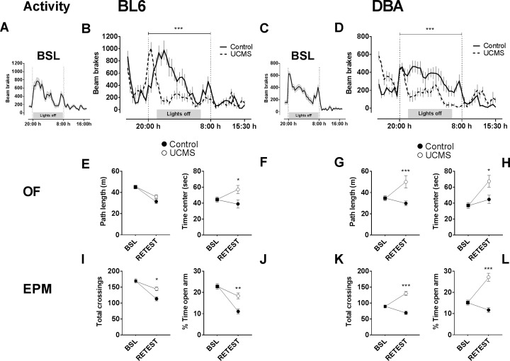
In the overnight activity baseline test (Fig 2A and 2C), prior to UCMS-exposure, BL6 mice were generally more active overnight when compared to DBA mice [F(1, 58) = 13.74, p < 0.001]. UCMS exposure decreased overnight activity in BL6 mice [F(1, 28) = 14.43, p < 0.001; Fig 2B] and DBA mice [F(1, 28) = 20.97, p < 0.001; Fig 2D] in comparison to control animals.
Fig 2. Effect of UCMS on overnight activity, exploratory and anxiety-like behavior.
(A-D) In the overnight activity test, at baseline testing BL6 mice (A) were more active than DBA mice (C), but showed similar activity patterns. Groups exposed to UCMS (C, D) showed decreased overnight activity. In the open-field test, (E) BL6 mice exposed to UCMS (white circles) do not differ from CTRLS (black circles) on path length. (G) DBA exposed to UCMS showed increased distance travelled. (F) UCMS-exposed BL6 mice and (H) UCMS-exposed DBA mice spent significantly more time in the center. In the elevated plus maze, total number of arm entries were recorded as well as time spent in the open arms. (I, J) BL6 mice exposed to UCMS and (K, L) UCMS-exposed DBA mice show increased beam crossings and time spent in the open arms. Values are expressed as mean +/- SEM. *, p < 0.05; ** p < 0.01; *** p < 0.001; UCMS vs. CTRL.
In the open field test (Fig 2E–2H), a significant decrease of distance travelled over time was observed in BL6 mice [F(1,28) = 10.66, p = 0.003; Fig 2E], but not in DBA mice [F(1,28) = 3.304 p = 0.079; Fig 2G]. Distance travelled was increased in UCMS DBA mice [F(1,28) = 10.48, p = 0.003; Fig 2G], but not in UCMS BL6 mice [F(1,28) = 0.049, p = 0.826; Fig 2E]. Post-hoc comparisons confirmed increased distance travelled following UCMS-exposure in DBA mice (t56 = 3.702, p < 0.001). Furthermore, UCMS-exposure lead to altered anxiety-like behavior in both BL6 mice [F(1,28) = 5.655, p = 0.024; Fig 2F] and DBA mice [F(1,28) = 4.945, p < 0.034; Fig 2H] as measured by time spent in the center. Both BL6 mice (t56 = 2.69, p < 0.05) and DBA mice (t56 = 2.812, p < 0.05) spent more time in the center of the open field following UCMS exposure in comparison to controls.
In the elevated plus maze (Fig 2I–2L), activity from baseline was generally decreased in BL6 mice [F(1,28) = 56.6, p < 0.001; Fig 2I], not in DBA mice [F(1,28) = 3.902, p < 0.058; Fig 2K]. Both BL6 mice [F(1, 28) = 5.092, p = 0.032; Fig 2I] and DBA mice [F(1, 28) = 22.02, p < 0.001; Fig 2K] showed increased arm entries following UCMS-exposure. Moreover, UCMS-exposed animals generally spent more time in the open arm, regardless of strain [BL6; F(1,28) = 12.97, p = 0.001; Fig 2J and DBA; F(1,28) = 22.47, p < 0.001; Fig 2L].
Sociability/preference for social novelty (SPSN)
During acclimation phase, DBA mice under UCMS regimen were more active than their controls (path length 7.79 ± 2.51 m and 18.32 ± 4.63 m for CTRL and UCMS, respectively. t28 = 7.738, p < 0.001). In contrast, UCMS regimen had no effect on path length in BL6 (path length 12.07 ± 3.42 m and 12.49 ± 2.43 m for CTRL and UCMS, respectively. t28 = 0.391, p = 0.698). This specific increase in exploratory behavior in UCMS DBA was also observed during the other phases (data not shown).
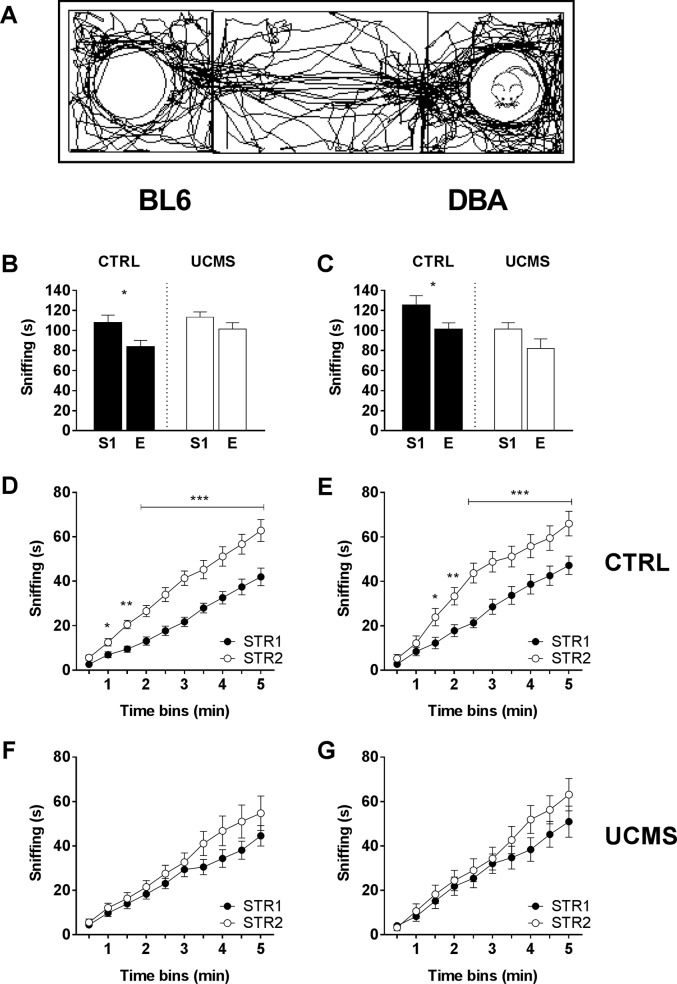
When presented with an unknown stranger mouse (Fig 3A), CTRL BL6 mice show increased sniffing behavior towards the stranger mouse, while UCMS BL6 are indifferent (for factor side: F(1, 28) = 8.662, p < 0.01; for factor condition: F(1, 28) = 4.775, p = 0.037; Fig 3B). Similarly, CTRL DBA show more interest towards a stranger mouse, while UCMS DBA are indifferent (for factor side: F(1, 28) = 5.534, p = 0.026; for factor condition: F(1, 28) = 9.92, p < 0.01; Fig 3C). These results suggest that stress decreases explorative sniffing behavior towards a stranger mouse and affects sociability in both strains similarly.
Fig 3. Sociability and preference for social novelty (SPSN).
(A) Representative track plot of single sociability trial. (B) During sociability trials, BL6 CTRLS significantly preferred the STR1 (S1) chamber over the empty (E) chamber, whereas UCMS mice showed no preference. (C) Similar results were observed for DBA mice, where the UCMS group showed decreased sociability when compared to CTRLS. (D-F) During preference for social novelty trials, CTRL mice preferred the novel STR2 during social novelty exploration (first 5 min.). UCMS mice showed no clear preference for either STR1 or STR2. Values are expressed as mean +/- SEM. *, p < 0.05; ** p < 0.01; *** p < 0.001.
During preference for social novelty testing, CTRL animals showed a preference towards a novel stranger. When adding up the time animals spent sniffing either stranger mouse, both CTRL group show a clear increase in sniffing time towards STR2 compared to STR1 (BL6: F(1,14) = 15.15, p < 0.01; DBA: F(1,14) = 6.886 p = 0.02; Fig 3D and 3E). In contrast, exposure to UCMS reduced the preference for social novelty in both BL6 [F(1,14) = 1.323, p = 0.269; Fig 3F] and DBA [F(1,14) = 0.665 p = 0.428; Fig 3G]. Taken together these results suggest similar behavioral effects of UCMS exposure across strains. Both UCMS-treated BL6 mice and UCMS-treated DBA mice show less sociability behavior. Moreover, both UCMS-treated BL6 mice and UCMS-treated DBA mice failed to (socially) discriminate between a novel and familiar stranger mouse.
Contextual threat conditioning and discrimination
Context threat acquisition
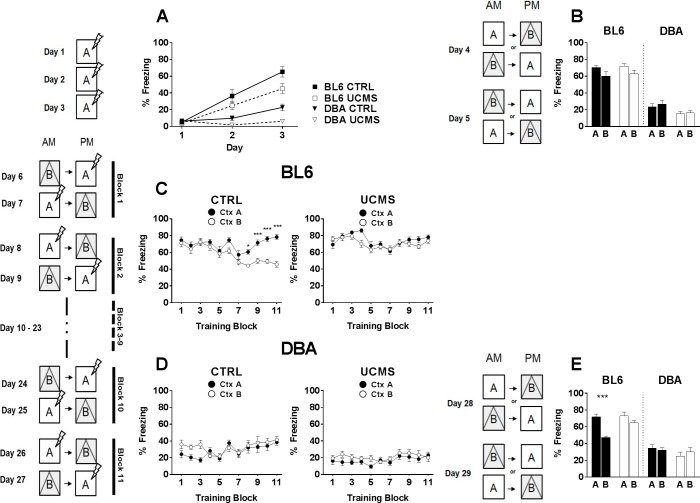
Contextual threat conditioning induced freezing behavior in all mice (Fig 4A). RM-ANOVA indicated a main effect of within-subjects factor acquisition [F(1,44) = 63.91, p < 0.001]. However, main effects of between-subjects factors strain [F(1,44) = 50.52, p < 0.001] and UCMS-exposure [F(1,44) = 9.223, p < 0.01] suggest that groups did not freeze similarly. We observed that BL6 animals in general displayed more robust freezing behavior than DBA. This difference has been reported before [62,63] and has been linked to differences in persistence of hippocampal long-term potentiation [53]. In both strains, UCMS animals displayed reduced freezing behavior over 3 days of threat conditioning (BL6: F(1,22) = 4.628, p = 0.043; DBA: F(1,22) = 9.424, p < 0.01).
Fig 4. Contextual discrimination threat conditioning.
(A) During context acquisition conditioning, mice exposed to UCMS showed less freezing behavior over time when compared to non-exposed controls. (B) Equivalent freezing behavior between UCMS-exposed and control groups was observed during generalization tests. (C) During context discrimination training, BL6 CTRL mice learned to distinguish context A from context B, whereas UCMS exposed mice did not. (D) DBA mice overall showed little freezing behavior and failed to distinguish context A from B, regardless of UCMS exposure. (E) A second generalization test revealed increased contextual discrimination behavior in unexposed BL6 controls. Values are expressed as mean +/- SEM. *, p < 0.05; ** p < 0.01; *** p < 0.001.
Generalization test
Fear generalization towards different contexts was tested on days 4 and 5 (Fig 4B). Animals were exposed to two highly similar contexts to evaluate fear generalization in the absence of shocks. In the BL6 strain, two-way ANOVA indicated an effect of context [F(1,44) = 5.663, p = 0.021], but not of condition [F(1,44) = 0.341, p < 0.001]. In the DBA strain, two-way ANOVA indicated an effect of condition [F(1,44) = 7.534, p < 0.01], but not of context [F(1,44) = 0.337, p = 0.564].
Discrimination training
Following the generalization test, mice were trained to discriminate between context A and context B for 22 consecutive days. In context A, mice received a shock after 3 min, while context B was without shock. Freezing in either context during the initial 3 min was plotted. CTRL BL6 learned to discriminate between contexts (RM ANOVA for freezing over blocks [F(10,220) = 4.494, p < 0.001]; for context [F(1,22) = 30.56, p < 0.001]; Fig 4C). In contrast, UCMS BL6 were unable to learn to discriminate and displayed consistent levels of freezing throughout the 22 days (RM ANOVA for freezing over blocks [F(10,220) = 1.701, p = 0.081]; for context [F(1,22) = 1.969, p = 0.174]; Fig 4C). Furthermore, CTRL DBA (Fig 4D) showed increased freezing over time [F(10,220) = 5.214, p = 0.040], but were unable to learn to discriminate [F(1,22) = 1.375, p = 0.253]. UCMS DBA (Fig 4D) overall showed very little freezing [F(10,220) = 0.875, p = 0.556] and never displayed context discrimination [F(1,22) = 1.429, p = 0.245].
Generalization test
Fear generalization between contexts was again tested on days 28 and 29 (Fig 4E). In the BL6 strain, two-way ANOVA indicated an effect of both context [F(1,44) = 22.26, p < 0.001] and condition [F(1,44) = 7.658, p < 0.01]. Post hoc analysis confirmed CTRL BL6 mice (p < 0.001) learned to discriminate between contexts, whereas UCMS BL6 mice (p = 0.499) failed to do so. DBA CTRL and UCMS mice showed similar behavioral patterns (two-way ANOVA for context [F(1,44) = 0.075, p = 0.786]; for condition [F(1,44) = 1.782, p = 0.189]). CTRL and UCMS mice failed to discriminate between contexts.
Biomarkers
In order to minimize circadian fluctuation, basal corticosterone concentrations were collected in the morning when circulating corticosterone levels are low [64]. Efforts were made to collect samples under stress-free conditions. In week 10, CORT analysis revealed no differences in concentrations of plasma corticosterone levels following exposure to UCMS in BL6 mice (Mann-Whitney test, p = 0.10) and DBA mice (Mann-Whitney test, p > 0.99) when compared to controls (see Table 2). CORT analysis conducted in week 17 revealed significantly higher plasma corticosterone levels in UCMS BL6 when compared to CTRL BL6 (Mann-Whitney test, *p = 0.016; Table 2). UCMS DBA mice did not display significantly higher concentrations of plasma corticosterone levels in comparison to CTRL DBA (Mann-Whitney test, p = 0.547; Table 2).
Table 2. Summary of blood plasma corticosterone quantification.
|
Avg. blood plasma CORT levels (ng/ml) in week 10 (n = 3/group) |
Avg. blood plasma CORT levels (ng/ml) in week 17 (n = 5/group) | ||
|---|---|---|---|
| BL6 | Control | 348 ± 15.2 | 247 ± 45.7 |
| UCMS | 657 ± 258.2 | 323 ± 43.3* | |
| DBA | Control | 320 ± 136.7 | 289 ± 143.5 |
| UCMS | 457 ± 124.8 | 354 ± 190 | |
UCMS increased significantly corticosterone levels in blood plasma (Mann-Whitney test * P<0.05)
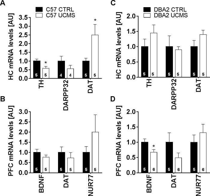
Furthermore, qRT-PCR analysis was performed to check for hippocampal and prefrontal cortical changes in expression of genes involved in dopaminergic neurotransmission and neuronal plasticity. Differences in hippocampal expression of genes related to dopaminergic neurotransmission were observed between BL6 groups, but not between DBA groups (Fig 5B and 5C). In terms of altered dopaminergic neurotransmission, UCMS BL6 mice showed significantly decreased tyrosine hydroxylase (TH) mRNA levels (Mann-Whitney test, *p = 0.027) and significantly increased dopamine transporter (DAT) mRNA levels (Mann-Whitney test, *p = 0.045) when compared to CTRL. Moreover, analysis of PFC gene expression (Fig 5B and 5C) showed significantly decreased Brain-Derived Neurotrophic Factor (BDNF) mRNA levels in UCMS DBA mice relative to unexposed CTRL (Mann-Whitney test, *p = 0.030). Expression levels of other genes related to dopaminergic signaling (such as D1R, D2R, COMT and VMAT2) were assessed in HC and PFC, with no differences observed across groups (data not shown).
Fig 5. qRT-PCR assays.
(A) Hippocampal gene expression analysis revealed significantly decreased TH mRNA levels (n = 5) and DAT (n = 5) mRNA levels of UCMS exposed BL6 mice, relative to unexposed controls. (C) No such differences were observed between DBA groups. (B) PFC gene expression analysis of BL6 groups. (D) PFC gene expression analysis revealed decreased BDNF (n = 5) mRNA levels in DBA2 mice, relative to unexposed controls (n = 6). Values are expressed as mean +/- SEM. *, p < 0.05; ** p < 0.01; *** p < 0.001.
Discussion
In this study we compared the effect of chronic mild stress on exploration, social and cognitive behavior in two inbred mouse strains, C57BL/6j and DBA2 (referred to as BL6 and DBA, respectively). We observed that UCMS had a similar effect on exploratory and social behavior in both mouse strains, but distinct effects on discrimination learning in contextual threat conditioning. Furthermore, we found a differential effect on genes associated with dopaminergic neurotransmission in the two mouse strains.
BL6 and DBA groups have been shown to have strain-specific activity patterns in the overnight activity test [65], and in exploration and anxiety tests [66]. We observe that upon exposure to UCMS both BL6 and DBA mice strains showed comparable changes in behavioral patterns in these particular tests. Stress-exposed mice have been reported to exhibit reduced nocturnal activity and reduced anticipation to morning light phase onset [67]. Animals selected for high reactivity to stress have been shown to have lower activity levels during the dark period as well as a shift in peak activity [64]. Disturbances in circadian rhythm and sleep pattern are also associated with stress-induced deregulation of the HPA axis and the pathophysiology of stress-related disorders [25,68]. Therefore, the changes we observed are in accordance with other stress models and with clinical observations and validate our UCMS procedure.
Exploration in the open field as well as the elevated plus maze is considered to have conflict resolution aspects, such as center exploration in the open field and entering open arms in the elevated plus maze. We observed that exposure to UCMS increased the time spent in the center in the open field and increased the number of entries in the open arms of the elevated plus maze. We interpret the observed effects of increased center activity and open arm visits as due to changes in arousal and increased reactivity to novelty rather than reduced anxiety [69]. Results from other tests support this interpretation. We observed increased exploratory activity specifically in DBA in four independent tests: in the first light phase of the 24h activity test, in the open field, elevated plus maze as well as in the SPSN test, while BL6 showed hyperactivity in elevated plus maze and a trend in the open field and 24h activity. Similar increases in center time and open arm visits have been reported before [46,69], but other studies reported reductions [70–73]. We attribute these contradictory results as being caused by differences in housing condition, testing history and strain background, factors that have all been shown to influence exploration in open field and elevated plus maze [74].
In the current study, UCMS-exposed BL6 and UCMS-exposed DBA mice exhibited reduced sociability behavior, a behavior that has been associated with models for autism spectrum disorder [75], schizophrenia [11,76], anxiety [77] and depression [78]. This effect of chronic stress on social behavior in rodents has been reported before [79]. In addition, neuropeptides of the corticotrophin-releasing factor family that coordinate stress response have been shown to affect social behavior as well [80]. In addition to reduced sociability behavior we found that chronically stressed animals of both strains failed to (socially) discriminate between a novel and familiar mouse during the social novelty phase. These results are consistent with a report by Van Kooij et al. [81] showing that chronic restraint stress has detrimental effects on sociability and social memory in rodents. Social recognition memory consolidation depends on a functional network involving the PFC, HC, anterior cingulate, and amygdala [82–84]. Tanimizu and colleagues speculated that the PFC-HC network is crucial in social recognition and discrimination. Our observations indicate that in particular social discrimination (familiar versus novel mouse) is impaired when animals are exposed to chronic mild stress. This impairment in discrimination was also observed in contextual threat conditioning to conceptually very similar contexts. We observed that after initial threat conditioning to a specific context, BL6 discriminated between highly similar contexts, but UCMS had no effect on the discrimination. In contrast, DBA animals were unable to distinguish between the conditioned context and a highly similar context showing similar freezing responses to either context. This indicates that the contexts were similar enough to create a high generalization effect. Overall, we observed that the freezing level was much higher in BL6 than in DBA, however, this might not per se reflect memory impairment, but simply an increased activity level in DBA. Indeed, freezing levels in DBA were higher after conditioning, indicating that DBA are able to form an aversive contextual representation.
During discrimination training, animals are repeatedly exposed to the aversive and the neutral context and their behavior becomes over time more context specific. This process of discriminative learning has been shown to depend on a functional PFC-HC network [85,86]. The interplay of both PFC and HC is crucial in a series of adaptive learning paradigms such as reversal learning, threat extinction, context discrimination and generalization [87,88]. Stress-related disorders such as MDD have been linked to reduced mnemonic discrimination performance [89]. It has been argued that this propensity to form or recall information in an unspecific, more generalized way, could in part be due to reduced hippocampal function and connectivity [90]. UCMS mice were expected to perform worse in this complex cognitive task. Indeed, we confirmed that UCMS-exposure significantly diminished discrimination ability in BL6 mice compared to controls. These results are consistent with previous findings showing that chronic stress significantly impairs cognitive performance in discriminative fear conditioning [91], novel object recognition [44] and spatial memory [17,92,93]. However, our data indicate that these effects are strain-specific. Despite extensive conditioning, DBA mice exerted less context-evoked freezing behaviour when compared to BL6 mice. Moreover, neither UCMS-exposed DBA mice nor control DBA mice could successfully discriminate between contexts. When compared to BL6 mice of the standard genetic background, mice of the genetically unrelated DBA strain typically show more resilience to helplessness induced by an unavoidable stressor [94]. For example, exposure to an inescapable shock has been shown to reduce activity in a Y maze in BL6 mice, but not in DBA mice [95]. A possible explanation might be that DBA mice have altered HC-PFC functioning and therefore perform poorly on learning and memory tasks, and that chronic stress has very little influence on cognitive performance. Studies have shown DBA mice to have reduced persistence of hippocampal long-term potentiation (LTP) when compared to BL6 mice, as well as reduced levels of signalling proteins such as protein kinase C (PKC) [63,96]. Furthermore, DBA mice exert reduced levels of cAMP response element-binding protein (CREB), a transcription factor associated with long-term memory formation, in prefrontal cortex and hippocampus when compared to BL6 mice [97]. These differences manifest themselves in cognitive performances where strain-dependent differences have been observed in various spatial memory and contextual conditioning tasks. Specifically, DBA mice are outperformed by BL6 mice in Barnes maze [98], Morris water maze [99] and contextual fear conditioning [63]. Together, our data suggest that chronic exposure to stress could lead to impairments in HC-dependent social and cognitive behavior, comparable to what is seen in various stress-related psychopathologies [78,90,100,101].
Across the many gene sets involved in these divergent behavioural responses to chronic stress, we chose to focus on genes involved in neural plasticity and dopaminergic neurotransmission in HC and PFC. In addition to its role in anhedonia, motivation and the regulation of circadian rhythm, dopamine might play an important role in stress-induced social and cognitive impairments [43,102,103]. Specifically, stress-induced disturbances in neural plasticity have been associated with a hypodopaminergic state, which leads to disturbances in neural plasticity persistence [23,104]. We quantified the expression of tyrosine hydroxylase which catalyses the L-tyrosine to L-DOPA conversion. In the hippocampus of BL6 mice, UCMS induced a significant downregulation of TH expression. We also measured a downregulation of DARPP32, a post-synaptic inhibitor of PPP1CA, which has been linked to learning and memory defects [105,106]. DARPP32 is activated upon D1R activation [107], and a downregulation of this gene is suggestive of post-synaptic DAergic signal modulation. In addition, we observed an upregulation of presynaptic DA transporter (DAT) in response to UCMS in the HC of BL6. The upregulation of DAT, together with reduction in TH might reflect reduced hippocampal availability of DA as it has been shown that an increase in DA reuptake can induce a reduction of DA catalysis by TH in DA neurons [108]. Reduction in DA neuromodulation in HC has been linked to impaired learning and memory, in particular pattern separation [109]. Unexpectedly, UCMS did not affect TH, DARPP32, nor DAT in the HC of DBA mice.
Nevertheless, DBA mice have reportedly reduced responsiveness to DA releasing drugs such as cocaine and d-amphetamine [110–112], and altered D1/D2 receptor balance in the hippocampus [113]. Thus, the observed differences might be due to the involvement of stress-induced expression changes of different genes in the brain of DBA and BL6 mice [50], as DBA and BL6 mice show opposite differences in brain dopamine functioning under stressful conditions [114]. Specifically, DBA animals have an increased number of neurons positive for dopamine transporter (DAT) and tyrosine hydroxylase (TH) in HC and PFC [113,115]. These different DA systems might be differentially involved in liability to chronic stress and the observed social and cognitive impairments, underlining the importance of combining different inbred strains with different behavioral test batteries to study gene-environment interactions involved in the pathological outcomes of stress exposure [116].
In PFC, expression of transcription factors (NUR77) involved in the development and differentiation of dopamine neurons was similarly, but insignificantly, increased across strains [117]. Furthermore, the neurotrophin family member BDNF was significantly downregulated in DBA, not in BL6 mice. BL6 mice did show a similar, albeit not significant, expression pattern. Although inconsistencies in UCMS expression data have been reported, the lack of robust down-regulation of classic biomarkers for stress-induced neural dysfunctions may be related to the procedural design used in the current study [118,119]. Both UCMS and control mice were repeatedly exposed to mild foot shocks during discrimination training, a regimen comparable to the chronic stress procedure itself. Exposure to a single acute stressor has been shown to be sufficient for altered expression levels of the investigated cellular targets involved in learning and memory [120–122]. Therefore, due to similar stress-induced changes in gene expression levels of control animals, changes related to the UCMS-procedure itself might not have been apparent.
Taken together, our findings indicate that a chronic mild stress regimen lead to explorative, social and cognitive impairments. Stress-induced cognitive impairments could be related to altered dopaminergic neurotransmission in Bl6 mice, but not in DBA mice. We propose that applying UCMS to females of different inbred strains is a good model to study the impact of a genetic background and environmental factors in humans where some individuals show more susceptibility to developing stress-related disorder then others. Our results also highlight the importance of studying different strains in multiple test batteries, to help researchers in choosing appropriate strains for the analysis of the neural and genetic basis of stress-induced impairments in the anxiety, social and cognitive domain.
Acknowledgments
This research was financially supported by the Flemish Research Fund (FWO) (grant No. G.0746.13).
Data Availability
All relevant data are within the manuscript.
Funding Statement
Fonds Wetenschappelijk Onderzoek - Vlaanderen (G.0746.13) (www.FWO.be) sponsored the PhD project of MvB.
References
- 1.Finsterwald C, Alberini CM. Stress and glucocorticoid receptor-dependent mechanisms in long-term memory: From adaptive responses to psychopathologies. Neurobiology of Learning and Memory. 2014. doi: 10.1016/j.nlm.2013.09.017 [DOI] [PMC free article] [PubMed] [Google Scholar]
- 2.McEwen BS, Sapolsky RM. Stress and cognitive function. Curr Opin Neurobiol. 1995;5: 205–216. doi: 10.1016/0959-4388(95)80028-X [DOI] [PubMed] [Google Scholar]
- 3.Kupferberg A, Bicks L, Hasler G. Social functioning in major depressive disorder [Internet]. Neuroscience and Biobehavioral Reviews. Elsevier Ltd; 2016. pp. 313–332. doi: 10.1016/j.neubiorev.2016.07.002 [DOI] [PubMed] [Google Scholar]
- 4.WHO WHO. WHO | Global Burden of Disease. WHO; 2004; Available: http://www.who.int/healthinfo/global_burden_disease/GBD_report_2004update_full.pdf [Google Scholar]
- 5.Keilp JG, Gorlyn M, Oquendo MA, Burke AK, Mann JJ. Attention deficit in depressed suicide attempters. Psychiatry Res. 2008;159: 7–17. doi: 10.1016/j.psychres.2007.08.020 [DOI] [PMC free article] [PubMed] [Google Scholar]
- 6.Herrera-Guzmán I, Gudayol-Ferré E, Herrera-Guzmán D, Guàrdia-Olmos J, Hinojosa-Calvo E, Herrera-Abarca JE. Effects of selective serotonin reuptake and dual serotonergic-noradrenergic reuptake treatments on memory and mental processing speed in patients with major depressive disorder. J Psychiatr Res. Ars Médica, Barcelona; 2009;43: 855–863. doi: 10.1016/j.jpsychires.2008.10.015 [DOI] [PubMed] [Google Scholar]
- 7.Snyder HR. Major depressive disorder is associated with braod impairments on neuropsychological measures of evecutive function: a meta-analysis and review. Psychol Bull. 2014;139: 81–132. doi: 10.1037/a0028727.Major [DOI] [PMC free article] [PubMed] [Google Scholar]
- 8.Gonda X, Pompili M, Serafini G, Carvalho AF, Rihmer Z, Dome P. The role of cognitive dysfunction in the symptoms and remission from depression. Ann Gen Psychiatry. BioMed Central; 2015;14: 27 doi: 10.1186/s12991-015-0068-9 [DOI] [PMC free article] [PubMed] [Google Scholar]
- 9.Kleim B, Ehlers A. Reduced autobiographical memory specificity predicts depression and posttraumatic stress disorder after recent trauma. J Consult Clin Psychol. 2008;76: 231–42. doi: 10.1037/0022-006X.76.2.231 [DOI] [PMC free article] [PubMed] [Google Scholar]
- 10.O’Leary OF, Cryan JF. Towards translational rodent models of depression. Cell Tissue Res. 2013;354: 141–53. doi: 10.1007/s00441-013-1587-9 [DOI] [PubMed] [Google Scholar]
- 11.Ellenbroek BA, Ellenbroek BA, Cools AR, Cools AR. Animal models for the negative symptoms of schizophrenia. Behav Pharmacol. 2000;11: 223–33. Available: http://www.ncbi.nlm.nih.gov/pubmed/11103877 [DOI] [PubMed] [Google Scholar]
- 12.Yang RJ, Mozhui K, Karlsson R-M, Cameron HA, Williams RW, Holmes A. Variation in Mouse Basolateral Amygdala Volume is Associated With Differences in Stress Reactivity and Fear Learning. Neuropsychopharmacology. 2008;33: 2595–2604. doi: 10.1038/sj.npp.1301665 [DOI] [PubMed] [Google Scholar]
- 13.Bian Y, Pan Z, Hou Z, Huang C, Li W, Zhao B. Learning, memory, and glial cell changes following recovery from chronic unpredictable stress. Brain Res Bull. 2012; doi: 10.1016/j.brainresbull.2012.04.008 [DOI] [PubMed] [Google Scholar]
- 14.Sahin TD, Karson A, Balci F, Yazir Y, Bayramg??rler D, Utkan T. TNF-alpha inhibition prevents cognitive decline and maintains hippocampal BDNF levels in the unpredictable chronic mild stress rat model of depression. Behav Brain Res. 2015; doi: 10.1016/j.bbr.2015.05.062 [DOI] [PubMed] [Google Scholar]
- 15.Gumuslu E, Mutlu O, Sunnetci D, Ulak G, Celikyurt IK, Cine N, et al. The effects of tianeptine, olanzapine and fluoxetine on the cognitive behaviors of unpredictable chronic mild stress-exposed mice. Drug Res (Stuttg). 2013;63: 532–539. doi: 10.1055/s-0033-1347237 [DOI] [PubMed] [Google Scholar]
- 16.Duman RS, Aghajanian GK, Sanacora G, Krystal JH. Synaptic plasticity and depression: new insights from stress and rapid-acting antidepressants. Nat Med. Nature Publishing Group; 2016;22: 238–249. doi: 10.1038/nm.4050 [DOI] [PMC free article] [PubMed] [Google Scholar]
- 17.Park M, Kim C-H, Jo S, Kim EJ, Rhim H, Lee CJ, et al. Chronic Stress Alters Spatial Representation and Bursting Patterns of Place Cells in Behaving Mice. Sci Rep. 2015;5: 16235 doi: 10.1038/srep16235 [DOI] [PMC free article] [PubMed] [Google Scholar]
- 18.Campus P, Maiolati M, Orsini C, Cabib S. Altered consolidation of extinction-like inhibitory learning in genotype-specific dysfunctional coping fostered by chronic stress in mice. Behav Brain Res. 2016; doi: 10.1016/j.bbr.2016.08.014 [DOI] [PubMed] [Google Scholar]
- 19.Wang M, Perova Z, Arenkiel BR, Li B. Synaptic Modifications in the Medial Prefrontal Cortex in Susceptibility and Resilience to Stress. J Neurosci. 2014;34: 7485–7492. doi: 10.1523/JNEUROSCI.5294-13.2014 [DOI] [PMC free article] [PubMed] [Google Scholar]
- 20.Lee BH, Kim YK. The roles of BDNF in the pathophysiology of major depression and in antidepressant treatment. Psychiatry Investig. 2010;7: 231–235. doi: 10.4306/pi.2010.7.4.231 [DOI] [PMC free article] [PubMed] [Google Scholar]
- 21.Kunii Y, Hyde TM, Ye T, Li C, Kolachana B, Dickinson D, et al. Revisiting DARPP-32 in postmortem human brain: changes in schizophrenia and bipolar disorder and genetic associations with t-DARPP-32 expression. Mol Psychiatry. 2014;19: 192–199. doi: 10.1038/mp.2012.174 [DOI] [PubMed] [Google Scholar]
- 22.Moreines JL, Owrutsky ZL, Grace AA. Involvement of Infralimbic Prefrontal Cortex but not Lateral Habenula in Dopamine Attenuation After Chronic Mild Stress. Neuropsychopharmacology. 2017;42: 904–913. doi: 10.1038/npp.2016.249 [DOI] [PMC free article] [PubMed] [Google Scholar]
- 23.Reverses K, Belujon P, Grace AA. Restoring Mood Balance in Depression: Biol Psychiatry. 2014;76: 927–936. doi: 10.1016/j.biopsych.2014.04.014 [DOI] [PMC free article] [PubMed] [Google Scholar]
- 24.Lee JC, Wang LP, Tsien JZ. Dopamine rebound-excitation theory: Putting brakes on PTSD. Front Psychiatry. 2016;7 doi: 10.3389/fpsyt.2016.00163 [DOI] [PMC free article] [PubMed] [Google Scholar]
- 25.Balbo M, Leproult R, Van Cauter E. Impact of Sleep and Its Disturbances on Hypothalamo-Pituitary-Adrenal Axis Activity. Int J Endocrinol. 2010;2010: 1–16. doi:10.1155/2010/759234 [DOI] [PMC free article] [PubMed] [Google Scholar]
- 26.Lu B, Pang PT, Woo NH. The yin and yang of neurotrophin action. Nat Rev Neurosci. 2005;6: 603–614. doi: 10.1038/nrn1726 [DOI] [PubMed] [Google Scholar]
- 27.Filho CB, Jesse CR, Donato F, Giacomeli R, Del Fabbro L, da Silva Antunes M, et al. Chronic unpredictable mild stress decreases BDNF and NGF levels and Na+,K+-ATPase activity in the hippocampus and prefrontal cortex of mice: Antidepressant effect of chrysin. Neuroscience. 2015;289: 367–380. doi: 10.1016/j.neuroscience.2014.12.048 [DOI] [PubMed] [Google Scholar]
- 28.Bath KG, Schilit A, Lee FS. Stress effects on BDNF expression: Effects of age, sex, and form of stress. Neuroscience. 2013;239: 149–156. doi: 10.1016/j.neuroscience.2013.01.074 [DOI] [PubMed] [Google Scholar]
- 29.Gumuslu E, Mutlu O, Sunnetci D, Ulak G, Celikyurt IK, Cine N, et al. The antidepressant agomelatine improves memory deterioration and upregulates CREB and BDNF gene expression levels in unpredictable chronic mild stress (UCMS)-exposed mice. Drug Target Insights. 2014;2014: 11–21. doi: 10.4137/DTI.S13870 [DOI] [PMC free article] [PubMed] [Google Scholar]
- 30.Duman RS, Monteggia LM. A Neurotrophic Model for Stress-Related Mood Disorders. Biological Psychiatry. 2006. pp. 1116–1127. doi: 10.1016/j.biopsych.2006.02.013 [DOI] [PubMed] [Google Scholar]
- 31.Hünnerkopf R, Strobel A, Gutknecht L, Brocke B, Lesch KP. Interaction between BDNF Val66Met and Dopamine Transporter Gene Variation Influences Anxiety-Related Traits. Neuropsychopharmacology. 2007;32: 2552–2560. doi: 10.1038/sj.npp.1301383 [DOI] [PubMed] [Google Scholar]
- 32.Rossato JI, Bevilaqua LRM, Izquierdo I, Medina JH, Cammarota M. Dopamine Controls Persistence of Long-Term Memory Storage. Science (80-). 2009;325: 1017–1020. doi: 10.1126/science.1172545 [DOI] [PubMed] [Google Scholar]
- 33.Perreault ML, Jones-Tabah J, O’Dowd BF, George SR. A physiological role for the dopamine D5 receptor as a regulator of BDNF and Akt signalling in rodent prefrontal cortex. Int J Neuropsychopharmacol. 2013; 1–7. doi: 10.1017/S1461145712000685 [DOI] [PMC free article] [PubMed] [Google Scholar]
- 34.Hamid AA, Pettibone JR, Mabrouk OS, Hetrick VL, Schmidt R, Vander Weele CM, et al. Mesolimbic dopamine signals the value of work. Nat Neurosci. 2015;19: 117–126. doi: 10.1038/nn.4173 [DOI] [PMC free article] [PubMed] [Google Scholar]
- 35.Gunaydin LA, Grosenick L, Finkelstein JC, Kauvar I V., Fenno LE, Adhikari A, et al. Natural neural projection dynamics underlying social behavior. Cell. 2014;157: 1535–1551. doi: 10.1016/j.cell.2014.05.017 [DOI] [PMC free article] [PubMed] [Google Scholar]
- 36.da Silva WCN, Köhler CC, Radiske A, Cammarota M. D1/D5 dopamine receptors modulate spatial memory formation. Neurobiol Learn Mem. Elsevier Inc.; 2012;97: 271–5. doi: 10.1016/j.nlm.2012.01.005 [DOI] [PubMed] [Google Scholar]
- 37.Shivarama Shetty M, Gopinadhan S, Sajikumar S. Dopamine D1/D5 receptor signaling regulates synaptic cooperation and competition in hippocampal CA1 pyramidal neurons via sustained ERK1/2 activation. Hippocampus. 2015;0: n/a-n/a. doi: 10.1002/hipo.22497 [DOI] [PMC free article] [PubMed] [Google Scholar]
- 38.Felten A, Montag C, Markett S, Walter NT, Reuter M. Genetically determined dopamine availability predicts disposition for depression. Brain Behav. 2011;1: 109–18. doi: 10.1002/brb3.20 [DOI] [PMC free article] [PubMed] [Google Scholar]
- 39.Blood AJ, Iosifescu D V, Makris N, Perlis RH, Kennedy DN, Dougherty DD, et al. Microstructural abnormalities in subcortical reward circuitry of subjects with major depressive disorder. PLoS One. 2010;5: e13945 doi: 10.1371/journal.pone.0013945 [DOI] [PMC free article] [PubMed] [Google Scholar]
- 40.Nestler EJ, Carlezon W a. The mesolimbic dopamine reward circuit in depression. Biol Psychiatry. 2006;59: 1151–9. doi: 10.1016/j.biopsych.2005.09.018 [DOI] [PubMed] [Google Scholar]
- 41.Tye KM, Mirzabekov JJ, Warden MR, Ferenczi EA, Tsai H-C, Finkelstein J, et al. Dopamine neurons modulate neural encoding and expression of depression-related behaviour. Nature. 2012;493: 537–541. doi: 10.1038/nature11740 [DOI] [PMC free article] [PubMed] [Google Scholar]
- 42.Chaudhury D, Walsh JJ, Friedman AK, Juarez B, Ku SM, Koo JW, et al. Rapid regulation of depression-related behaviours by control of midbrain dopamine neurons. Nature. Nature Publishing Group; 2013;493: 532–6. doi: 10.1038/nature11713 [DOI] [PMC free article] [PubMed] [Google Scholar]
- 43.Belujon P, Grace AA. Regulation of dopamine system responsivity and its adaptive and pathological response to stress. Proc R Soc B Biol Sci. 2015;282: 20142516 doi: 10.1098/rspb.2014.2516 [DOI] [PMC free article] [PubMed] [Google Scholar]
- 44.Elizalde N, Gil-Bea FJ, Ramírez MJ, Aisa B, Lasheras B, Del Rio J, et al. Long-lasting behavioral effects and recognition memory deficit induced by chronic mild stress in mice: Effect of antidepressant treatment. Psychopharmacology (Berl). 2008; doi: 10.1007/s00213-007-1035-1 [DOI] [PubMed] [Google Scholar]
- 45.Mineur YS, Belzung C, Crusio WE. Effects of unpredictable chronic mild stress on anxiety and depression-like behavior in mice. Behav Brain Res. 2006;175: 43–50. doi: 10.1016/j.bbr.2006.07.029 [DOI] [PubMed] [Google Scholar]
- 46.Rössler AS, Joubert C, Chapouthier G. Chronic mild stress alleviates anxious behaviour in female mice in two situations. Behav Processes. 2000;49: 163–165. doi: 10.1016/S0376-6357(00)00080-2 [DOI] [PubMed] [Google Scholar]
- 47.Willner P. The chronic mild stress (CMS) model of depression: History, evaluation and usage. 2016; doi: 10.1016/j.ynstr.2016.08.002 [DOI] [PMC free article] [PubMed] [Google Scholar]
- 48.Zhu S, Wang J, Zhang Y, Li V, Kong J, He J, et al. Unpredictable chronic mild stress induces anxiety and depression-like behaviors and inactivates AMP-activated protein kinase in mice. Brain Res. 2014;1576: 81–90. doi: 10.1016/j.brainres.2014.06.002 [DOI] [PubMed] [Google Scholar]
- 49.Darcet F, Gardier AM, Gaillard R, David DJ, Guilloux JP. Cognitive dysfunction in major depressive disorder. A translational review in animal models of the disease. Pharmaceuticals. 2016. doi: 10.3390/ph9010009 [DOI] [PMC free article] [PubMed] [Google Scholar]
- 50.Mozhui K, Karlsson RM, Kash TL, Ihne J, Norcross M, Patel S, et al. Strain differences in stress responsivity are associated with divergent amygdala gene expression and glutamate-mediated neuronal excitability. J Neurosci. 2010;30: 5357–5367. doi: 10.1523/JNEUROSCI.5017-09.2010 [DOI] [PMC free article] [PubMed] [Google Scholar]
- 51.Moy SS, Nadler JJ, Young NB, Nonneman RJ, Segall SK, Andrade GM, et al. Social approach and repetitive behavior in eleven inbred mouse strains. Behav Brain Res. 2008;191: 118–129. doi: 10.1016/j.bbr.2008.03.015 [DOI] [PMC free article] [PubMed] [Google Scholar]
- 52.Cho W-H, Han J-S, Doyère V, Micheau J, W-h C. Differences in the Flexibility of Switching Learning Strategies and CREB Phosphorylation Levels in Prefrontal Cortex, Dorsal Striatum and Hippocampus in Two Inbred Strains of Mice. 2016; doi: 10.3389/fnbeh.2016.00176 [DOI] [PMC free article] [PubMed] [Google Scholar]
- 53.Lenselink AM, Rotaru DC, Li KW, Van Nierop P, Rao-Ruiz P, Loos M, et al. Strain differences in presynaptic function: Proteomics, ultrastructure, and physiology of hippocampal synapses in DBA/2J and C57Bl/6J MICE. J Biol Chem. 2015;290: 15635–15645. doi: 10.1074/jbc.M114.628776 [DOI] [PMC free article] [PubMed] [Google Scholar]
- 54.Campus P, Maiolati M, Orsini C, Cabib S. Altered consolidation of extinction-like inhibitory learning in genotype-specific dysfunctional coping fostered by chronic stress in mice. Behav Brain Res. 2016;315: 23–35. doi: 10.1016/j.bbr.2016.08.014 [DOI] [PubMed] [Google Scholar]
- 55.D’Este L, Casini A, Puglisi-Allegra S, Cabib S, Renda TG. Comparative immunohistochemical study of the dopaminergic systems in two inbred mouse strains (C57BL/6J and DBA/2J). J Chem Neuroanat. 2007;33: 67–74. doi: 10.1016/j.jchemneu.2006.12.005 [DOI] [PubMed] [Google Scholar]
- 56.Callaerts-Vegh Z, Beckers T, Ball SM, Baeyens F, Callaerts PF, Cryan JF, et al. Concomitant deficits in working memory and fear extinction are functionally dissociated from reduced anxiety in metabotropic glutamate receptor 7-deficient mice. J Neurosci. 2006;26: 6573–82. doi: 10.1523/JNEUROSCI.1497-06.2006 [DOI] [PMC free article] [PubMed] [Google Scholar]
- 57.Naert A, Callaerts-Vegh Z, D’Hooge R. Nocturnal hyperactivity, increased social novelty preference and delayed extinction of fear responses in post-weaning socially isolated mice. Brain Res Bull. 2011;85: 354–362. doi: 10.1016/j.brainresbull.2011.03.027 [DOI] [PubMed] [Google Scholar]
- 58.Nadler JJ, Moy SS, Dold G, Trang D, Simmons N, Perez a, et al. Automated apparatus for quantitation of social approach behaviors in mice. Genes Brain Behav. 2004;3: 303–14. doi: 10.1111/j.1601-183X.2004.00071.x [DOI] [PubMed] [Google Scholar]
- 59.Naert A, Gantois I, Laeremans A, Vreysen S, Van den Bergh G, Arckens L, et al. Behavioural alterations relevant to developmental brain disorders in mice with neonatally induced ventral hippocampal lesions. Brain Res Bull. 2013;94: 71–81. doi: 10.1016/j.brainresbull.2013.01.008 [DOI] [PubMed] [Google Scholar]
- 60.Nakashiba T, Cushman JD, Pelkey KA, Renaudineau S, Buhl DL, Mchugh TJ, et al. Young Dentate Granule Cells Mediate Pattern Separation, whereas Old Granule Cells Facilitate Pattern Completion. Cell. Elsevier Inc.; 2012;149: 188–201. doi: 10.1016/j.cell.2012.01.046 [DOI] [PMC free article] [PubMed] [Google Scholar]
- 61.Goddyn H, Callaerts-Vegh Z, Stroobants S, Dirikx T, Vansteenwegen D, Hermans D, et al. Deficits in acquisition and extinction of conditioned responses in mGluR7 knockout mice. Neurobiol Learn Mem. 2008;90: 103–11. doi: 10.1016/j.nlm.2008.01.001 [DOI] [PubMed] [Google Scholar]
- 62.Holmes a, Wrenn CC, Harris a P, Thayer KE, Crawley JN. Behavioral profiles of inbred strains on novel olfactory, spatial and emotional tests for reference memory in mice. Genes Brain Behav. 2002;1: 55–69. Available: http://www.ncbi.nlm.nih.gov/pubmed/12886950 [DOI] [PubMed] [Google Scholar]
- 63.André JM, Cordero K a, Gould TJ. Comparison of the performance of DBA/2 and C57BL/6 mice in transitive inference and foreground and background contextual fear conditioning. Behav Neurosci. 2012;126: 249–57. doi: 10.1037/a0027048 [DOI] [PMC free article] [PubMed] [Google Scholar]
- 64.Touma C, Fenzl T, Ruschel J, Palme R, Holsboer F, Kimura M, et al. Rhythmicity in mice selected for extremes in stress reactivity: Behavioural, endocrine and sleep changes resembling endophenotypes of major depression. PLoS One. 2009;4 doi: 10.1371/journal.pone.0004325 [DOI] [PMC free article] [PubMed] [Google Scholar]
- 65.Loos M, Koopmans B, Aarts E, Maroteaux G, Van Der Sluis S, Verhage M, et al. Sheltering behavior and locomotor activity in 11 genetically diverse common inbred mouse strains using home-cage monitoring. PLoS One. 2014;9 doi: 10.1371/journal.pone.0108563 [DOI] [PMC free article] [PubMed] [Google Scholar]
- 66.Podhorna J, Brown RE. Strain differences in activity and emotionality do not account for differences in learning and memory performance between C57BL/6 and DBA/2 mice. Genes Brain Behav. 2002;1: 96–110. doi: 10.1034/j.1601-183X.2002.10205.x [DOI] [PubMed] [Google Scholar]
- 67.Logan RW, Edgar N, Gillman AG, Hoffman D, Zhu X, McClung CA. Chronic Stress Induces Brain Region-Specific Alterations of Molecular Rhythms that Correlate with Depression-like Behavior in Mice. Biol Psychiatry. 2015;78: 249–258. doi: 10.1016/j.biopsych.2015.01.011 [DOI] [PMC free article] [PubMed] [Google Scholar]
- 68.Haridas S, Kumar M, Manda K. Melatonin ameliorates chronic mild stress induced behavioral dysfunctions in mice. Physiol Behav. 2013;119: 201–207. doi: 10.1016/j.physbeh.2013.06.015 [DOI] [PubMed] [Google Scholar]
- 69.Schweizer MC, Henniger MSH, Sillaber I. Chronic mild stress (CMS) in mice: Of anhedonia, “anomalous anxiolysis” and activity. PLoS One. 2009;4 doi: 10.1371/journal.pone.0004326 [DOI] [PMC free article] [PubMed] [Google Scholar]
- 70.Zhu S, Shi R, Wang J, Wang J-F, Li X-M. Unpredictable chronic mild stress not chronic restraint stress induces depressive behaviours in mice. Neuroreport. 2014;25: 1151–1155. doi: 10.1097/WNR.0000000000000243 [DOI] [PubMed] [Google Scholar]
- 71.Chang CH, Grace AA. Amygdala-ventral pallidum pathway decreases dopamine activity after chronic mild stress in rats. Biol Psychiatry. 2014;76: 223–230. doi: 10.1016/j.biopsych.2013.09.020 [DOI] [PMC free article] [PubMed] [Google Scholar]
- 72.Dhillon SS. THE HORMONAL CONTROL OF NEUROPEPTIDE Y AND GONADOTROPIN-RELEASING HORMONE HYPOTHALAMIC NEURONS The Hormonal Control of Neuropeptide Y and Gonadotropin-. 2010; [Google Scholar]
- 73.Jung Y-H, Hong S-I, Ma S-X, Hwang J-Y, Kim J-S, Lee J-H, et al. Strain Differences in the Chronic Mild Stress Animal Model of Depression and Anxiety in Mice. Biomol Ther (Seoul). 2014;22: 453–459. doi: 10.4062/biomolther.2014.058 [DOI] [PMC free article] [PubMed] [Google Scholar]
- 74.Crabbe JC. Genetics of Mouse Behavior: Interactions with Laboratory Environment. Science (80-). 1999;284: 1670–1672. doi: 10.1126/science.284.5420.1670 [DOI] [PubMed] [Google Scholar]
- 75.Chadman KK. Fluoxetine but not risperidone increases sociability in the BTBR mouse model of autism. Pharmacol Biochem Behav. Elsevier Inc.; 2011;97: 586–594. doi: 10.1016/j.pbb.2010.09.012 [DOI] [PubMed] [Google Scholar]
- 76.Karlsson R-M, Tanaka K, Saksida LM, Bussey TJ, Heilig M, Holmes A. Assessment of Glutamate Transporter GLAST (EAAT1)-Deficient Mice for Phenotypes Relevant to the Negative and Executive/Cognitive Symptoms of Schizophrenia. Neuropsychopharmacology. 2009;34: 1578–1589. doi: 10.1038/npp.2008.215 [DOI] [PMC free article] [PubMed] [Google Scholar]
- 77.Kelly AM, Goodson JL. Social functions of individual vasopressin-oxytocin cell groups in vertebrates: What do we really know? Frontiers in Neuroendocrinology. 2014. pp. 512–529. doi: 10.1016/j.yfrne.2014.04.005 [DOI] [PubMed] [Google Scholar]
- 78.Sandi C, Haller J. Stress and the social brain: behavioural effects and neurobiological mechanisms. Nat Rev Neurosci. 2015;16: 290–304. doi: 10.1038/nrn3918 [DOI] [PubMed] [Google Scholar]
- 79.Beery AK, Kaufer D. Stress, social behavior, and resilience: Insights from rodents. Neurobiol Stress. Elsevier Inc; 2015;1: 116–127. doi: 10.1016/j.ynstr.2014.10.004 [DOI] [PMC free article] [PubMed] [Google Scholar]
- 80.Rotzinger S, Lovejoy DA, Tan LA. Behavioral effects of neuropeptides in rodent models of depression and anxiety. Peptides. Elsevier Inc.; 2010;31: 736–756. doi: 10.1016/j.peptides.2009.12.015 [DOI] [PubMed] [Google Scholar]
- 81.van der Kooij MA, Fantin M, Kraev I, Korshunova I, Grosse J, Zanoletti O, et al. Impaired Hippocampal Neuroligin-2 Function by Chronic Stress or Synthetic Peptide Treatment is Linked to Social Deficits and Increased Aggression. Neuropsychopharmacology. 2014;39: 1148–1158. doi: 10.1038/npp.2013.315 [DOI] [PMC free article] [PubMed] [Google Scholar]
- 82.Kogan JH, Frankland PW, Silva AJ. Long-term memory underlying hippocampus-dependent social recognition in mice. Hippocampus. 2000;10: 47–56. doi: 10.1002/(SICI)1098-1063(2000)10:1<47::AID-HIPO5>3.0.CO;2-6 [DOI] [PubMed] [Google Scholar]
- 83.Hitti FL, Siegelbaum SA. The hippocampal CA2 region is essential for social memory. Nature. 2014;508: 88–92. doi: 10.1038/nature13028 [DOI] [PMC free article] [PubMed] [Google Scholar]
- 84.Tanimizu T, Kenney JW, Okano E, Kadoma K, Frankland PW, Kida S. Functional connectivity of multiple brain regions required for the consolidation of social recognition memory. J Neurosci. 2017; 3451–16. doi: 10.1523/JNEUROSCI.3451-16.2017 [DOI] [PMC free article] [PubMed] [Google Scholar]
- 85.Nakashiba T, Cushman JD, Pelkey K a, Renaudineau S, Buhl DL, McHugh TJ, et al. Young dentate granule cells mediate pattern separation, whereas old granule cells facilitate pattern completion. Cell. Elsevier Inc.; 2012;149: 188–201. doi: 10.1016/j.cell.2012.01.046 [DOI] [PMC free article] [PubMed] [Google Scholar]
- 86.Zelikowsky M, Bissiere S, Hast TA, Bennett RZ, Abdipranoto A, Vissel B, et al. Prefrontal microcircuit underlies contextual learning after hippocampal loss. Proc Natl Acad Sci. 2013;110: 9938–9943. doi: 10.1073/pnas.1301691110 [DOI] [PMC free article] [PubMed] [Google Scholar]
- 87.Korzus E. Prefrontal cortex in learning to overcome generalized fear. J Exp Neurosci. 2015;2015: 53–56. doi: 10.4137/JEN.S26227 [DOI] [PMC free article] [PubMed] [Google Scholar]
- 88.Yassa M a, Stark CEL. Pattern separation in the hippocampus. Trends Neurosci. Elsevier Ltd; 2011;34: 515–25. doi: 10.1016/j.tins.2011.06.006 [DOI] [PMC free article] [PubMed] [Google Scholar]
- 89.Camfield DA, Fontana R, Wesnes KA, Mills J, Croft RJ. Effects of aging and depression on mnemonic discrimination ability. Aging, Neuropsychol Cogn. Routledge; 2017;0: 1–20. doi: 10.1080/13825585.2017.1325827 [DOI] [PubMed] [Google Scholar]
- 90.Besnard A, Sahay A. Adult Hippocampal Neurogenesis, Fear Generalization, and Stress. Neuropsychopharmacology. Nature Publishing Group; 2016;41: 24–44. doi: 10.1038/npp.2015.167 [DOI] [PMC free article] [PubMed] [Google Scholar]
- 91.Snyder JS, Cameron HA. Reduced adult neurogenesis sometimes alters behavioural and endocrine discriminative fear conditioning. Figshare. 2013; doi: 10.6084/M9.FIGSHARE.884597.V4 [Google Scholar]
- 92.Song L, Che W, Min-wei W, Murakami Y, Matsumoto K. Impairment of the spatial learning and memory induced by learned helplessness and chronic mild stress. Pharmacol Biochem Behav. 2006;83: 186–193. doi: 10.1016/j.pbb.2006.01.004 [DOI] [PubMed] [Google Scholar]
- 93.Bian Y, Pan Z, Hou Z, Huang C, Li W, Zhao B. Learning, memory, and glial cell changes following recovery from chronic unpredictable stress. Brain Res Bull. 2012;88: 471–476. doi: 10.1016/j.brainresbull.2012.04.008 [DOI] [PubMed] [Google Scholar]
- 94.Cabib S, Campus P, Colelli V. Learning to cope with stress: Psychobiological mechanisms of stress resilience. Rev Neurosci. 2012;23: 659–672. doi: 10.1515/revneuro-2012-0080 [DOI] [PubMed] [Google Scholar]
- 95.Shanks N, Anisman H. Strain-specific effects of antidepressants on escape deficits induced by inescapable shock. Psychopharmacology (Berl). 1989;99: 122–128. doi: 10.1007/BF00634465 [DOI] [PubMed] [Google Scholar]
- 96.Nguyen P V. Comparative plasticity of brain synapses in inbred mouse strains. J Exp Biol. 2006;209: 2293–303. doi: 10.1242/jeb.01985 [DOI] [PubMed] [Google Scholar]
- 97.Cho W-H, Han J-S. Differences in the Flexibility of Switching Learning Strategies and CREB Phosphorylation Levels in Prefrontal Cortex, Dorsal Striatum and Hippocampus in Two Inbred Strains of Mice. Front Behav Neurosci. 2016;10 doi: 10.3389/fnbeh.2016.00176 [DOI] [PMC free article] [PubMed] [Google Scholar]
- 98.O'Leary TP, Savoie V, Brown RE. Learning, memory and search strategies of inbred mouse strains with different visual abilities in the Barnes maze. Behav Brain Res. Elsevier B.V.; 2011;216: 531–542. doi: 10.1016/j.bbr.2010.08.030 [DOI] [PubMed] [Google Scholar]
- 99.Bel’nik AP, Ostrovskaia RU, Poletaeva II. Genotype-dependent characteristics of behavior in mice in cognitive tests.The Effects of noopept. Neurosci Behav Physiol. 2009;39: 721–728. Available: http://www.ncbi.nlm.nih.gov/pubmed/18592707 [DOI] [PubMed] [Google Scholar]
- 100.Gurvits T V, Shenton ME, Hokama H, Ohta H, Lasko NB, Gilbertson MW, et al. Magnetic resonance imaging study of hippocampal volume in chronic, combat-related posttraumatic stress disorder. Biol Psychiatry. 1996;40: 1091–9. doi: 10.1016/S0006-3223(96)00229-6 [DOI] [PMC free article] [PubMed] [Google Scholar]
- 101.Petrik D, Lagace DC, Eisch AJ. The neurogenesis hypothesis of affective and anxiety disorders: are we mistaking the scaffolding for the building? Neuropharmacology. Elsevier Ltd; 2012;62: 21–34. doi: 10.1016/j.neuropharm.2011.09.003 [DOI] [PMC free article] [PubMed] [Google Scholar]
- 102.Gunaydin LA, Deisseroth K. Dopaminergic dynamics contributing to social behavior. Cold Spring Harb Symp Quant Biol. 2014;79: 221–227. doi: 10.1101/sqb.2014.79.024711 [DOI] [PubMed] [Google Scholar]
- 103.Korshunov KS, Blakemore LJ, Trombley PQ. Dopamine: A Modulator of Circadian Rhythms in the Central Nervous System. Front Cell Neurosci. 2017;11 doi: 10.3389/fncel.2017.00091 [DOI] [PMC free article] [PubMed] [Google Scholar]
- 104.Takamura N, Nakagawa S, Masuda T, Boku S, Kato A, Song N, et al. The effect of dopamine on adult hippocampal neurogenesis. Prog Neuropsychopharmacol Biol Psychiatry. Elsevier Inc.; 2014;50: 116–24. doi: 10.1016/j.pnpbp.2013.12.011 [DOI] [PubMed] [Google Scholar]
- 105.Haege S, Galetzka D, Zechner U, Haaf T, Gamerdinger M, Behl C, et al. Spatial learning and expression patterns of PP1 mRNA in mouse hippocampus. Neuropsychobiology. 2010;61: 188–196. doi: 10.1159/000297736 [DOI] [PubMed] [Google Scholar]
- 106.Yang H, Hou H, Pahng A, Gu H, Nairn AC, Tang Y-P, et al. Protein Phosphatase-1 Inhibitor-2 Is a Novel Memory Suppressor. J Neurosci. 2015;35: 15082–15087. doi: 10.1523/JNEUROSCI.1865-15.2015 [DOI] [PMC free article] [PubMed] [Google Scholar]
- 107.Nishi A, Snyder GL, Greengard P. Bidirectional regulation of DARPP-32 phosphorylation by dopamine. J Neurosci. 1997;17: 8147–8155. [DOI] [PMC free article] [PubMed] [Google Scholar]
- 108.Masoud ST, Vecchio LM, Bergeron Y, Hossain MM, Nguyen LT, Bermejo MK, et al. Increased expression of the dopamine transporter leads to loss of dopamine neurons, oxidative stress and l-DOPA reversible motor deficits. Neurobiol Dis. Elsevier Inc.; 2015;74: 66–75. doi: 10.1016/j.nbd.2014.10.016 [DOI] [PMC free article] [PubMed] [Google Scholar]
- 109.Du H, Deng W, Aimone JB, Ge M, Parylak S, Walch K, et al. Dopaminergic inputs in the dentate gyrus direct the choice of memory encoding. Proc Natl Acad Sci. 2016;113: E5501–E5510. doi: 10.1073/pnas.1606951113 [DOI] [PMC free article] [PubMed] [Google Scholar]
- 110.Cabib S. Abolition and Reversal of Strain Differences in Behavioral Responses to Drugs of Abuse After a Brief Experience. Science (80-). 2000;289: 463–465. doi: 10.1126/science.289.5478.463 [DOI] [PubMed] [Google Scholar]
- 111.Freet CS, Arndt A, Grigson PS. Compared with DBA/2J mice, C57BL/6J mice demonstrate greater preference for saccharin and less avoidance of a cocaine-paired saccharin cue. Behav Neurosci. 2013;127: 474–84. doi: 10.1037/a0032402 [DOI] [PMC free article] [PubMed] [Google Scholar]
- 112.Zocchi a, Orsini C, Cabib S, Puglisi-Allegra S. Parallel strain-dependent effect of amphetamine on locomotor activity and dopamine release in the nucleus accumbens: an in vivo study in mice. Neuroscience. 1998;82: 521–8. Available: http://www.ncbi.nlm.nih.gov/pubmed/9466458 [DOI] [PubMed] [Google Scholar]
- 113.Ng GY, O’Dowd BF, George SR. Genotypic differences in brain dopamine receptor function in the DBA/2J and C57BL/6J inbred mouse strains. [Internet]. 1994. November 15 pp. 349–64. Available: http://www.ncbi.nlm.nih.gov/pubmed/7895774 [DOI] [PubMed] [Google Scholar]
- 114.Puglisi-Allegra S, Kempf E, Cabib S. Role of genotype in the adaptation of the brain dopamine system to stress. Neurosci Biobehav Rev. 1990;14: 523–528. doi: 10.1016/S0149-7634(05)80078-8 [DOI] [PubMed] [Google Scholar]
- 115.D’Este L, Casini A, Puglisi-Allegra S, Cabib S, Renda TG. Comparative immunohistochemical study of the dopaminergic systems in two inbred mouse strains (C57BL/6J and DBA/2J). J Chem Neuroanat. 2007;33: 67–74. doi: 10.1016/j.jchemneu.2006.12.005 [DOI] [PubMed] [Google Scholar]
- 116.Ventura R, Cabib S, Puglisi-Allegra S. Genetic susceptibility of mesocortical dopamine to stress determines liability to inhibition of mesoaccumbens dopamine and to behavioral “despair” in a mouse model of depression. Neuroscience. 2002;115: 999–1007. doi: 10.1016/S0306-4522(02)00581-X [DOI] [PubMed] [Google Scholar]
- 117.Eells JB, Wilcots J, Sisk S, Guo-Ross SX. NR4A gene expression is dynamically regulated in the ventral tegmental area dopamine neurons and is related to expression of dopamine neurotransmission genes. J Mol Neurosci. 2012;46: 545–553. doi: 10.1007/s12031-011-9642-z [DOI] [PMC free article] [PubMed] [Google Scholar]
- 118.Allaman I, Papp M, Kraftsik R, Fiumelli H, Magistretti PJ, Martin JL. Expression of brain-derived neurotrophic factor is not modulated by chronic mild stress in the rat hippocampus and amygdala. Pharmacol Rep. 2008;60: 1001–1007. Available: http://www.ncbi.nlm.nih.gov/pubmed/19211996 [PubMed] [Google Scholar]
- 119.Chiba S, Numakawa T, Ninomiya M, Richards MC, Wakabayashi C, Kunugi H. Chronic restraint stress causes anxiety- and depression-like behaviors, downregulates glucocorticoid receptor expression, and attenuates glutamate release induced by brain-derived neurotrophic factor in the prefrontal cortex. Prog Neuro-Psychopharmacology Biol Psychiatry. Elsevier Inc.; 2012;39: 112–119. doi: 10.1016/j.pnpbp.2012.05.018 [DOI] [PubMed] [Google Scholar]
- 120.Ahmed T, Frey JU, Korz V. Long-term effects of brief acute stress on cellular signaling and hippocampal LTP. J Neurosci. 2006;26: 3951–8. doi: 10.1523/JNEUROSCI.4901-05.2006 [DOI] [PMC free article] [PubMed] [Google Scholar]
- 121.Lin Y, Ter Horst GJ, Wichmann R, Bakker P, Liu A, Li X, et al. Sex differences in the effects of acute and chronic stress and recovery after long-term stress on stress-related brain regions of rats. Cereb Cortex. 2009;19: 1978–1989. doi: 10.1093/cercor/bhn225 [DOI] [PMC free article] [PubMed] [Google Scholar]
- 122.Stepanichev M, Dygalo NN, Grigoryan G, Shishkina GT, Gulyaeva N. Rodent Models of Depression: Neurotrophic and Neuroinflammatory Biomarkers. 2014;2014 doi: 10.1155/2014/932757 [DOI] [PMC free article] [PubMed] [Google Scholar]
Associated Data
This section collects any data citations, data availability statements, or supplementary materials included in this article.
Data Availability Statement
All relevant data are within the manuscript.


