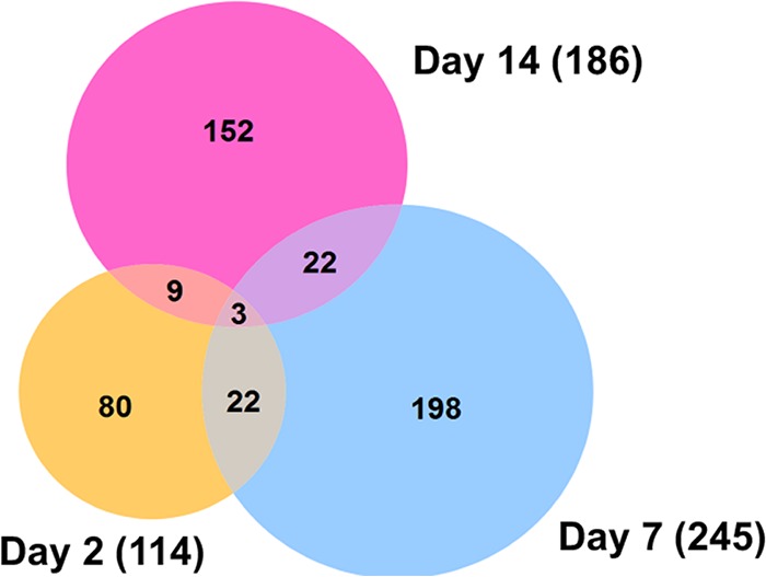
FIG 6 .

Venn diagram representing the number of differentially expressed lincRNAs at three different time points post-ZIKV infection (fold change of >2 and P value of <0.05). The majority of altered lincRNAs were found at 7 dpi, and 56 out of these lincRNAs showed significant alteration at least at two time points.
