Abstract
Nanoparticles provide new fields for life medical science application, including targeted-drug delivery and cancer treatment. To maximize the delivery efficiency of nanoparticle, one must understand the uptake mechanism of nanoparticle in cells, which may determine their ultimate fate and localization in cells. Recently, the proposed-cancer stem cell (CSC) theory has been attracted great attention and regarded as new targets for the new nanodrug developmet and cancer therapies. The interaction between nanoparticles and cancer cells has been extensively studied, but the uptake mechanism of nanoparticles in CSCs has received little attention. Here, we use the pharmacological inhibitors of major endocytic pathways to study the silica nanoparticle (SiNP) uptake mechanisms in the human breast adenocarcinoma cell line (MCF-7) and MCF-7-derived breast cancer stem cells (BCSCs). The results demonstrate that the uptake of SiNPs, particularly amino-functionalized SiNPs, in MCF-7 cells is strongly affected by the actin depolymerization, whereas BCSCs more strongly inhibit the amino-functionalized SiNP uptake after the scavenger receptor disruption. These findings indicate a distinct endocytic mechanism of functionalized SiNPs in BCSCs, which is significant for designing ideal nanosized drug delivery systems and improving the selectivity for CSC-targeted therapy.
Introduction
Nanoparticles (NPs) are vital tools in the developing field of biology and nanomedicine; they provide novel ideas for life medical science application, including drug delivery in cancer treatment1–3 and gene therapy4,5. These NPs enable specific modifications to bind to the targeted cell plasma membranes and enter into cytoplasm or nuclear with longer circulation half-lives and reduced toxicity of the normal tissue. To improve the therapeutic efficacy of nanomedicine, a thorough understanding of NPs uptake mechanisms in cells is required to strengthen the delivery efficiency6. Especially, understanding the uptake mechanisms by which NPs are delivered and entered into cell can supply delivery strategies with high targeting efficiency and minimal side effect7.
Breast cancer has different subtypes, is regarded as malignant neoplasms with a multidrug-resistant property and high lethality rate worldwide8. The multidrug-resistant of a cancer is considered related to small populations of cancer stem cells (CSCs) in the tumors. The proposed-CSC theory indicates that a small population of tumor cells has the ability of self-renewal, cancer-initiating, differentiation and metastasis. CSCs have higher chemotherapeutic resistant ability than most differentiated cancer cells due to the higher expression of drug resistance and anti-apoptotic genes than differentiated cells9. If so, a very small number of CSCs can preferentially survive from chemotherapy, even in the case where an apparently suppression of the tumors was observed. This hypothesis is consistent with the studies that chemotherapies that efficiently suppress the tumor reformation rarely inhibit metastasis. In this, CSC-targeted therapy is destined to be a core to development effective anticancer therapeutics. Nanomedicine has an enormous potential in the exploration of CSC-targeted drugs, development of novel gene-specific drugs, controlled drug delivery and release and diagnostic modalities10,11. However, the efficiency of nano-based therapy targeted to CSCs is far lower than those targeted to cancer cells12. To maximze the efficiency of NP delivery to CSCs, we must understand the uptake mechanisms by which NPs are internalized by CSCs, which possiblely determines their final sub-cellular fate, localization in cells, and efficacy of the cancer treatment. In recent years, scientists have been investigating different mechanisms to understand the cellular internalization processes of NPs with different sizes, shapes, surface charges, and surface chemistry in living cancer cells13, which includes clathrin-mediated (CME) and caveolae- and clathrin-independent endocytic mechanism, and phagocytosis. However, the cellular internalization processes of NPs into CSCs are not clear. Understanding the mechanisms of NP cellular internalization may be significant to develop ways to let NPs enter to the nucleus or other organelles for high curative effect or directly deliver nanomedicine to the lesion site by specific surface modification. Recently, inorganic-based nanocarriers (such as silica nanoparticles, SiNPs) have major breakthroughs on the morphology control, temporal control, and surface modification, which provided a great potential for the drug delivery14. It has reported that the surface of SiNPs can be easily functionalized with a specific group for targeted release of drugs or genes, which highlight SiNP as potential vehicle for therapeutic applications in biomedical science15. In our work, the major endocytic pathways are investigated to understand the carboxyl- and amino-functionalized SiNP uptake mechanisms in MCF-7 and MCF-7-derived CSCs (BCSCs) using seven pharmacological inhibitors. The inhibitors examined in this work are as follows: genistein, which inhibits tyrosine kinases in caveolae-mediated endocytosis16; chlorpromazine (CPZ), an inhibitor of the clathrin disassembly and receptor recycling to the plasma membrane during CME17; nocodazole, a microtubule-disturbing agent18; cytochalasin D, disturbs the actin filaments in cells18; Dynasore, which is an inhibitor of dynamin function7; Nystain, which interacts with cholesterol7; and Poly-I, which is an inhibitor of scavenger receptor19. Specially, we determined whether carboxyl- and amino-functionalized SiNPs showed different effects on the cell uptake. The cellular internalization of SiNPs was determined by confocal microscope and transmission electron microscope (TEM) imaging. The inhibition of SiNP uptake rate in the MCF-7 and BCSCs was quantified by flow cytometry relative to the control groups in the absence of pharmacological inhibitors. Importantly, these SiNPs have different uptake mechanisms in MCF-7 and BCSCs, which provide a potential platform for the CSC targeting drug delivery. The result has significance in the design and functionalization of nano-based drug delivery for CSC-targeted therapy.
Results
Characterization of Dye-Loaded Functionalized-SiNPs
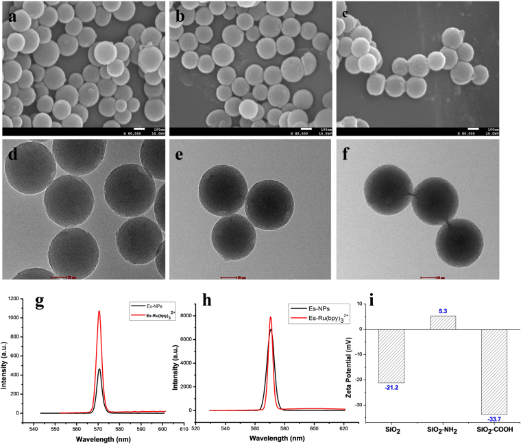
SiNPs with different surface groups were successfully prepared, and the average size and shape were determined by TEM and SEM by measuring the size of approximately 100 ± 10 nm (Fig. 1a–f). The amino- and carboxyl-functionalized SiNPs were characterized by IR (Figure S1). Because SiNPs were encapsulated with Ru(bpy)3 2+, their emission wavelengths were verified using a fluorescent spectrometer. SiNPs that were excited at 572 nm had the peak of emission at 570 nm (Fig. 1g and h). Figure 1i shows that the zeta potential of the unfunctionalized SiNPs (SiNPs-OH) was approximately −21.2 mV, i.e., in the absence of an -NH2 group and -COOH. However, when functionalized with the -NH2 group or -COOH group, the zeta potential was approximately 5.3 mV and −33.7 mV, respectively.
Figure 1.
Representative SEM and TEM images of SiNPs with the (a and d) -OH group, (b and e) -NH2 group, and (c and f) -COOH group; (g and h) Fluorescence spectra of SiNPs; (i) zeta potential of SiNPs.
BCSCs Expressed Markers of Various Stem Cells
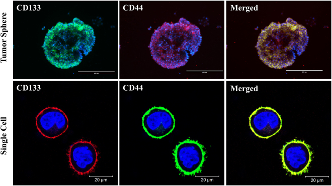
The tumor spheres and single BCSC (digested from the tumor sphere), which were cultured and passaged with completed culture medium, could maintain a stable morphological phenotype. Immunofluorescence staining shows that the tumor-sphere-derived BCSCs could stably express cancer stem cell markers CD44 and CD133 (Fig. 2).
Figure 2.
Immunofluorescence detection of the stemness marker expression in the tumor sphere and single BCSC.
In vivo Tumorigenesis of BCSCs
As shown in Fig. 3, palpable tumors were visible after a week in the BCSC-injected mice, whereas a bubble growth was observed in MCF-7-injected mice. Additionally, the tumor formation of the enriched BCSCs was faster, which resulted in a sharper tumor growth than that observed after the injection of MCF-7 cells. These data suggest that the BCSC can initiate breast cancer in nude mice.
Figure 3.
Tumor formation in nude mice after the injection of (a) BCSC and (b) MCF-7.
Interactions of SiNPs with MCF-7 cells and BCSCs
Cells were treated with SiNPs-OH, SiNPs-NH2, and SiNPs-COOH at non-cytotoxic concentrations (25, 50, 100, and 200 μg/mL) for 4 and 24 h, respectively (Figure S2). As shown in the flow cytometry, the median fluorescence intensity (MFI) of SiNP-treated cells increased with a time- and dose-dependent manner. Compared to the MFI of non-treated cells, there was a significant difference between control and treatment groups after 4 h and 24 h incubation (Fig. 4). Figure 4 further illustrates that the cellular uptake of SiNPs-NH2 in both MCF-7 and BCSCs was significantly higher than those of SiNPs-OH and SiNPs-COOH. In addition, the cell internalization generally increased with the dose.
Figure 4.
MFI of at least 10,000 (a and b) MCF-7 cells or (c and d) BCSCs, which was analyzed by FCM without or with SiNPs-OH, SiNPs-NH2, and SiNPs-COOH treatment for 4 and 24 h. The data are expressed as the mean ± SD; n = 5 of one of 3 independent experiments. *p < 0.05; **p < 0.01.
SiNPs Localization in Lysosome
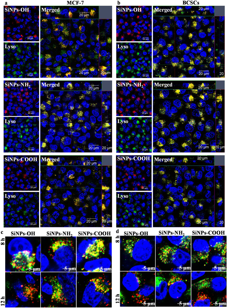
To determine the subcellular localization of SiNPs, the cells were respectively treated with SiNPs-OH, SiNPs-NH2, and SiNPs-COOH and subsequently stained with organelle-specific fluorescent markers, which including Hoechst 33342, LysoTracker Green to visualize inside the cells by z-stack imaging. The merged images reveal that SiNPs-OH, SiNPs-NH2, and SiNPs-COOH co-localized with lysosomes at 4h (Fig. 5a and b). Figure 5c and d showed that the SiNPs firstly entered the cells and localized in lysosomes, and then the SiNPs escaped into the cytoplasm. The overlap rate of SiNPs with lysotracker reduced to around 70% and 49% after 8 h and 12 h incubation (Figure S6b and S6c), respectively. Magnification images and 3D images of the co-localization of SiNPs-OH, SiNPs-NH2, and SiNPs-COOH with lysosome are shown in Figures S3 to S6. The serial z-section images of the cells show the increase in fluorescence intensity from the surface of the cells, which indicates that the NPs were internalized by the cells and not simply bound to their surface.
Figure 5.
Z-stack images of the SiNPs-OH, SiNPs-NH2, and SiNPs-COOH co-localized with lysosome in (a) MCF-7 and (b) BCSCs. Lysosome escape of SiNPs in (c) MCF-7 and (d) BCSCs.
Analysis of the Cellular Uptake and Location of SiNPs by TEM
To determine whether the SiNPs are taken up by the cells, and if they are, where the SiNPs localize in the cell, TEM images were recorded for MCF-7 and BCSCs that were treated with 200 μg/mL NPs for 24 h. As shown in Fig. 6, most cells were intact, and SiNPs were present in the lysosomes and cytoplasm. Not much cell debris was observed by TEM at this concentration. The nucleus can be identified, and the images consistently indicate that NPs were not present in the nucleus.
Figure 6.
TEM images of (a–d) MCF-7 and (e–h) BCSC cellular uptake of (b and f) SiNPs-OH, (c and g) SiNPs-NH2 and (d and h) SiNPs-COOH after 24 h of incubation (scale bar: 5.0 μm). The magnified images of the area are indicated by the white box (scale bar: 500 nm).
A Distinct Mechanism of SiNP Uptake in BCSCs
To understand the major endocytic pathways, the cells were treated with seven inhibitors: CPZ, nystatin, Poly-I, dynasore hydrate, cyto D, nocodazole, and genistein. Prior to the use of inhibitors, the energy-dependent internalization of SiNPs was studied as a common negative control. We conducted these experiments at 4 °C, which sharply decreased the BCSC and MCF-7 uptake of SiNPs-OH, SiNPs-NH2, and SiNPs-COOH (concentration: 200 μg/mL), relative to the uptake and transport at 37 °C (Fig. 7a and b). The uptake inhibition of SiNPs-OH, SiNPs-NH2, and SiNPs-COOH was studied using flow cytometry. As shown in Fig. 7c, cyto D dramatically reduced uptake (>75%) in all treatment groups in MCF-7 and produced a much smaller effect in BCSCs (around 30%). Confocal results indicated that the actin of BCSCs demonstrated a thinner actin network with stress fibers, whereas in the present F-actin inhibitor (cyto D), the actin filaments and stress fibers were destroyed. Consequently, the uptake of SiNPs by cells was inhibited in the present of cyto D (Figure S7). In addition, negative internalization scores were obtained for CPZ in MCF-7 and BCSCs, which suggests low internalization after the treatment with this inhibitor. After the treatment with nystatin, nocodazole, genistein, and dynasore hydrate, the internalization value was similar to the positive control, which indicates no significant inhibition of SiNP internalization with these inhibitors (Fig. 7c–h). However, the BCSC uptake of SiNPs was sharply reduced because of the inhibition of the scavenger receptor via the Poly-I treatment. Similar results were obtained for PLAG nanoparticles and nanoliposomes (Figure S8).
Figure 7.
Percentage of cellular uptake of SiNPs-OH, SiNPs-NH2, and SiNPs-COOH in MCF-7 and BCSCs in the presence of different endocytic inhibitors. (a) and (b) Energy-dependent internalization of NPs; (c) cyto D; (d) CPZ; (e) nystatin; (f) nocodazole; (g) Dynasore; (h) Genistein; (i) poly I. The values are the mean ± SD from three independent experiments. *p < 0.05; **p < 0.01 BCSCs versus MCF-7 in each group of SiNPs. #p < 0.05 SiNPs-OH versus SiNPs-NH2 versus SiNPs-COOH in each cells type.
As shown in Fig. 8, the internalization of SiNPs-NH2 in BCSCs was significantly inhibited after the Poly-I treatment. For the cyto D treatment, notably few SiNPs-NH2 were observed in BCSCs, although the uptake of SiNPs-NH2 in BCSCs was sharply reduced. Conversely, the uptake of SiNPs-NH2 in MCF-7 was arrested by the treatment with cyto D but not Poly-I. This finding is consistent with the flow cytometry result in Fig. 7. To understand the role of scavenger receptor in the cellular uptake of SiNPs, we analyzed the expression of the scavenger receptor in BCSCs. The results indicated that the level of scavenger receptor was up-regulated after the SiNP treatment compared with MCF-7 (Figure S11). In addition, SiNPs-NH2 adsorb the proteins on to the surface when incubated in medium, forming an protein-SiNP s-NH2 complex (Figure S10), which are redirected to scavenger receptors (Fig. 9a and b). Thus, the SiNPs-NH2 mimic the complex structure that Poly-I forms to bind to scavenger receptors, which is activated for endocytosis. To confirm this finding, we conjugated the anti-scavenger receptor class B number 1(SCARB1) to the surface of SiNPs to study the efficiency of uptake in BCSCs. As shown in Fig. 9c and d, a significant uptake was observed after antiSR-SiNPs (100 μg/mL) at 1 h incubation compared to SiNPs, indicating a specific binding of SR-SiNPs to scavenger receptor, which promoted the uptake efficiency of NPs in a short time. This finding showed that scavenger receptor plays an important role in uptake of SiNPs in BCSCs, suggesting a rational design approach to target NPs to cancer stem cells.
Figure 8.
TEM images of the internalization inhibition of SiNPs-NH2 in MCF-7 and BCSCs after the Poly-I and cyto D treatments (scale bar: 5.0 μm). The magnified images of the area are indicated by the white box (scale bar, 500 nm).
Figure 9.
(a) BSA and (b) scavenger receptor content of protein-SiNP complexes. Protein-SiNP complexes formed from SiNPs-NH2 bind to scavenger receptors while complexes formed from SiNPs-COOH bind to native protein receptors. (c) Confocal images of the SiNPs-NH2 and SR-SiNPs-NH2 co-localized with lysosome in BCSCs after 1 h incubation. (d) MFI of at least 10,000 BCSCs, which was analyzed by FCM without or with SiNPs-NH2 and SR-SiNPs-NH2 treatment for 1 and 24 h. *p < 0.05; **p < 0.01 BCSCs versus MCF-7 in each group of SiNPs. ##p < 0.01 SiNPs-OH versus SiNPs-NH2 versus SiNPs-COOH in each cells type.
Nanoparticles can localize the tumor site through the leaky blood vessel, which called the enhanced permeation and retention (EPR) effect. Two major methods are reported when nanocarriers target to tumour cells by EPR effect: passive targeting and active targeting. NPs whose surface is tailored (such as positive and negative charges) for prolonged blood circulation times, the concept is referred to as passive targeting. Targeting ligands, which bind to specific receptors on the tumour cells and endothelium, can be attached on the nanocarrier surface, this is called active targeting. Both passive and active nano-based drug delivery enhance the delivery efficiency of non-soluble or easily degradable therapeutic agents, and increase the therapeutic efficacy with minimal side effects through the different uptake mechanism (Figure S9).
Discussion
Currently, increasingly many in vitro experiments to understand the endocytic mechanisms of NPs are typically conducted in either suspension cells or adherent cancer cells in nanomedicine-related fields for cancer treatment21–23. However, mechanisms by which NPs internalize the CSCs have been poorly defined. CSCs can more strongly initiate tumors and resist drugs than other cancer cells, which results in more inefficiency in CSC-targeted drug delivery. Therefore, we hypothesized that different internalization and transport processes were responsible for the cellular uptake in CSCs and cancer cell line. To test this hypothesis, a systematic study was present to understand the endocytic mechanisms of fluorescent-dye-loaded SiNPs-OH, SiNPs-NH2, and SiNPs-COOH in BCSCs and MCF-7, which provides an insight into the NP interaction with BCSCs, which may be different from other cancer cell lines. Figure 4 shows that the chemical groups play a prominent role in the uptake of NPs, where positively charged SiNPs-NH2 are taken up more readily than their negatively charged counterparts (SiNPs-OH and SiNPs-COOH) of identical diameters (Fig. 1). This result is perhaps not surprising considering that CSCs display a net negative surface charge because there are negatively charged extracellular plasma membrane protein moieties24. Therefore, the positively charged SiNPs-NH2 are expected to electrostatically interact with the plasma membrane, and this interaction may promote the SiNPs-NH2 association with the cells and induce the cell uptake via cellular endocytic mechanisms. Similarly, with the internalization of poly(ethylene glycol)-D, L-polylactide (PEG-PLA) NPs with a positive charge were twice as high as the internalization of PEG-PLA (negatively charged) NPs in polarized epithelial Madin-Darby Canine Kidney (MDCK) cells25. Since the NP uptake into cells can experience two major processes (phagocytosis and pinocytosis)26, we performed several studies using metabolic inhibitors. Phagocytosis mainly occurs in a small population of mammiferous cell types such as macrophages and monocytes. Pinocytosis is a common process has two subcategories: macropinocytosis and micropinocytosis. Macropinocytosis plays a major role in the non-specific uptake of soluble macromolecules. However, micropinocytosis happens for smaller particles in all cell types27. First, we showed that the internalization of SiNPs by CSCs was sensitive to temperature (Fig. 7a and b), which confirms the involvement of energy-dependent active processes. Then, we studied the cellular uptake mechanisms of SiNPs-OH, SiNPs-NH2, and SiNPs-COOH in MCF-7 and BCSCs by using seven endocytic inhibitors. Initially, we concentrated on three major pincytic uptake pathways: macropincytosis, clathrin-dependent, and caveolea/lipid raft-dependent micropinocytosis via treatment with nocodazole, CPZ and nystatin. There is no any uptake suppression was observed in MCF-7 and BCSCs for SiNP-OH, SiNP-NH2, and SiNP-COOH (Fig. 7d–f). The cytoskeleton plays a signigicant role in various cellular activities, including the formation of endocytosis vesicles in the endocytic progress. To investigate the function of the cytoskeletal reorganization on the SiNP uptaken in cells, we chose two inhibitors: cytochalasin D (cyto D) and nocodazole, which disturb the F-actin organization and microtubule formation, respectively28. As shown in Fig. 7c, for MCF-7, cyto D inhibited the endocytosis of SiNPs-OH, SiNPs-NH2, and SiNPs-COOH by approximately 80%, 85%, and 80% relative to the control, respectively. This result suggests that actin filaments are strongly involved in the endocytic precess of SiNPs and particularly SiNPs-NH2 in the MCF-7 cell line. However, only approximately 20% reduction for SiNPs uptake in BCSCs was observed, indicating a less F-actin-dependent uptake of SiNP for BCSCs. The result can be explained by a different inhibition efficiency induced by cyto D in different cell lines. It has reported that microtubules play a key role in the process of endocytosis, and nacodazole disrupts the microtubule. As shown in Fig. 7f, only 5% inhibition rate of SiNPs-COOH was obtained relative to the control group in MCF-7, and no inhibition of uptake of other SiNPs was observed in BCSC. Thus, microtubules did not involve in the uptake of SiNPs in MCF-7 and BCSCs. It has reported that the clathrin-dependent pathway can regulate the uptake of SiNPs29. In this study, chlorpromazine (CPZ), which is an inhibitor of clathrin-mediated encytosis, was used to investigate the internalization of SiNPs. The data showed that the SiNP uptake by MCF-7 and BCSCs slightly decreased after the clathrin inhibition (Fig. 7d). However, the non-uptake inhibition was obtained for SiNPs in the cells treated with nystatin, which indicated that the uptake of SiNPs is irrelevant to the lipid raft-dependent uptake mechanisms (Fig. 7e). Lately, the dynamin-dependent endocytic mechanism has been widely studied as a possible clathrin-independent endocytic pathway into cells30. However, our results did not have any significant changes of cellular uptake after treatment with dynasore, which disrupts the dynamin-dependent pathways. These results indicate that macropinocytosis, clathrin-dependent endocytosis, and lipid raft-dependent endocytosis are not the major entry mechanisms of SiNPs in MCF-7 and BCSCs. To evaluate whether a more specific endocytic mechanism is involved for the SiNP uptake in MCF-7 and BCSCs, we discussed another two uptake pathways in MCF-7 and BCSCs, including the scavenger receptors and membrane-bound G-protein coupled receptor (GPCR)-mediated uptake. Scavenger receptors have been reported to be involved in AuNP uptake through binding to various ligands, such as low-density lipoproteins and polysaccharides31. Poly-I, which is a widely accepted inhibitor of scavenger receptors, supressed the SiNP enter into BCSCs (Figs 7i and 8), which confirms the function of scavenger receptors in the endocytic process of SiNP in different cell lines. In this work, the uptake inhibition was significantly inhibited for SiNPs in breast cancer stem cells treated with Poly-I, but no similar results was observed in MCF-7 cells. All results with different endocytic pathways inhibitors in MCF-7 and BCSCs are summarized in Table 1.
Table 1.
Summary of the uptake inhibition of SiNPs in the presence of endocytic inhibitors.
| Inhibitor | Function | SiNP-OH | SiNP-NH2 | SiNP-COOH | |||
|---|---|---|---|---|---|---|---|
| MCF-7 | BCSC | MCF-7 | BCSC | MCF-7 | BCSC | ||
| cyto D | Inhibits F-actin polymerization | ++ | + | ++ | + | ++ | + |
| Nocodazole | Disruption of microtubules | − | − | − | − | − | − |
| CPZ | Inhibits clathrin | + | + | + | + | + | + |
| Dynasore | Inhibits dynamin-GTPase | − | − | − | − | − | − |
| Nystatin | Lipid-raft inhibitor | − | − | − | − | − | − |
| Poly I | Scavenger receptor inhibitor | − | ++ | − | ++ | − | ++ |
| Genistein | Inhibitor of several tyrosine kinases | − | − | − | − | − | − |
+p < 0.05, ++p < 0.01 through unpaired t-test between control and inhibitor-treated groups. − no significant inhibition.
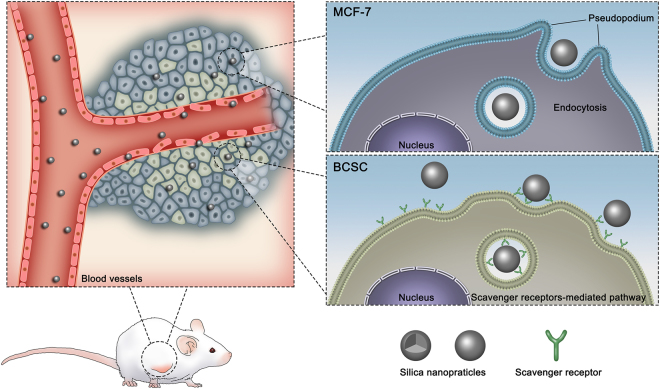
Overall, a large range of endocytic pathways, i.e., macropinocytosis, clathrin-mediated endocytosis, and caveplae/lipid raft-mediated endocytosis, are not involved in the uptake for SiNPs in MCF-7 and BCSCs. However, for the MCF-7 cell line, strongly mediated uptake was observed only via actin filament endocytosis. For BCSCs, both actin filament endocytosis and scavenger receptor-induced uptake were primary for all SiNPs. The actin is a major part of the cytoskeleton and a large number of evidence showed that F-actin plays a vital role in the process of internalization. Recent microscopy studies provide strong evidence that formation and organization of actin occurs at endocytic vesica formation sites, where create protrusions that encompass extracellular materials18,32–35. In this work, we evaluated how the scavenger receptor facilitates the SiNPs uptake into BCSCs through inhibited the expression of F-actin. The results indicated that the actin of BCSCs demonstrated a thinner actin network with stress fibers, whereas in the present F-actin inhibitor (cyto D), the actin filaments and stress fibers were destroyed. Consequently, the uptake of SiNPs by cells was inhibited in the present of cyto D. It has reported that several proteins (including SCAR/WASP) are directly participated in mediating actin organization and could thus control forces production during actin organization to promote specific steps in the endocytosis36–38. The expression of the scavenger receptor in BCSCs was up-regulated after the SiNP treatment compared with MCF-7 (Figure S11). In addition, both cationic and anionic SiNPs adsorb the proteins on to the surface when incubated in medium, forming an anionic protein-SiNP complex (Figure S10). We determined that protein-SiNP-COOH complexes bind to albumin receptors on the cell surface. Protein-SiNP-NH2 complexes are redirected to scavenger receptors (Fig. 9a and b). Thus, the SiNPs mimic the complex structure that Poly-I forms to bind to scavenger receptors, which is activated for endocytosis. Indeed, the reduction in density of gold nanoparticles reportedly decreases the Poly-I-dependent inhibition of NP uptake19,31. In addition, the chemical groups of SiNPs play a crucial role in terms of dictating their interaction with the cells. The positively charged SiNPs-NH2 more efficiently associate with the cells than the negatively charged SiNPs-OH and SiNPs-COOH and consequently induce the cell uptake via cellular endocytic mechanisms. A schematic model to illustrate different endocytic mechanisms of SiNPs in breast cancer cells and BCSCs is shown in Fig. 10.
Figure 10.
Schematic model to illustrate different endocytic mechanisms of SiNPs in breast cancer cells and BCSCs.
Conclusion
In conclusion, SiNPs can internalize in notably different cells through different endocytic pathways. This study highlights the complexity and interaction among the endocytic mechanisms by which different cell lines can uptake SiNPs and suggests that the SiNPs with the same functionalization may be uptaken via different endocytic mechanisms in MCF-7 and BCSCs. We suggest that for MCF-7 cells, the uptake of SiNPs was inhibited by treatment with Cyto D, indicating a F-actin-dependent pathway. In contrast, SiNPs are internalized by BCSCs via a different route that involves the scavenger-receptor-mediated endocytosis pathway. Importantly, positively charged SiNPs have higher cell uptake than negatively charged SiNPs in both MCF-7 and BCSCs, which is important in designing drug carriers that can cross the mucosal barriers and enable a noninvasive delivery of biological therapeutics. In addition, the presence of different endocytic mechanisms for functionalized SiNPs in tumor cells and CSCs provides a valuable platform to design nanocarriers of cancer-stem-cell-targeted therapeutics.
Methods
Materials
Tetraethyl orthosilicate (TEOS) and tris(2,2′-bipyridyl)dichlororuthenium(II) hexahydrate (Rubpy) was obtained from Acros Organics. Triton X-100 and 3-aminopropyl trimethoxysilane (APTES) were obtained from Acros. Carboxyethylsilanetriol sodium salt (CTES) (25 wt.% in water) was bought from Sigma, USA. Genistein, nystatin, chlorpromazine hydrochloride (CPZ), cyto D, nocodazole, dynasore and Poly-I were obtained from Sigma-Aldrich. The cell culture reagents were purchased from HyClone. The used reagents are of analytical grade. Anti-CD44-FITC, CD133-PE, and anti-Scavenger Receptor were obtained from Invitrogen.
Synthesis of Dye-loaded Silica Nanoparticles
SiNPs were synthesized by a water-in-oil (W/O) microemulsion method. Briefly, cyclohexane (7.5 mL), Triton X-100 (1.77 mL) and hexanol (1.8 mL) were mixed into the round-bottom flask with string for 10 min to compose the microemulsion system; then, H2O (340 μL), Ru(bpy)3Cl2 dye (80 μL of 0.1 M), and TEOS (100 μL) were added. Then the mixture was added 100 μL of aqueous ammonia (28–30 wt%) and stirred for 24 h at room temperature to complete the hydrolysis reaction. Finally, acetone was introduced to break the microemulsion; After centrifugation, the SiNPs were obtained and sequentially washed three times with acetone and deionized water.
Synthesis of Functionalized Silica Nanoparticles
The amino-group-functionalized SiNPs (SiNPs-NH2) were synthesized according to the reported method20. Briefly, 50 mg SiNPs was dispersed to 20 mL toluene by magnetic stirring; then, 50 μL APTES was added during the nitrogen protection; the reaction was continued for 24 h stirring. Subsequently, the SiNPs-NH2 were washed ethanol and deionized water for three times. The carboxyl group functionalized SiNPs (SiNPs-COOH) was synthesized according to the reported method20. In brief, 50 μL APTES and 0.02 mg succinic anhydride evenly dispersed in 20 mL dimethyl formamide for 3 h of stirring at 37 °C. Then, 20 mL SiNPs-NH2 by dimethyl formamide ultrasonic dispersion and 2 mL deionized water were added into the system and continuously stirred for 5 h. Finally, the SiNPs-COOH were washed with ethanol and deionized water for three times.
Characterization of Functionalized SiNPs
The diameter and morphology of the SiNPs were studied using a scanning electron microscope (SEM, Hitachi), a transmission electron microscope (TEM, Philips CM12). The size of SiNPs was studied by dynamic light scattering (DLS). The DLS values and zeta potential were tested through a DLS Particle Size Analyzer (Malvern ZetasizerNano ZS, UK). The fluorescence spectra were obtained at room temperature using a fluorescence spectrophotometer (Hitachi F-4600, Japan). The surface groups of carboxyl and amino were characterized by infrared spectroscopy (IR, ThermoFisher).
Isolation of MCF-7-derived Breast Cancer Stem Cells
Breast cancer cell line (MCF-7) was purchased from ATCC and cultured in Dulbecco’s Modified Eagle’s Medium (DMEM, with additional L-Glutamine) in present with 10% fetal bovine serum (FBS, Gibco) and 1% penicillin/streptomycin (Gibio) at the condition of 37 °C and 5% CO2. The culture medium was changed every three days.
BCSCs were isolated from the MCF-7 cell line and grown in a DMEM/F12 medium (Gibco) in presence of 20 ng/mL EGF, 10 ng/mL bFGF, 2% B27, 1% N2 (Invitrogen, USA), and 1% penicillin/streptomycin. Then, the 5,000 cells were seeded and incubated in ultra-low-attachment 6-well plates (Corning, USA) to form the tumor sphere.
Tumor Sphere Formation and Passage Assay
After gentle centrifugation, the tumor spheres were concentrated, and then digested with trypsin-EDTA and mechanically separated with a pipette. The digested single cells at a density of 5000 cells/well were washed with PBS and centrifuged to remove the trypsin-EDTA and re-seeded in a medium supplemented with growth factors to re-form tumor spheres. The re-formed spheres must be passaged every 10–12 days when the diameter reached to 200 μm.
Identification of BCSCs
For the surface marker analysis by confocal microscopy, the tumor spheres were digested from the culture plates using trypsin-EDTA (Invitrogen). Single MCF-7 and BCSCs were washed with PBS and stained with CD44-FITC and CD133-PE antibodies according to the manufacture. The results were monitored using a laser scanning confocal microscope (LSCM, ZEISS).
In vivo Tumorigenesis of BCSCs
BCSCs and MCF-7 cells were suspended in 100 μL sterile PBS at the density of 5000 cells and subcutaneously injected in the back of the nude mice. The mice were measured every five days for tumor growth for up to 30 days. All animal experiments were carried followed with the laboratory animal care and the guide for the care and use of laboratory animals. All experimental protocols were authorized by the administration office committee of laboratory animal of Hebei University.
In vitro Cytotoxicity of SiNPs
The viability of cells cultured with SiNPs was observed by 4,5-(dimethyl-2-thiazolyl)-2,5-diphenyl-2-H-tetrazolium bromide (MTT) assay23. SiNP solutions (20, 50, 100, and 200 μg/mL) incubated with cells for 24, 48, and 72 h, and then the medium was added 10 μL of MTT solution. The optical density (OD) of the medium at 570 nm was recorded with a microplate reader. The data are expressed as follows: (ODsample − ODblank)/(ODcontrol − ODblank) × 100%.
Treatments with Chemical Inhibitors
For the experiment of endocytic pathways suppression, 5 × 10⁴ cells were pre-treated with different endocytic inhibitors for 30 min at the following concentrations: genistein (10 μg/mL); Dynasore (20 μM); cyto D (5 μg/mL); polyinosinic acid potassium salt (Poly-I, 10 μg/mL); nocodazole (10 μg/mL); nystatin (10 μg/mL); CPZ (10 μg/mL). After these pre-treatments, SiNPs were introduced to the cell culture medium and incubated for 4 h. Then, the cells were washed three times with PBS and stored in darkness at 4 °C before the measurement.
Flow Cytometry Analysis of SiNP Uptake
The cells (5 × 10⁴ cells/well) were cultured on 6-well plates and added SiNPs when they had reached 70–80% confluence. The medium was removed, and the uptake medium containing 200 μg/mL SiNPs was introduced into cells and continued to culture for 4 and 24 h. The cellular uptake and inhibition of SiNPs were determined by flow cytometry (BD CantoII, USA).
SiNPs Uptake Using a Confocal Microscope
The cellular uptake and localization of fluorescent-dye-loaded SiNPs was imaged using a confocal microscope (ZEISS, LSM880, Germany). The cells (4 × 10⁴ cells/well) were treated with 200 µg/mL SiNPs, and then were fixed in 4% PFA for 15 min at room temperature and washed with PBS for 3 times. The nucleus was stained with Hoest33342. The cells were mounted with a cover slip for the confocal microscope.
Analysis of the Cellular Uptake and Location of SiNPs by TEM
4 × 10⁴ cells were incubated with 200 μg/mL SiNPs for 24 h. Then, the cells were first fixed in 2% buffered in 0.1 M sodium cacodylatefor 2 h. The samples were post-fixed with 2% OsO4 in the dark for 2 h. The samples were dehydrated through alcohol with gradient concentrations (10, 50, 70, 90, 95, and 100%) and further acetone dehydration (90, 96, and 100%). Finally, the samples were treated with propylene oxide: resin (1:1) overnight. Then, the ultra-thin sections (80 nm) was obtained and viewed under a TEM at 80 kV (Philips Tecnai 12 BioTWIN).
Western Blot
BCSCs and MCF-7 cells (2 × 10⁴ cells/well) were treated with SiNPs overnight in a normal cell culture medium. Then, the cells were rinsed 3 times with cold PBS and lysed in protein lysate. The collected protein from each sample was separated by 10% SDS-PAGE gel and transferred onto nitrocellulose membranes, which were blocked in a buffer contained 5% BSA in a TBST solution for 2 h and hybridized with anti-scavenger receptor overnight at 4 °C, and then incubated with peroxidase-conjugated secondary antibody and interacted with a chemoluminescence test kit (Thermo Fisher Scientific). β-Actin was used as the loading control.
Elisa assay
The SiNPs were incubated with cells in normal culture medium with 10% FBS for 24 h to evaluate the ability of protein absorption. Then, the zeta potential of protein-SiNPs was measured by DLS Particle Size Analyzer. The concentration of BSA and scavenger receptor was tested by using Bio-rad protein assay (Bio-rad) and elisa assay (Beyotime Biotechnology, China) according to the protocol.
Statistical Analysis
Each experiment was performed three times in quadruplicate. The statistical analysis was analyzed using Student’s t-test and analysis of variance (one-way ANOVA) followed by Bonferroni post hoc test for multiple comparisons, and the results were presented as the mean ± SD. Statistical significance was accepted at a level of p < 0.05.
Data Availability
All data generated or analysed during this study are included in this published article (and its Supplementary Information files).
Electronic supplementary material
Acknowledgements
This work was supported by Natural Science Foundation Project (31500812, 31470961, 21471044), Key Program of Natural Science Foundation of Hebei Provience (B2017201226, B2016201031), Natural Science Foundation of Hebei province (B2017201230, B2015201097, B2016201031,), Science and Technology Research Project of Higher Education Institutions in Hebei Province (QN2015230), Fund Program for the Scientific Activities of Selected Returned Overseas Professionals (CG2015003009), Hebei Province “Hundred Talents Program” (BR2-202), Hebei Province “Three Three Three Talents Program” (A201401002).
Author Contributions
Jinchao Zhang and Dandan Liu planned the project. Jiadong Sun, Yajing Liu, and Min Ge performed the cellular experiments. Dandan Liu analyzed data and wrote the paper. Wentong Sun and Guoqiang Zhou synthesized and characterized the nanoparticles. Jinchao Zhang and Xing-Jie Liang prepared Figs 1–9.
Competing Interests
The authors declare that they have no competing interests.
Footnotes
Jiadong Sun and Yajing Liu contributed equally to this work.
Electronic supplementary material
Supplementary information accompanies this paper at 10.1038/s41598-017-16591-z.
Publisher's note: Springer Nature remains neutral with regard to jurisdictional claims in published maps and institutional affiliations.
Contributor Information
Dandan Liu, Email: leo-liudan@163.com.
Xing-Jie Liang, Email: liangxj@nanoctr.cn.
Jinchao Zhang, Email: jczhang6970@163.com.
References
- 1.Tasciotti E, et al. Mesoporous silicon particles as a multistage delivery system for imaging and therapeutic applications. Nat. Nanotechnol. 2008;3:151–157. doi: 10.1038/nnano.2008.34. [DOI] [PubMed] [Google Scholar]
- 2.Sun Q, et al. Integration of nanoassembly functions for an effective delivery cascade for cancer drugs. Adv. Mater. 2014;26:7615–7621. doi: 10.1002/adma.201401554. [DOI] [PubMed] [Google Scholar]
- 3.Qiu LP, et al. A cell-targeted, size-photocontrollable, nuclear-uptake nanodrug delivery system for drug-resistant cancer therapy. Nano Lett. 2015;15:457–463. doi: 10.1021/nl503777s. [DOI] [PMC free article] [PubMed] [Google Scholar]
- 4.Liu X, et al. Fusogenic reactive oxygen species triggered charge-reversal vector for effective gene delivery. Adv. Mater. 2016;28:1743–1752. doi: 10.1002/adma.201504288. [DOI] [PubMed] [Google Scholar]
- 5.Zhang JX, Sun HL, Ma PX. Host–guest interaction mediated polymeric assemblies: multifunctional nanoparticles for drug and gene delivery. ACS Nano. 2010;4:1049–1059. doi: 10.1021/nn901213a. [DOI] [PMC free article] [PubMed] [Google Scholar]
- 6.Saha K, et al. Surface functionality of nanoparticles determines cellular uptake mechanisms in mammalian cells. Small. 2013;9:300–305. doi: 10.1002/smll.201201129. [DOI] [PMC free article] [PubMed] [Google Scholar]
- 7.Iversen T-G, Skotland T, Sandvig K. Endocytosis and intracellular transport of nanoparticles: Present knowledge and need for future studies. Nano Today. 2011;6:176–185. doi: 10.1016/j.nantod.2011.02.003. [DOI] [Google Scholar]
- 8.Pece S, et al. Biological and molecular heterogeneity of breast cancers correlates with their cancer stem cell content. Cell. 2010;140:62–73. doi: 10.1016/j.cell.2009.12.007. [DOI] [PubMed] [Google Scholar]
- 9.Baker M. Cancer stem cells tracked. Nature. 2012;488:13–14. doi: 10.1038/488013a. [DOI] [PubMed] [Google Scholar]
- 10.Rao W, et al. Augmented cancer destruction by nanodrug surface-decorated with chitosan to target CD44+ cancer cells. ACS Nano. 2015;9:5725–5740. doi: 10.1021/nn506928p. [DOI] [PubMed] [Google Scholar]
- 11.Liu Q, et al. Targeted delivery of miR-200c/DOC to inhibit cancer stem cells and cancer cells by the gelatinases-stimuli nanoparticles. Biomaterials. 2013;34:7191–7203. doi: 10.1016/j.biomaterials.2013.06.004. [DOI] [PubMed] [Google Scholar]
- 12.Zuo ZQ, et al. Promoting tumor penetration of nanoparticles for cancer stem cell therapy by TGF-beta signaling pathway inhibition. Biomaterials. 2016;82:48–59. doi: 10.1016/j.biomaterials.2015.12.014. [DOI] [PubMed] [Google Scholar]
- 13.Bannunah AM, Vllasaliu D, Lord J, Stolnik S. Mechanisms of nanoparticle internalization and transport across an intestinal epithelial cell model: effect of size and surface charge. Mol. Pharmaceutics. 2014;11:4363–4373. doi: 10.1021/mp500439c. [DOI] [PubMed] [Google Scholar]
- 14.Vivero-Escoto JL, Slowing II, Trewyn BG, Lin VS-Y. Mesoporous silica nanoparticles for intracellular controlled drug delivery. Small. 2010;6:1952–1967. doi: 10.1002/smll.200901789. [DOI] [PubMed] [Google Scholar]
- 15.Slowing II, Vivero-Escoto JL, Wu C-W, Lin VS-Y. Mesoporous silicananoparticles as controlled release drug delivery and gene transfection carriers. Adv. Drug Deliv. Rev. 2008;60:1278–1288. doi: 10.1016/j.addr.2008.03.012. [DOI] [PubMed] [Google Scholar]
- 16.Pelkmans L, Püntener D, Helenius A. Local Actin Polymerization and dynamin recruitment in SV40-induced internalization of caveolae. Science. 2002;296:535–539. doi: 10.1126/science.1069784. [DOI] [PubMed] [Google Scholar]
- 17.Wang LH, Rothberg KG, Anderson RG. Mis-assembly of clathrin lattices on endosomes reveals a regulatory switch for coated pit formation. J. Cell Biol. 1993;123:1107–1117. doi: 10.1083/jcb.123.5.1107. [DOI] [PMC free article] [PubMed] [Google Scholar]
- 18.Fujimoto LM, Roth JE, Heuser JE, Schmid SL. Actin assembly plays a variable, but not obligatory role in receptor-mediated endocytosis in mammalian cells. Traffic. 2000;1:161–171. doi: 10.1034/j.1600-0854.2000.010208.x. [DOI] [PubMed] [Google Scholar]
- 19.Patel PC, et al. Scavenger receptors mediate cellular uptake of polyvalent oligonucleotide-functionalized gold nanoparticles. Bioconjugate Chem. 2010;21:2250–2256. doi: 10.1021/bc1002423. [DOI] [PMC free article] [PubMed] [Google Scholar]
- 20.Graf C, et al. Surface functionalization of silica nanoparticles supports colloidal stability in physiological media and facilitates internalization in cells. Langmuir. 2012;28:7598–7613. doi: 10.1021/la204913t. [DOI] [PubMed] [Google Scholar]
- 21.Peer D, et al. Nanocarriers as an emerging platform for cancer therapy. Nat. Nanotechnol. 2007;2:751–760. doi: 10.1038/nnano.2007.387. [DOI] [PubMed] [Google Scholar]
- 22.Fowler R, et al. Nanoparticle transport in epithelial cells: pathway switching through bioconjugation. Small. 2013;9:3282–3294. doi: 10.1002/smll.201202623. [DOI] [PubMed] [Google Scholar]
- 23.Jayanth P, Labhasetwar V. Biodegradable nanoparticles for drug and gene delivery to cells and tissue. Adv. Drug Deliver. Rev. 2003;55:329–347. doi: 10.1016/S0169-409X(02)00228-4. [DOI] [PubMed] [Google Scholar]
- 24.Nangia S, Sureshkumar R. Effects of nanoparticle charge and shape anisotropy on translocation through cell membranes. Langmuir. 2012;28:17666–17671. doi: 10.1021/la303449d. [DOI] [PubMed] [Google Scholar]
- 25.Harush-Frenkel O, Rozentur E, Benita S, Altschuler Y. Surface charge of nanoparticles determines their endocytic and transcytotic pathway in polarized MDCK cells. Biomacromolecules. 2008;9:435–443. doi: 10.1021/bm700535p. [DOI] [PubMed] [Google Scholar]
- 26.He B, et al. The transport pathways of polymer nanoparticles in MDCK epithelial cells. Biomaterials. 2013;34:4309–4326. doi: 10.1016/j.biomaterials.2013.01.100. [DOI] [PubMed] [Google Scholar]
- 27.Zhang SL, Gao HJ, Bao G. Physical principles of nanoparticle cellular endocytosis. ACS Nano. 2015;9:8655–8671. doi: 10.1021/acsnano.5b03184. [DOI] [PMC free article] [PubMed] [Google Scholar]
- 28.Xia T, Kovochich M, Liong M, Zink JI, Nel AE. Cationic polystyrene nanosphere toxicity depends on cell-specific endocytic and mitochondrial injury pathways. ACS Nano. 2008;2:85–96. doi: 10.1021/nn700256c. [DOI] [PubMed] [Google Scholar]
- 29.Bale SS, et al. Nanoparticle-mediated cytoplasmic delivery of proteins to target cellular machinery. ACS Nano. 2010;4:1493–1500. doi: 10.1021/nn901586e. [DOI] [PubMed] [Google Scholar]
- 30.Mayor S, Richard EP. Pathways of clathrin-independent endocytosis. Nat. Rev. Mol. Cell Bio. 2007;8:603–612. doi: 10.1038/nrm2216. [DOI] [PubMed] [Google Scholar]
- 31.Devika CB, Chan WCW. Elucidating the mechanism of cellular uptake and removal of protein-coated gold nanoparticles of different sizes and shapes. Nano letters. 2007;7:1542–1550. doi: 10.1021/nl070363y. [DOI] [PubMed] [Google Scholar]
- 32.Wang X, Galletta BJ, Cooper JA, Carlsson AE. Actin-Regulator Feedback Interactions during Endocytosis. Biophys. J. 2016;110:1430–1443. doi: 10.1016/j.bpj.2016.02.018. [DOI] [PMC free article] [PubMed] [Google Scholar]
- 33.Qualmann B, Kessels MM, Kelly RB. Molecular links between endocytosis and the actin cytoskeleton. J. Cell Biol. 2000;150:F111–116. doi: 10.1083/jcb.150.5.F111. [DOI] [PMC free article] [PubMed] [Google Scholar]
- 34.Pollard TD, Cooper JA. Actin, a central player in cell shape and movement. Science. 2009;326:1208–1212. doi: 10.1126/science.1175862. [DOI] [PMC free article] [PubMed] [Google Scholar]
- 35.Romer W, et al. Actin dynamics drive membrane reorganization and scission in clathrin-independent endocytosis. Cell. 2010;140:540–53. doi: 10.1016/j.cell.2010.01.010. [DOI] [PubMed] [Google Scholar]
- 36.Fleischer CC, Payne CK. Nanoparticle Surface Charge Mediates the Cellular Receptors Used by Protein-Nanoparticle Complexes. J. Phys. Chem. B. 2012;116:8901–8907. doi: 10.1021/jp304630q. [DOI] [PMC free article] [PubMed] [Google Scholar]
- 37.Uruno T, et al. Activation of Arp2/3 complex-mediated actin polymerization by cortactin. Nat. Cell Biol. 2001;3:259–266. doi: 10.1038/35060051. [DOI] [PubMed] [Google Scholar]
- 38.Taunton J, et al. Actin-dependent propulsion of endosomes and lysosomes by recruitment of N-WASP. J. Cell Biol. 2000;148:519–530. doi: 10.1083/jcb.148.3.519. [DOI] [PMC free article] [PubMed] [Google Scholar]
Associated Data
This section collects any data citations, data availability statements, or supplementary materials included in this article.
Supplementary Materials
Data Availability Statement
All data generated or analysed during this study are included in this published article (and its Supplementary Information files).


