. 2017 Nov 10;6:e29626. doi: 10.7554/eLife.29626

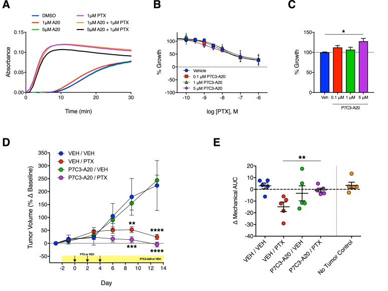
Figure 8. Antitumoral efficacy of PTX is maintained in the presence of P7C3-A20.

(A) Tubulin polymerization curves corresponding to addition of P7C3-A20 with and without PTX (n = 3 independent experiments). (B) Concentration-dependent anti-proliferation of PTX (48 hr) in MDA-MB-231 breast cancer cells pretreated (1 hr) with vehicle or P7C3-A20 (0.1–5 μM). (C) Effects of P7C3-A20 treatment only on growth of MDA-MB-231 cells. *p<0.05 vs. Veh by one-way ANOVA followed by Dunnett’s post-hoc test, n = 3 independent experiments. (D) Timecourse of changes in MDA-MB-231 tumor volumes in female athymic nude mice treated with P7C3-A20 (20 mg/kg/day, i.p.) or vehicle and PTX (11.7 mg/kg, i.p.) as indicated. ****p<0.0001, ***p<0.001, **p<0.01 vs. Veh/Veh by two-way mixed-effect ANOVA with Dunnett’s post-hoc test, n = 8–9 tumors/group. (E) Mechanical AUCs from tumored mice. Control mice lacking tumors were tested concurrently with the tumored mice. **p<0.01 by one-way ANOVA followed by Sidak’s post-hoc test, n = 5 mice/group.

Figure 8.

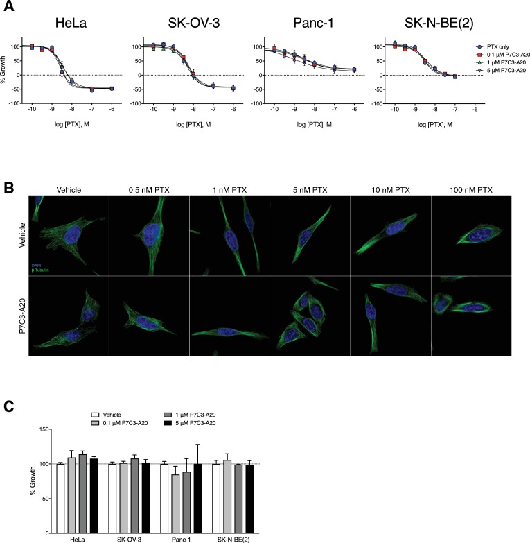
Figure 8—figure supplement 1. P7C3-A20 does not alter the anti-proliferative or microtubule-stabilizing capacity of PTX in vitro.

Figure 8—figure supplement 1.

(A) Effect of various concentrations of P7C3-A20 on concentration-response curves for PTX to inhibit growth of several cancer cell lines with distinct genetic backgrounds. P7C3-A20 did not alter the potency or efficacy of PTX to inhibit growth. Cells were treated for 48 hr and cell growth was determined with the SRB assay. (B) P7C3-A20 did not alter the formation of perinuclear microtubule bundles by PTX. Immunofluorescent images of MDA-MB-231 cells pretreated with P7C3-A20 (5 μM) or vehicle for 1 hr followed by various concentrations of PTX or vehicle for an additional 4 hr and then stained for β-tubulin. (C) Effect of P7C3-A20 on the growth of various cancer cell lines. P7C3-A20 had no effect on the growth of the cell lines tested. Cells were incubated with P7C3-A20 (0.1–5 μM) or vehicle for 48 hr. Growth was monitored with the SRB assay.
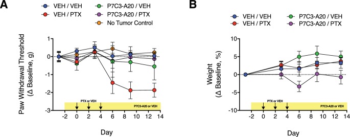
Figure 8—figure supplement 2. Effect of P7C3-A20 on PTX-induced mechanical allodynia and body weight in mice with implanted MDA-MB-231 tumor xenografts.

Figure 8—figure supplement 2.

(A) Athymic nude mice received bilateral injections into both flanks with MDA-MB-231 cells. When tumors reached at least 250 mm3, mice were treated with P7C3-A20 (20 mg/kg/day; i.p.) or vehicle beginning on day −2. PTX (11.7 mg/kg, i.p.) or vehicle was injected on days 0, 2, and 4. Paw withdrawal threshold to mechanical stimulation of the hindpaw were measured on indicated days before and after PTX treatment. P7C3-A20 abolished the development of PTX-induced mechanical allodynia in tumored mice. The xenografts did not alter mechanical paw withdrawal threshold as indicated by the lack of difference between the responses from mice with or without implanted tumors. (B) Neither P7C3-A20 nor PTX affected the body weight of tumored mice. Data are expressed as mean ± SEM, n = 5 mice/group.