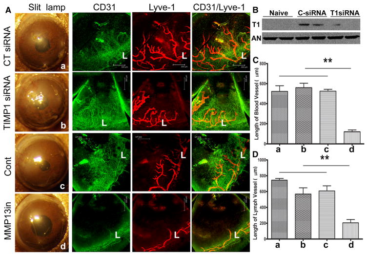
Fig. 5.
Effects of MMP13 and TIMP1 on angiogenesis in the sutured mouse corneas. A. B6 mouse corneas were subconjunctivally injected CT siRNA or TIMP1 siRNA twice 24 and 4 h or MMP13 inhibitor 4 h prior to suture. Three non-penetrating sutures were placed to the treated corneas. At 7 days post suture, the corneas were photographed, excised, and subjected to whole mount staining with CD31 for blood (green) and LYVE1 for lymph (red) vessels, respectively. L: limbus. B. Quantitation of blood vessels from the edge of the limbal region to the tips of vessels in the infected corneas. C. Quantitation of lymph vessels from the edge of the limbal region to the tip of vessels in the infected corneas. The results are representative of two independent experiments (N = 3 each) and indicated p values were generated using unpaired Student’s t test. **p < 0.01, *p < 0.05. (Color figure online)

