Abstract
Elevation of tendon core temperature during severe activity is well known. However, its effects on tenocyte function have not been studied in detail. The present study tested a hypothesis that heat stimulation upregulates tenocyte catabolism, which can be modulated by the inhibition or the enhancement of gap junction intercellular communication (GJIC). Tenocytes isolated from rabbit Achilles tendons were subjected to heat stimulation at 37 °C, 41 °C or 43 °C for 30 min, and changes in cell viability, gene expressions and GJIC were examined. It was found that GJIC exhibited no changes by the stimulation even at 43 °C, but cell viability was decreased and catabolic and proinflammatory gene expressions were upregulated. Inhibition of GJIC demonstrated further upregulated catabolic and proinflammatory gene expressions. In contrast, enhanced GJIC, resulting from forced upregulation of connexin 43 gene, counteracted the heat-induced upregulation of catabolic and proinflammatory genes. These findings suggest that the temperature rise in tendon core could upregulate catabolic and proinflammatory activities, potentially leading to the onset of tendinopathy, and such upregulations could be suppressed by the enhancement of GJIC. Therefore, to prevent tendon injury at an early stage from becoming chronic injury, tendon core temperature and GJIC could be targets for post-activity treatments.
Electronic supplementary material
The online version of this article (doi:10.1007/s12079-017-0397-3) contains supplementary material, which is available to authorized users.
Keywords: FLIP, Gap junction, Heat stimulation, Intercellular communication, Tendinopathy, Tenocytes
Introduction
Tendons are joint connective tissues, transmitting muscle force to bones during joint movements. Structural and mechanical integrity of tendons are maintained by a balanced activity between anabolism and catabolism of cells in tendons, called tenocytes. It has been commonly believed that one of major effectors of tenocyte functions is mechanical loading, possibly via mechanotransduction events to convert extrinsic mechanical cues to intracellular chemical signals at an individual cell level (Wang 2006). In addition, it is believed that the coordination of cellular activities through gap junction intercellular communication (GJIC) contributes to tenocyte responses to mechanical loadings (Wall and Banes 2005; Waggett et al. 2006).
Connexins are constitutive proteins of gap junctions. A unit of six connexin proteins, connexon (also known as hemichannel) on the surface of the plasma membrane of a cell binds to another connexon of the adjacent cell to form a gap junction. As intercellular communications, small molecules (nucleotides and miRNAs) as well as ions are transported between neighbouring cells through a central pore of the junction with a diameter of approximately 1 nm (Levin 2007). It has recently been demonstrated that the permeability of gap junctions between tenocytes is changed in response to mechanical loading (Maeda et al. 2012, 2017; Maeda and Ohashi 2015). Accordingly, it is suggested that the alterations in GJIC also influence behaviours of tenocytes as an organised multicellular system.
Chronic injury of tendon, generally termed as “tendinopathy”, is known to occur frequently in tendons subjected to a high level of repetitive mechanical loading, such as equine digital flexor tendon and human Achilles tendon (Patterson-Kane et al. 2012). When these tendons are under such severe activities, the temperature of tendon core is increased to the level between 43 °C and 45 °C (Wilson and Goodship 1994; Farris et al. 2011). Thus, in addition to the effects of overloading, effects of high temperature on tenocyte functions potentially have a significant impact on the onset of tendinopathy. So far, however, there has been only a limited amount of knowledge on how tenocyte functions are affected by the high temperature. For example, tenocyte survival rate was approximately 80% and 50% by the exposure of the cells to 45 °C for 30 and 60 min, respectively (Birch et al. 1997). It was also exhibited that tenocyte viability was dependent on both temperature and duration of heating treatment, and the synthesis of proMMP (matrix metalloproteinase) -9 from tenocytes were increased by a 10 min exposure of the cells to 40 °C (Hosaka et al. 2006). Detailed information of how tenocyte functions, such as the balance of anabolism and catabolism, are changed by the exposure to high temperature, and how these responses are related to GJIC remain to be investigated.
Therefore, the present study has been performed to test the hypothesis that the exposure of tenocytes to high temperature induces catabolic and proinflammatory responses, which can be modulated by the suppression or the enhancement of GJIC.
Materials and methods
Sample preparation
Tenocytes were isolated from the Achilles tendons of Japanese white rabbits (approximately 3-month-old) and cultured in DMEM supplemented with 10% of fetal bovine serum, 100 U ml−1 of penicillin, 100 μl ml−1 of streptomycin and 4 mM of L-glutamine (DMEM + 10%FBS, all from Gibco, USA). The Achilles tendon dissected in a sterile condition was minced and incubated for about 5 days. The cells migrating out from the tissue to the culture dish were collected by trypsinization. The cells were used in subsequent experiments described below between passage 1 and 4.
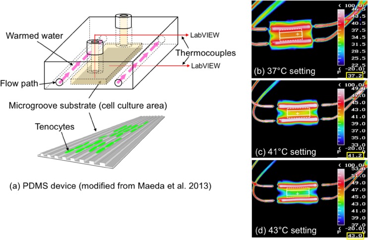
For the experiments, cell culture devices were made from polydimethylsiloxane (PDMS, Dow Corning), which was modified from the device that we developed previously (Maeda et al. 2013). The new device also consisted of two parts: an upper part and a microgroove membrane. The upper part was modified to equip flow paths for circulating warmed water, which enabled to control the temperature of cell culture area (Fig. 1). The microgroove membrane was fabricated through photolithography and soft-lithography, and the depth, the width and the spacing of microgrooves were all 10 μm. Microgrooves covered a rectangular area of 10 × 15 mm (white rectangular region with broken line in Fig. 1b-d). Isolated primary tenocytes were seeded on the microgroove membrane at a density of 200,000 cells ml−1 and were kept in an incubator at 37 °C and 5% CO2 overnight prior to the heating experiments. During heating experiments, the temperature of culture medium in the device and that of the microgroove substrate were monitored using thermocouples (Type-k, 20 μm in diameter, class 0.25) connected to LabVIEW software (National Instruments, USA).
Fig. 1.
Experimental device for the application of heat stress. a A schematic of PDMS device used for the experiments. The device was modified from the original reported in Maeda et al. 2013. b-d The temperature distribution around the cell culture area when the temperature monitored in the cell culture area was 37 °C b, 41 °C c or 43 °C d. Images were captured from the bottom of the PDMS device using a thermographic camera (TH7102WV, NEC Sanei Co., Japan). Rectangular region (white solid line) was selected for FLIP and cell viability analysis. For gene expression analysis, cells were collected from the entire microgroove substrate (rectangular region with white broken line). The temperature at the cross (+) mark in white is indicated in the right bottom of each image (enclosed by yellow rectangles)
Heating experiments
The device was taken out from the incubator and connected to a thermostat bath with a pumping function. The temperature of the bath was first set to let the temperature of the cell culture area reach and keep at 37 °C. To provide heat stress at 37 °C, 41 °C or 43 °C, the temperature of the bath was adjusted to reach the temperature of the cell culture area at a designated level and this was maintained for 30 min, followed by cooling down to 37 °C. Tenocytes in the device was analysed immediately after the heating experiment (0 h) or at the end of a 24-h post-incubation at 37 °C in the incubator (24 h).
Cell viability
Cell viability was assessed at the end of the 24-h post-incubation period by fluorescently labelling viable and dead cells using calcein-AM and ethidium homodimer (both from Thermo Fisher Scientific), respectively. Both dyes were dissolved in DMEM + 10%FBS at 5 μM and introduced to the cells in the device. The cells were incubated in the incubator for 1 h, followed by washing with DMEM + 10%FBS. Viable cells and dead cells were visualised on a fluorescence microscope using excitation wavelength at 488 nm and 545 nm, respectively. Due to the temperature distribution, the viability was examined within a central rectangular area (approximately 5 × 15 mm) of the microgroove pattern where the temperature distribution was uniform at the designated level (white rectangular region with solid line in Fig. 1b-d). A total of 10 small regions (520 × 440 μm each) was selected for imaging viable and dead cells, covering the rectangular area. In each of these regions, the number of each type of cells was manually counted, and cell viability was determined by dividing the number of viable cells by the total number of cells. An average viability of the ten regions represented the viability of the device, and three separate experiments were performed for the viability assay.
Quantitative evaluation of GJIC
To evaluate GJIC quantitatively, FLIP (fluorescence loss in photobleaching) protocol (Maeda et al. 2012) was employed with a spinning disk confocal laser scanning microscope system (Crest Optics, Italy and Molecular Device Japan, Japan) as described previously (Maeda et al. 2017). Briefly, tenocytes in the device were loaded with fluorescent tracer molecules, calcein-AM (Dojindo, Japan) at 5 μM before the FLIP protocol by incubating the cells at 37 °C/5% CO2 for 20 min. The device was mounted on the microscope, and tenocytes were visualised with a ×20 objective lens. One cell was selected as a target in a row of three or four tenocytes aligned and connected. The protocol consisted of a series of 100 consecutive steps of photobleaching of the target cell with a high-power laser at a wavelength of 488 nm and acquiring confocal fluorescence intensity of all the cells in the row with an excitation at 480 nm LED light immediately after each photobleaching. As the photobleaching was carried out every 3 s, the total duration of FLIP protocol was 300 s. To evaluate intracellular and intercellular diffusion coefficients from data obtained, temporal changes in the fluorescence intensity of the cells was obtained using ImageJ (version 1.48, NIH). Mathematical model of molecular transport (Maeda and Ohashi 2015), based on one-dimensional diffusion theory, was applied to the data to estimate intracellular diffusion coefficient (D C) and intercellular diffusion coefficient (D GJ) using MATLAB Optimization Toolbox (Mathworks, USA).
With a single device, FLIP experiment was performed up to 7 time in different locations within the device during about a 1 h period. In each experimental conditions, the number of the devices used was between 2 to 5. Some of the image sets obtained were excluded from subsequent analysis because of insufficient quality.
Real-time quantitative PCR
For samples assigned to qPCR, total RNA was extracted from the cells in the device in accordance with protocols supplied with RNeasy Plus Micro Kit (Qiagen, Germany). Twenty nanogram of total RNA was reverse-transcribed to cDNA using AffinityScript QPCR cDNA Synthesis Kit (Agilent technologies, USA), which was then used for qPCR analysis.
In the present study, type I collagen alpha 2 chain (Col1a2), matrix metalloproteinase 1 (MMP-1), interleukin-1β (IL-1β), interleukin-6 (IL-6), connexin 43 (Cx43), heat shock protein 47 (Hsp47) and caspase 3 (Casp3) were selected as genes of interest. Type I collagen and MMP-1 was a marker representing tenocyte anabolism and catabolism, respectively. The expressions of IL-1β and IL-6, both known as proinflammatory cytokines, have been reported to be upregulated in tenocytes when tendon is subjected to fatigue loading (Sun et al. 2008; Legerlotz et al. 2013). Cx43 is the predominant type of connexin expressed in rabbit Achilles tendon. Hsp47 was selected as a marker of heat-shock reaction and examined at 0 h. Casp3 was selected as a marker of cell apoptosis and examined at 24 h. Gapdh and 18 s rRNA (QuantumRNA Universal 18S Internal Standard Kit, Ambion, USA) were used as internal controls. Primer sequences for all the genes examined, except for 18 s rRNA, are listed in supplementary material. The amplification of cDNA was performed using Brilliant III Ultra-Fast SYBR Green QPCR Master Mix (Agilent) with the following thermal cycles: 3 min at 95 °C, followed by 40 cycles of 20 s at 60 °C and 20 s at 95 °C.
To evaluate the abundance of target mRNAs in samples, the expression level of each target gene was normalised to the level of internal controls, which was a geometrical mean of the level of Gapdh and 18 s rRNA expressions. To compare the expression levels among 37 °C, 41 °C and 43 °C, relative expression levels to the levels at 37 °C were calculated. It is noted that one RNA sample was obtained from a single experimental run in a single experimental condition.
Gap junction blocking experiment
To examine tenocyte responses to heat stress in the absence of GJIC, gap junction inhibitor, 18α glycyrrhetinic acid (18αGA, Sigma-Aldrich, Japan), was used. To introduce the inhibitor to tenocytes in the device prior to the heat stimulation, culture medium (DMEM + 10%FBS) in the device was replaced with the medium supplemented with 50 μM of 18αGA, followed by a 1 h incubation in the incubator. To confirm the effects of gap junction inhibition, FLIP analysis was performed to tenocytes at the end of the 1 h incubation period.
In addition, analyses of cell viability and gene expressions were performed at 24 h after the heat stimulation at 37 °C or 43 °C for 30 min. FLIP was also performed only to the cells subjected to 43 °C stimulation. Protocols for the analyses were the same as described above. As the control group to 18αGA-treated tenocytes, the vehicle of 18αGA, DMSO, was only supplemented to the culture medium at 0.5 μl ml−1.
Transfection of Gj1a gene for forced enhancement of Cx43 mRNA expression
A separate set of experiments were performed to increase Cx43 mRNA expression in isolated tenocytes, in an attempt to enhance GJIC. The oligonucleotide primers 5′-GAGAggatccATGGGTGACTGGAGTGCCTTAGGC-3′ and 5′-GAGActcgagCTAGATCTCTAGGTCATCAGGCCGAGG-3′ (Fasmac oligonucleotide service, Japan) were used for a PCR to amplify a cDNA clone of rabbit Gj1a1 cDNA (NCBI accession #NM_001198948). The PCR fragment was subcloned into the pLXRN vector (Clonetech, USA) after digestion by BamHI and XhoI. The DNA construct was co-transfected with pVSVG into the GP2–293 packaging line (Clontech) using PEI Max (PolyScience Inc., USA). Viral supernatants were collected 48 h later, clarified by filtration, and concentrated by polyethylene glycol precipitation. The concentrated virus was used to infect freshly isolated, primary tenocytes (P0) with 10 μg ml−1 polybrene (Sigma). Stable transfectants were selected in the presence of G418 (Nacalai tesque, Japan) at 400 μg ml−1 in the medium and used for subsequent experiments (denoted as pLX-Cx43 group). As control group, a portion of primary tenocytes were transfected with virus vector integrated with luciferase mRNA (denoted as Mock group), and the transfected cells were selected in the same fashion with pLX-Cx43 group. These cells were subjected to the heat stimulation at 37 °C or 43 °C for 30 min, followed by a 24 h post-incubation. FLIP was performed before and at 24 h after the 43 °C stimulation. Cell viability and gene expression analysis were also performed to examine effects of GJIC enhancement on tenocyte response to the heat stress.
Statistical analysis
For the assessment of statistically significant differences in the viability and gene expression data from the experiments with primary tenocytes treated at 37 °C, 41 °C and 43 °C, one-way ANOVA was used. This was followed by Tukey multiple comparison tests if statistical significance was detected. Non-parametric methods (Kruskal-Wallis for the analysis of variance) was used for the analysis of diffusion coefficients.
In the analysis of the data from gap junction blocking experiments and transfection experiments, we examined 1) if there is a significant difference between 37 °C and 43 °C in each of the control group and modified group (18αGA and pLX-Cx43), and 2) if there is a significant difference between control and modified groups at 43 °C. For the viability and gene expression data, t-tests with Holm’s correlation of significant criteria were used. For FLIP data, non-parametric Wilcoxon rank sum tests with Holm’s correlation of significant criteria were used. All the analyses were performed using a statistical computing language R (version 3.3.2, The R Foundation). Statistical significance was set at P < 0.05.
Results
Responses to heat stress in normal tenocytes
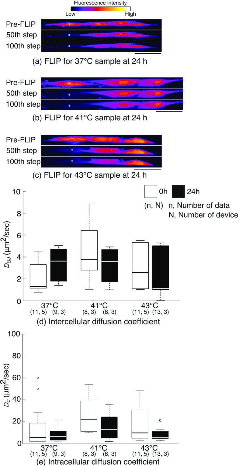
GJIC exhibited no significant alterations by the heat stimulations (Fig. 2). There were no remarkable differences among three experimental conditions (37 °C, 41 °C and 43 °C) in the extent of fluorescence decay in the cells adjacent to the target cells in FLIP experiments both at 0 h and 24 h (Fig. 2a-c for FLIP results for 37 °C, 41 °C and 43 °C sample at 24 h). At 0 h, although intercellular diffusion coefficient at 41 °C and 43 °C was higher than that at 37 °C, the differences were not statistically significant (Fig. 2d). Intracellular diffusion coefficient was also not changed significantly by the heating stimulations (Fig. 2e). At 24 h, both intercellular and intracellular diffusion coefficients in the three groups were approximately at an equal level.
Fig. 2.
a-c Representative fluorescence images during FLIP experiments performed at 24 h after the stimulation. Tenocytes were stimulated with 37 °C a, 41 °C b or 43 °C c heat stress. White asterisks indicate the location of target cell. Bars = 50 μm. c, d Intercellular and intracellular diffusion coefficients obtained from FLIP experiments. 0 h indicates an experimental group examined immediately after the end of 30 min heat stimulation, and 24 h examined at the end of 24-h post-incubation period. Open circles indicate the values identified by outliers based on statistical criterion and were included in statistical analyses.
It was clearly demonstrated that cell viability depended on the stimulating temperature (Table 1). The viability was 94.4 ± 3.0% (mean ± standard deviations), 89.9 ± 1.6% and 72.0 ± 13.0% at 24 h for 37 °C, 41 °C and 43 °C stimulation, respectively. The viability with 43 °C stimulation was significantly lower than that with 37 °C stimulation. The difference between 41 °C and 37 °C stimulations was not significant (P = 0.06).
Table 1.
Percentage of cell viability measured at 24 h after heat stimulations
| Primary tenocytes | 37 °C | 41 °C | 43 °C | |
| 94.4 ± 3.0 | 89.9 ± 1.6 | 72.0 ± 13.0* | ||
| 18αGA experiment | 37 °C (Vehicle) | 37 °C (18αGA) | 43 °C (Vehicle) | 43 °C (18αGA) |
| 91.3 ± 3.6 | 94.4 ± 2.2 | 70.5 ± 9.9* | 85.0 ± 2.4* | |
| Cx43 transfection | 37 °C (Mock) | 37 °C (pLX-Cx43) | 43 °C (Mock) | 43 °C (pLX-Cx43) |
| 94.1 ± 2.0 | 96.6 ± 1.5 | 75.0 ± 12.8 | 90.9 ± 2.7* |
Mean ± S.D.; number of experiments = 3; *P < 0.05 vs. corresponding 37 °C stimulation
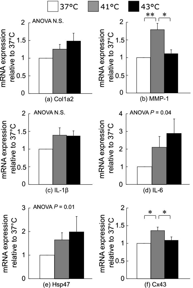
Expression profiles of selected genes demonstrated temperature-dependent significant alterations both at 0 and 24 h (Fig. 3). In the cells collected at 0 h, there were significant increases in MMP-1 and Cx43 gene expressions in the cells stimulated at 41 °C compared to those stimulated at 37 °C and 43 °C (Fig. 3b and f). However, no statistically significant differences were observed in Col1a2 and IL-1β gene expressions (Fig. 3a and c). There was an increasing trend in the expression of IL-6 gene expression with temperature, although differences between each temperature were not statistically significant (Fig. 3d). The expression of Hsp47 exhibited the same trend with IL-6 expression (Fig. 3e).
Fig. 3.
Results of qPCR analysis for the samples collected at 0 h (at the end of 30 min heat stimulation). a Col1a2, b MMP-1, c IL-1β, d IL-6, e Hsp47 and f Cx43. *P < 0.05 and **P < 0.01. The number of samples was 7 in all the three groups. Mean + SE
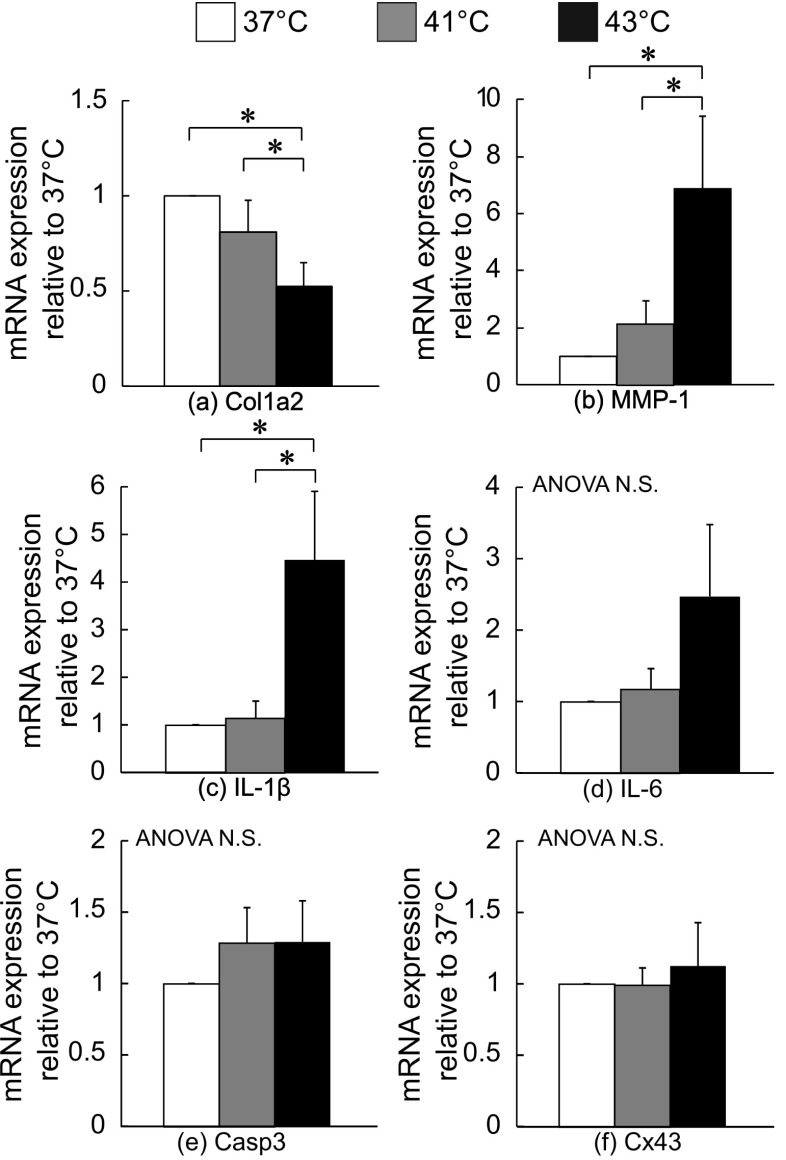
At 24 h after the heat stimulation, a much clear trend of the elevation of tenocyte catabolism as well as the inhibition of tenocyte anabolism was observed (Fig. 4). For Col1a2 mRNA, the expressions in the cells stimulated at 41 °C and 43 °C were significantly lower than that stimulated at 37 °C (Fig. 4a). In contrast, there was an increasing trend in the expression for MMP-1, IL-1β and IL-6 mRNA with the temperature. Indeed, the levels for MMP-1 in the cells stimulated at 43 °C was significantly higher than that at 37 °C and 41 °C (Fig. 4b). The expression of IL-1β in 43 °C was also significantly higher than that in 37 °C and 41 °C (Fig. 4c). There were no significant changes for IL-6, Casp3 and Cx43 mRNA expressions (Fig. 4d-f).
Fig. 4.
Results of qPCR analysis for the samples collected at 24 h (at the end of 24-h post-incubation period). a Col1a2, b MMP-1, c IL-1β, d IL-6, e Caps3 and f Cx43. *P < 0.05. The number of samples was 6 in all the three groups. Mean + SE
Effects of GJ blocking on tenocyte responses to heat stress
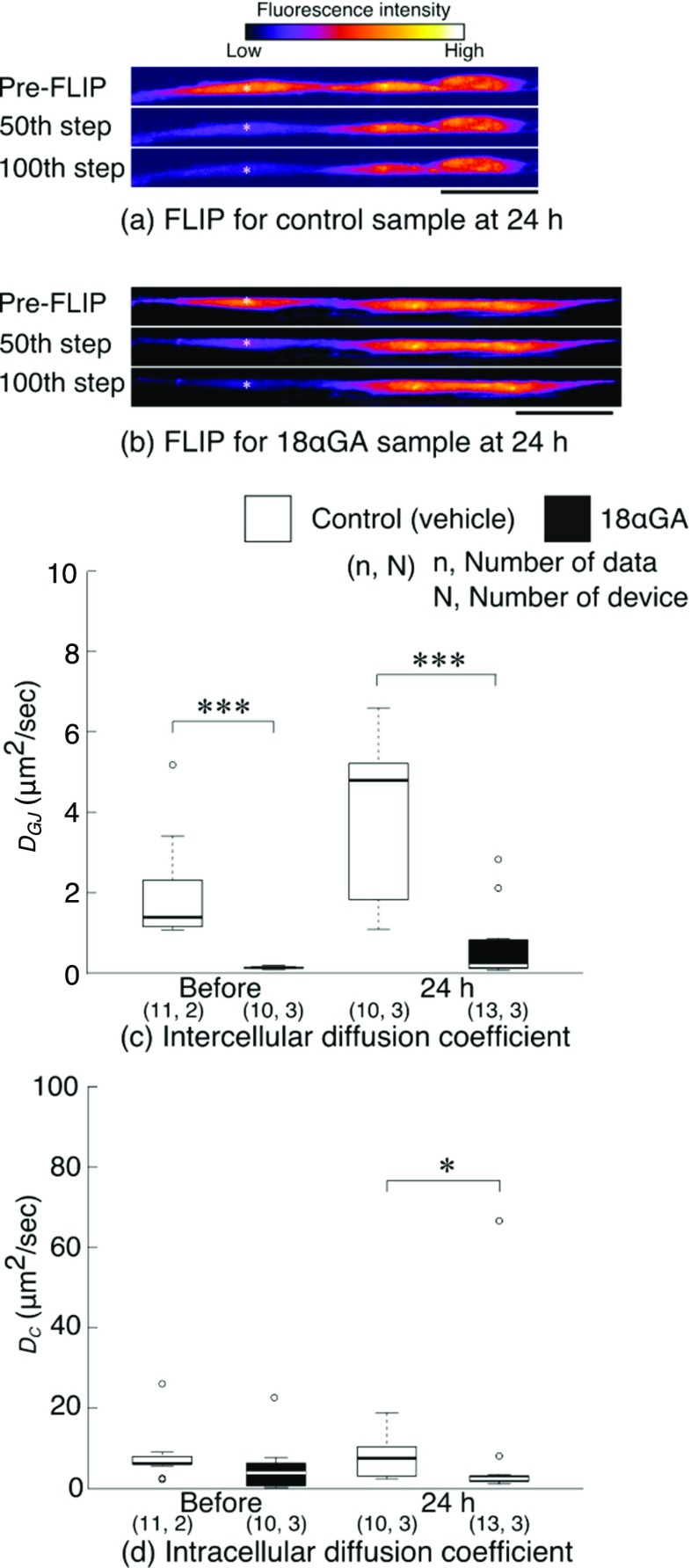
The supplementation of 18αGA remarkably suppressed intercellular molecular transport, which was evident from the fluorescence decay in FLIP experiment (Fig. 5a and b). This was also confirmed by a significantly low level of intercellular diffusion coefficient at 1 h after the supplementation (before the heat stimulation) (Fig. 5c). The suppressive effect on GJIC was also observed at 24 h after the heat stimulation. Intercellular diffusion coefficient for the control group was at a level similar with primary tenocytes (Fig. 2d). At 24 h, the coefficient was elevated, although the change was not statistically significant. The difference between the control and 18αGA groups was statistically significant both before and after the stimulation. On the other hand, there were no significant differences between the two groups in the levels of intracellular diffusion coefficient at both 0 and 24 h (Fig. 5d).
Fig. 5.
a, b Representative fluorescence images of FLIP experiments of a control and b 18αGA groups, performed at 24 h after the heat stimulation at 43 °C for 30 min. White asterisks indicate the location of target cell. Bars = 50 μm. a Control tenocytes treated with DMSO (vehicle) only. b Tenocytes treated with 18αGA. c, d Intercellular and intracellular diffusion coefficients obtained from FLIP experiments. Open circles indicate the values identified by outliers based on statistical criterion and were included in statistical analyses. *P < 0.05 and ***P < 0.001
Heat stimulation at 43 °C significantly reduced cell viability in the control (70.5 ± 9.9%) from the levels obtained by the stimulation at 37 °C for 30 min (91.3 ± 3.6%). Similar to the control group, 18αGA group also demonstrated a significant reduction of cell viability by 43 °C heat stimulation (85.0 ± 2.4% by 43 °C stimulation from 94.7 ± 2.2% by 37 °C stimulation). Although the viability in 18αGA group was higher than that of the control, the difference was not significant (P = 0.07).
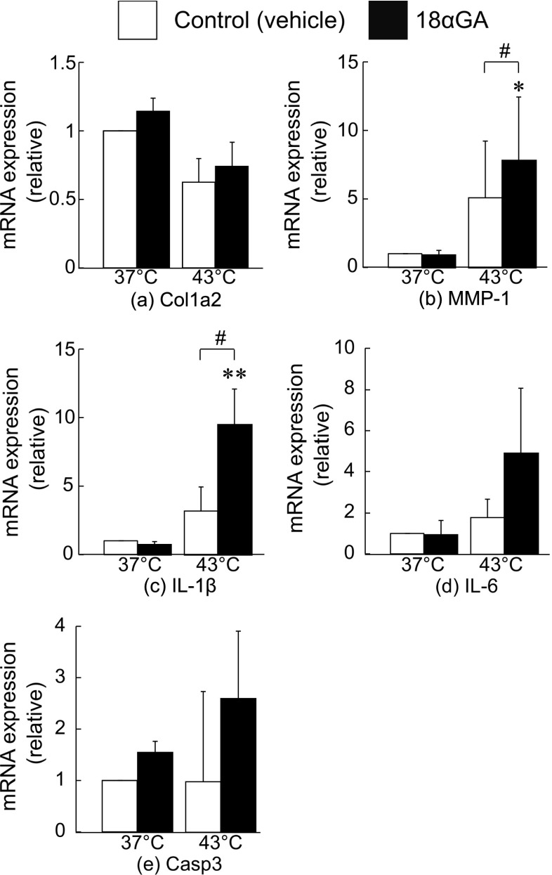
The inhibition of gap junction communication demonstrated stimulatory effects on heat-induced upregulation of catabolic gene expression (Fig. 6). With the stimulation at 37 °C, the expression level of genes examined in the control group were at similar levels to 18αGA group. For Col1a2 mRNA, the expression was downregulated by 43 °C stimulation in both the control and 18αGA groups, although the difference between the two groups was not statistically significant (Fig. 6a). In the control group, MMP-1 and IL-1β mRNA expression was upregulated by the stimulation at 43 °C. This was further and significantly upregulated when GJIC was inhibited (Fig. 6b and c). IL-6 expression also exhibited the same trend, although the changes were not statistically significant (Fig. 6d). No significant changes were also observed in the expressions of Casp3 gene (Fig. 6e).
Fig. 6.
Results of qPCR analysis for the samples collected at 24 h. a Col1a2, b MMP-1, c IL-1β, d IL-6 and e Casp3. *P < 0.05 vs 37 °C, **P < 0.01 vs 37 °C and # P < 0.05. The number of sample was 5 in all the four groups. Mean + SE
Effects of Cx43 gene transfection on tenocyte response to heat stress
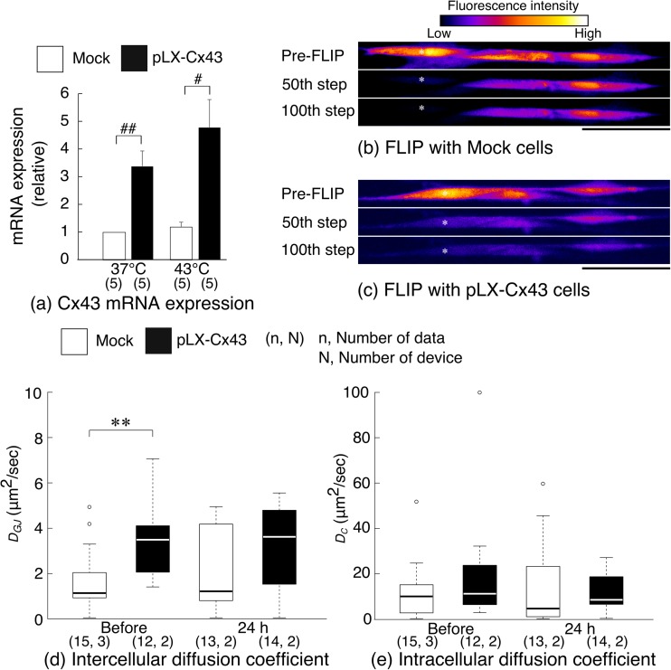
It was observed that Cx43 mRNA expression in pLX-Cx43 group was 3.8 times higher following 37 °C stimulation and 4.0 times higher following 43 °C stimulation than that in the mock control group. Indeed, the differences were statistically significant (Fig. 7a). The increased Cx43 mRNA expression was reflected in the level of GJIC; there were marked differences between the mock and pLX-Cx43 groups in the extent of fluorescence decay in the cells adjacent to the target cell in FLIP experiments (Fig. 7b and c). Indeed, the intercellular diffusion coefficient of pLX-Cx43 group before the 43 °C stimulation was significantly higher than that of the mock group (Fig. 7d). The elevated level of intercellular diffusion coefficient was also evident at 24 h. Because the coefficient in the mock group at 24 h exhibited a large variability, there was no statistically significant difference between the two groups at 24 h. For intracellular diffusion coefficient, there were no significant differences between the two groups both before and after the stimulations (Fig. 7e).
Fig. 7.
a Expression of Cx43 mRNA in mock and pLX-Cx43 tenocytes subjected to 37 °C or 43 °C stimulation. The expression was measured at the end of 24 h post-incubation. # P < 0.05 and ## P < 0.01. ( ) = number of sample. Mean + SE. b, c Representative fluorescence images during FLIP experiments of b the mock control and c pLX-Cx43 groups, performed at 24 h after the stimulation with 43 °C. White asterisks indicate the location of target cell. Bars = 50 μm. d, e Intercellular and intracellular diffusion coefficients obtained from FLIP experiments. Open circles indicate the values identified by outliers based on statistical criterion and were included in statistical analyses. **P < 0.01
Cell viability in the samples subjected to 37 °C stimulation was 94.1 ± 2.0% and 96.6 ± 1.5% in the mock control and pLX-Cx43 groups, respectively (Table 1). This was decreased to 75.0 ± 12.8% and 90.9 ± 2.7% by 43 °C stimulation in the mock control and pLX-Cx43 groups, respectively. The decrease in pLX-Cx43 group was only statistically significant. The difference between the mock and pLX-Cx43 groups in 43 °C stimulation was not significant (P = 0.10).
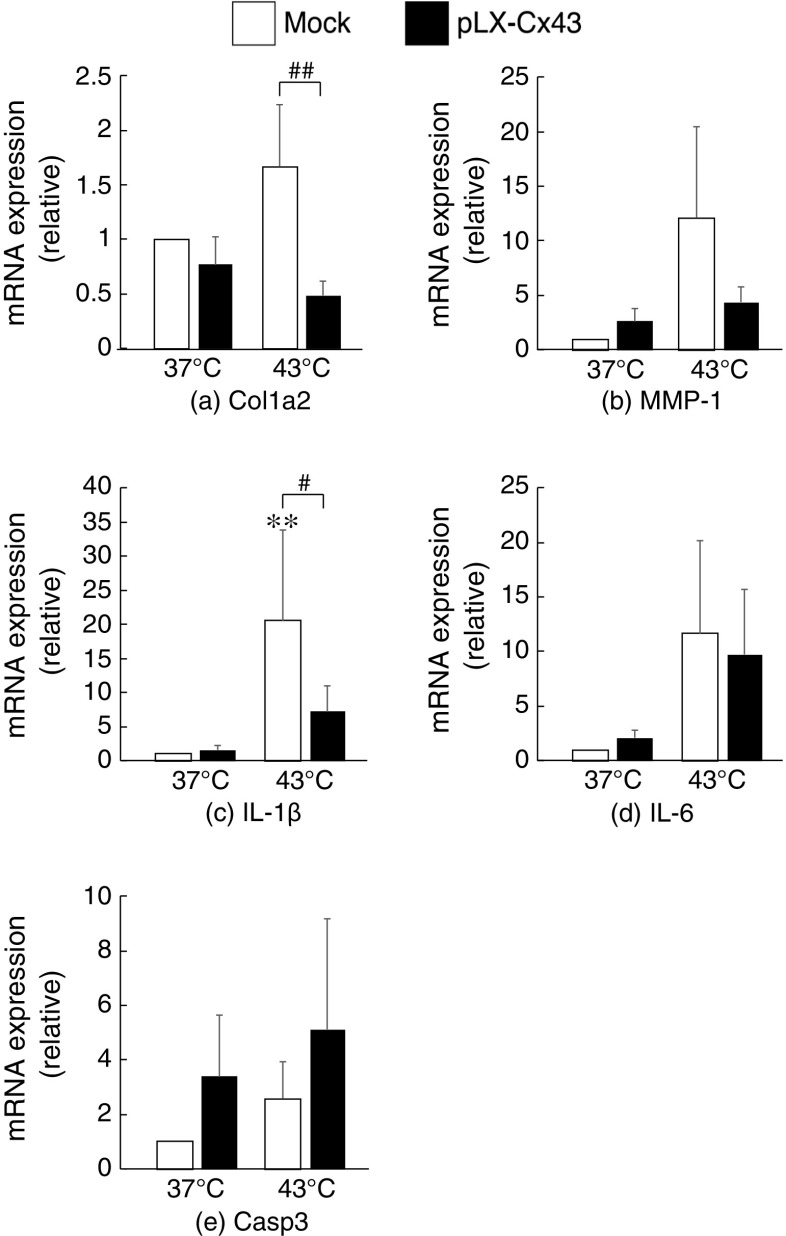
In the gene expression profiles, there were no significant differences between the mock and pLX-Cx43 groups in the case of the stimulation at 37 °C (Fig. 8). In contrast, the expressions of Col1a2, MMP-1 and IL-1β were approximately 70% lower in pLX-Cx43 group than the mock control group following the stimulation at 43 °C (Fig. 8a-c). Indeed, statistically significant difference was observed in Col1a2 and IL-1β. IL-6 expression exhibited a similar trend with MMP-1 expression, but the difference was not significant (Fig. 8d). Casp3 expression in pLX-Cx43 group was higher than the control group in both cases, although the differences were not significant (Fig. 8e).
Fig. 8.
Results of qPCR analysis for the samples collected at 24 h. a Col1a2, b MMP-1, c IL-1β, d IL-6 and e Caps3. **P < 0.01 vs 37 °C. # P < 0.05 and ## P < 0.01. The number of sample was 5 in all the four conditions. Mean + SE.
Discussion
The present study has tested the hypothesis that the application of heat stress to tenocytes induces catabolic and proinflammatory responses, and this can be modulated by GJIC. Results obtained showed that the heat stress at 43 °C for 30 min induced the death in a small population of normal primary tenocytes and upregulations of the expression of catabolic and proinflammatory genes from surviving cells. In the case that primary tenocytes were treated with non-specific GJIC inhibitor, 18αGA, the cell viability was slightly improved but catabolic and proinflammatory genes were further upregulated. In contrast, the enhanced GJIC, resulting from forced upregulation of Cx43 mRNA expression, improved cell viability and decreased the extent of the upregulation of catabolic and proinflammatory gene expressions. Accordingly, it has been confirmed that GJIC is involved in the regulation of catabolic and proinflammatory responses to the heat stress, and the modulation of GJIC alters tenocyte functions to the heat stress.
The trends of tenocyte responses to heat stress observed in the present study are essentially similar with those reported in the past studies (Birch et al. 1997; Hosaka et al. 2006). Namely, heat stimulation at above 40 °C resulted in cell death and catabolic gene expression. However, the alterations in GJIC by heat stress was different from those observed in the past studies. In the present study, there have been no evidence of significant changes in intercellular diffusion coefficient (the amount of GJIC) by heat stimulation (Fig. 2d). Stimulation of human fibroblasts at 43 °C resulted in a time-dependent decrease in cell viability and GJIC (Hamada et al. 2003). Treatment at 42 °C for 6 h increased GJIC in rabbit skeletal myoblasts but decreased in Cx43-transfected HeLa cells (Antanavičiute et al. 2014). Differences in GJIC response may reflect functional requirements in different anatomical locations.
For heat sensing mechanisms of cells, transient receptor potential vanilloid-1 (TRPV1) channel has been identified as a heat sensing ion channel (Caterina et al. 1997) and shown to open the channel by heat stimulation above a threshold temperature of approximately 43 °C (Hayes et al. 2000). In addition, the involvement of TRPV1 channel in the heat-induced gene expression has been reported for MMP-1 (Li et al. 2007; Lee et al. 2008) and IL-6 (Son et al. 2015). To examine the mechanism of the heat-induced upregulation of MMP-1 and IL-1β mRNAs in tenocytes, we have also examined if TRPV1 channels are involved in response to the heat stimulation at 43 °C. Although TRPV1 gene expression was confirmed, the use of TRPV1 inhibitor, capsazepin, in the experiments with normal primary tenocytes demonstrated no effects on the heat-induced upregulation of MMP-1 and IL-1β gene expressions (data not shown). Because these responses were essentially similar with results obtained with normal primary tenocytes, it was suggested that TRPV1 channels may play a minor role in tenocyte heat sensing and subsequent changes in cell functions. For other mechanisms, we have examined pathways involving heat shock proteins. Among a family of heat shock proteins, the synthesis of Hsp47 is reported to be upregulated at 42 °C, while the synthesis of other major heat shock proteins is upregulated at a higher temperature (e.g. 45 °C) (Nagata et al. 1986). Indeed, it was observed that Hsp47 gene expression was elevated in the cells treated with 43 °C stimulation compared to those treated at 37 °C. Although the difference in the Hsp47 expression level between 37 °C and 43 °C was not statistically significant (Fig. 3e), these findings may suggest that tenocyte heat sensing and associated changes in cell functions involved pathways including heat shock proteins. It has been also reported that the permeability of the plasma membrane is elevated at the temperature above 40 °C compared to at 37 °C, leading to passive ion leakage (Bischof et al. 1995). Thus, changes in intracellular concentration of ions due to the compromised plasma membrane could be another mechanism of evoking changes in tenocyte functions by heat stress.
According to previous studies, roles of GJIC are classified into two categories. One is that signals of cell injury or death are transferred to neighbouring cells via GJIC (Brosnan et al. 2001; García-Dorado et al. 2004). The other is that cells connected with gap junctions share molecules necessary to survive (Pitts 1998) and stabilise cellular calcium homeostasis, making the cells less vulnerable to external stress (Blanc et al. 1998). As for tendon, tenocytes within tendon explant subjected to mechanical loading upregulated collagen synthesis, which was abolished by the inhibition of GJIC between tenocytes (Banes et al. 1999). It has also been reported in tenocytes that GJIC through Cx32 demonstrated a stimulatory effect on collagen synthesis, whereas GJIC through Cx43 an inhibitory effect (Waggett et al. 2006). Cell apoptosis was induced in tenocytes subjected to cyclic strain when GJIC was blocked (Qi et al. 2011). In the present study, the application of heat stress at 43 °C to GJIC-inhibited tenocytes induced further upregulation of MMP-1 and IL-1β gene expressions but improved cell viability slightly (Fig. 6 and Table 1). The enhanced expression of Cx43 mRNA and associated elevation of GJIC counteracted the heat-induced upregulation of MMP-1 and IL-1β genes and the reduction of cell viability (Fig. 8 and Table 1). Accordingly, the closure of gap junctions and thus the inhibition of GJIC in tenocytes helped the cells to survive heat stress but induce a profound pro-catabolic effects, while GJIC enhancement helped the cells to survive and suppress their catabolism. Thus, it is indicated that the presence of GJIC is prerequisite for normal functioning of tenocytes, and plays a cytoprotective role to external stress when GJIC is enhanced.
It may be deemed counterintuitive that both GJ blocking and Cx43 overexpression and associated GJIC enhancement improved the viability of tenocytes following the stimulation at 43 °C. Indeed, GJ blocking has demonstrated to impair cell viability when extrinsic stress was applied to the cells (Hamada et al. 2003; Qi et al. 2011). It was also shown that GJ plays a minor role in the resistance to heat stress, but the formation of tight junctions protects cells against heat stress (Ning and Hahn 1994). Slight improvement of cell viability observed in GJ blocking experiments in the present study may indicate that the absence of GJIC play no role in the cell viability following heat stress, but there may be a mechanism of compensating GJIC loss by other cell-cell junctions such as tight junctions and desmosomes. In the case of enhanced GJIC, it was reported that a reduction of cell viability by damages from extrinsic stress was improved by Cx43 overexpression (Lin et al. 2003), and this was reportedly independent of GJ channel functions. Therefore, it is assumed that changes in GJIC have little effects on cell viability in heat stimulation, but instead associated changes in cell structure resulted in affecting the viability.
Type I collagen (Col1a2) gene expression was downregulated in response to 43 °C stimulation in all the experimental conditions, except for the mock control group in Gja1 transfection experiment. It has been demonstrated that mRNA expressions of type I collagen and MMP-1 are regulated in a reciprocal manner (Lavagnino et al. 2003; Lavagnino and Arnoczky 2005); when MMP-1 mRNA expression is upregulated, type I collagen mRNA expression is suppressed, and vice versa. The overall trend of the results obtained in the present study follows this reciprocal expression pattern. Because the mock control group exhibited upregulation of both Col1a2 and MMP-1 mRNA expressions in response to 43 °C stimulation, Col1a2 expression in pLX-CX43 group following 43 °C stimulation was seemed significantly downregulated in comparison to that in the mock control. We confirmed that Col1a2 mRNA was still robustly expressed even after 43 °C stimulation. Namely, an average Ct value for Col1a2 gene in pLX-Cx43 group following 43 °C stimulation was 24.2, suggesting that Col1a2 expression was still at a sufficient level even though the expression level was reduced to the half of the level in the mock control treated at 37 °C. Therefore, the manipulation of GJIC alone may have little effect on type I collagen expression, and the effect of the manipulation on collagen expression would become evident when the manipulation was in conjunction with stimulation in favor of collagen expression such as mechanical loading.
The implication of the present findings to the pathogenesis of tendinopathy is that heat stress may synergistically act with mechanical overloading to induce cell death and catabolism in overloaded tissue (Scott et al. 2005; Lavagnino et al. 2006; Egerbacher et al. 2008). Overloading could break not only extracellular matrix but also gap junctional network. Indeed, we have demonstrated in an in vitro study that the level of GJIC in tenocytes subjected to static strain with a physiological amplitude is higher than that of non-strained tenocytes or those under static strain with an over-physiological amplitude (Maeda et al. 2017). Thus, within tendon exposed to a large magnitude of cyclic loading, it is thought that GJIC is suppressed either by overstretching the cellular network or the removal of loading to the cells due to the rupture of load-bearing collagen fibers (Lavagnino et al. 2006). Therefore, a reduced level of GJIC may further enhance tenocyte catabolism, which is already upregulated by mechanical overloading and heat stress. This potentially leads to the onset of tendinopathy. To prevent tendon injury at an early stage from becoming chronic injury, tendon core temperature and GJIC could be targets for post-activity treatments.
It should be noted that regulation of cell functions with connexin expression does not necessarily require the establishment of GJIC (Lin et al. 2003). Even the formation of non-functional gap junctions or the formation of hemichannels improved the cell resistance to cytotoxic stress. This may suggest that GJIC plays a minor role in the stress resistance. In the present study, the elevation of GJIC was demonstrated in Cx43-transfected tenocytes using FLIP technique. Nonetheless, it is possible that a fraction of connexins synthesised form hemichannels that may mainly contribute to the inhibition of tenocyte catabolic responses and the improvement of cell viability.
In the present study, we have only examined changes in tenocyte functions at transcriptional level, and thus the findings obtained cannot directly be extrapolated to the tissue level. Type I collagen is a secreted protein undergoing several post-translational modifications related to maturation (cross-linking) or degradation (by MMPs). To understand if there is any increased catabolism in collagen, type I collagen synthesis is needed to be analysed at the protein level. Similarly, gene expression analysis of MMP-1 is not sufficient to predict the activity, and thus MMP-1 activity is also needed to be analysed using zymography. Moreover, TIMP-1, the main inhibitor of MMP-1 should also be analysed in terms of collagen turnover. The lack of these findings is the limitation of the present study, and we will examine in future study how the manipulations of GJIC changes tenocyte functions and responses to external stress, such as mechanical loading and heat stimulation, at protein level.
It is known that the opening and closure of GJ pores is regulated by changes in transjunctional voltage difference, cytoplasmic acidification, intracellular Ca2+ concentration, or chemical uncouplers (Bukauskas and Verselis 2004). In addition, it was reported that heat stress promoted Cx43 phosphorylation and resulted in the inhibition of GJIC (Hamada et al. 2003). Therefore, although there were no significant alterations in intercellular diffusion coefficient between each of the three temperatures of the stimulation in the experiment with normal primary tenocytes, these results may be from a combination of the gating of GJ pores due to Cx43 phosphorylation, changes in intracellular Ca2+ concentration as well as recruitment and internalization of Cx43 connexons. To understand the regulation of GJIC in detail, such gating mechanisms are also needed to investigate.
In conclusion, tenocytes from rabbit Achilles tendon underwent cell death and exhibited catabolic and proinflammatory responses from surviving cells by heat stress at 43 °C for 30 min. The inhibition of GJIC had a limited effect on improving these responses. However, forced expression of Cx43 and associated elevation of GJIC suppressed tenocyte catabolic responses to heat stress and improved the cell viability. Therefore, enhanced Cx43 GJIC in tendon may possess an inhibitory effect on catabolic and proinflammatory responses to against heat stress.
Electronic supplementary material
(DOCX 72 kb)
Acknowledgements
EM thanks Professor Takeo Matsumoto in Nagoya University for generous supports. The present study was supported in part by the Japan Society for the Promotion of Science (JSPS) KAKENHI Grants 25702022 and 16 K01346.
Abbreviations
- Casp
Caspase
- Col1a2
Type I collagen alpha 2 chain
- Cx
Connexin
- FLIP
Fluorescence loss in photobleaching
- Gapdh
Glyceraldehyde 3-phosphate dehydrogenase
- GJIC
Gap junction intercellular communication
- Hsp
Heat shock protein
- IL
Interluekin
- MMP
Matrix metalloproteinase
- PDMS
Polydimethylsiloxane
- TRPV1
Transient receptor potential vanilloid-1
- 18αGA
18α glycyrrhetinic acid
Compliance with ethical standards
Conflict of interests
The authors declare no conflicts of interest.
Footnotes
Electronic supplementary material
The online version of this article (doi:10.1007/s12079-017-0397-3) contains supplementary material, which is available to authorized users.
References
- Antanavičiute I, Mildažiene V, Stankevičius E, et al. Hyperthermia differently affects connexin43 expression and gap junction permeability in skeletal myoblasts and Hela cells. Mediat Inflamm. 2014;2014:748290. doi: 10.1155/2014/748290. [DOI] [PMC free article] [PubMed] [Google Scholar]
- Banes AJ, Weinhold P, Yang X et al (1999) Gap junctions regulate responses of tendon cells ex vivo to mechanical loading. Clin Orthop Relat Res 367S:S356–S370 [DOI] [PubMed]
- Birch HL, Wilson AM, Goodship AE. The effect of exercise-induced localised hyperthermia on tendon cell survival. J Exp Biol. 1997;200:1703–1708. doi: 10.1242/jeb.200.11.1703. [DOI] [PubMed] [Google Scholar]
- Bischof JC, Padanilam J, Holmes WH, et al. Dynamics of cell membrane permeability changes at supraphysiological temperatures. Biophys J. 1995;68:2608–2614. doi: 10.1016/S0006-3495(95)80445-5. [DOI] [PMC free article] [PubMed] [Google Scholar]
- Blanc EM, Bruce-Keller AJ, Mattson MP. Astrocytic gap junctional communication decreases neuronal vulnerability to oxidative stress-induced disruption of Ca2+ homeostasis and cell death. J Neurochem. 1998;70:958–970. doi: 10.1046/j.1471-4159.1998.70030958.x. [DOI] [PubMed] [Google Scholar]
- Brosnan CF, Scemes E, Spray DC. Cytokine regulation of gap junction connectivity. Am J Pathol. 2001;158:1565–1569. doi: 10.1016/S0002-9440(10)64110-7. [DOI] [PMC free article] [PubMed] [Google Scholar]
- Bukauskas FF, Verselis VK. Gap junction channel gating. Biochim Biophys Acta Biomembr. 2004;1662:42–60. doi: 10.1016/j.bbamem.2004.01.008. [DOI] [PMC free article] [PubMed] [Google Scholar]
- Caterina MJ, Schumacher MA, Tominaga M, et al. The capsaicin receptor: a heat-activated ion channel in the pain pathway. Nature. 1997;389:816–824. doi: 10.1038/39807. [DOI] [PubMed] [Google Scholar]
- Egerbacher M, Arnoczky SP, Caballero O, et al. Loss of homeostatic tension induces apoptosis in tendon cells: an in vitro study. Clin Orthop Relat Res. 2008;466:1562–1568. doi: 10.1007/s11999-008-0274-8. [DOI] [PMC free article] [PubMed] [Google Scholar]
- Farris DJ, Trewartha G, Polly McGuigan M. Could intra-tendinous hyperthermia during running explain chronic injury of the human Achilles tendon? J Biomech. 2011;44:822–826. doi: 10.1016/j.jbiomech.2010.12.015. [DOI] [PubMed] [Google Scholar]
- García-Dorado D, Rodríguez-Sinovas A, Ruiz-Meana M. Gap junction-mediated spread of cell injury and death during myocardial ischemia-reperfusion. Cardiovasc Res. 2004;61:386–401. doi: 10.1016/j.cardiores.2003.11.039. [DOI] [PubMed] [Google Scholar]
- Hamada N, Kodama S, Suzuki K, Watanabe M. Gap junctional intercellular communication and cellular response to heat stress. Carcinogenesis. 2003;24:1723–1728. doi: 10.1093/carcin/bgg135. [DOI] [PubMed] [Google Scholar]
- Hayes P, Meadows HJ, Gunthorpe MJ, et al. Cloning and functional expression of a human orthologue of rat vanilloid receptor-1. Pain. 2000;88:205–215. doi: 10.1016/S0304-3959(00)00353-5. [DOI] [PubMed] [Google Scholar]
- Hosaka Y, Ozoe S, Kirisawa R, et al. Effect of heat on synthesis of gelatinases and pro-inflammatory cytokines in equine tendinocytes. Biomed Res. 2006;27:233–241. doi: 10.2220/biomedres.27.233. [DOI] [PubMed] [Google Scholar]
- Lavagnino M, Arnoczky SP. In vitro alterations in cytoskeletal tensional homeostasis control gene expression in tendon cells. J Orthop Res. 2005;23:1211–1218. doi: 10.1016/j.orthres.2005.04.001. [DOI] [PubMed] [Google Scholar]
- Lavagnino M, Arnoczky SP, Tian T, Vaupel Z. Effect of amplitude and frequency of cyclic tensile strain on the inhibition of MMP-1 mRNA expression in tendon cells: an in vitro study. Connect Tissue Res. 2003;44:181–187. doi: 10.1080/03008200390215881. [DOI] [PubMed] [Google Scholar]
- Lavagnino M, Arnoczky SP, Egerbacher M, et al. Isolated fibrillar damage in tendons stimulates local collagenase mRNA expression and protein synthesis. J Biomech. 2006;39:2355–2362. doi: 10.1016/j.jbiomech.2005.08.008. [DOI] [PubMed] [Google Scholar]
- Lee YM, Li WH, Kim YK, et al. Heat-induced MMP-1 expression is mediated by TRPV1 through PKCa signaling in HaCaT cells. Exp Dermatol. 2008;17:864–870. doi: 10.1111/j.1600-0625.2008.00738.x. [DOI] [PubMed] [Google Scholar]
- Legerlotz K, Jones GC, Screen HRC, Riley GP. Cyclic loading of tendon fascicles using a novel fatigue loading system increases interleukin-6 expression by tenocytes. Scand J Med Sci Sports. 2013;23:31–37. doi: 10.1111/j.1600-0838.2011.01410.x. [DOI] [PMC free article] [PubMed] [Google Scholar]
- Levin M. Gap junctional communication in morphogenesis. Prog Biophys Mol Biol. 2007;94:186–206. doi: 10.1016/j.pbiomolbio.2007.03.005. [DOI] [PMC free article] [PubMed] [Google Scholar]
- Li WH, Lee YM, Kim JY, et al. Transient receptor potential vanilloid-1 mediates heat-shock-induced matrix metalloproteinase-1 expression in human epidermal keratinocytes. J Invest Dermatol. 2007;127:2328–2335. doi: 10.1038/sj.jid.5700880. [DOI] [PubMed] [Google Scholar]
- Lin JH-C, Yang J, Liu S, et al. Connexin mediates gap junction-independent resistance to cellular injury. J Neurosci. 2003;23:430–441. doi: 10.1523/JNEUROSCI.23-02-00430.2003. [DOI] [PMC free article] [PubMed] [Google Scholar]
- Maeda E, Ohashi T. Mechano-regulation of gap junction communications between tendon cells is dependent on the magnitude of tensile strain. Biochem Biophys Res Commun. 2015;465:281–286. doi: 10.1016/j.bbrc.2015.08.021. [DOI] [PubMed] [Google Scholar]
- Maeda E, Ye S, Wang W, et al. Gap junction permeability between tenocytes within tendon fascicles is suppressed by tensile loading. Biomech Model Mechanobiol. 2012;11:439–447. doi: 10.1007/s10237-011-0323-1. [DOI] [PubMed] [Google Scholar]
- Maeda E, Hagiwara Y, Wang JHC, Ohashi T. A new experimental system for simultaneous application of cyclic tensile strain and fluid shear stress to tenocytes in vitro. Biomed Microdevices. 2013;15:1067–1075. doi: 10.1007/s10544-013-9798-0. [DOI] [PubMed] [Google Scholar]
- Maeda E, Pian H, Ohashi T. Temporal regulation of gap junctional communication between tenocytes subjected to static tensile strain with physiological and non-physiological amplitudes. Biochem Biophys Res Commun. 2017;482:1170–1175. doi: 10.1016/j.bbrc.2016.12.007. [DOI] [PubMed] [Google Scholar]
- Nagata K, Saga S, Yamada KM. A major collagen-binding protein of chick embryo fibroblasts is a novel heat shock protein. J Cell Biol. 1986;103:223–229. doi: 10.1083/jcb.103.1.223. [DOI] [PMC free article] [PubMed] [Google Scholar]
- Ning S, Hahn GM. Formation of tight junctions and desmosomes protects MDCK cells against hyperthermic killing. J Cell Physiol. 1994;160:249–254. doi: 10.1002/jcp.1041600206. [DOI] [PubMed] [Google Scholar]
- Patterson-Kane JC, Becker DL, Rich T. The pathogenesis of tendon Microdamage in athletes: the horse as a natural model for basic cellular research. J Comp Pathol. 2012;147:227–247. doi: 10.1016/j.jcpa.2012.05.010. [DOI] [PubMed] [Google Scholar]
- Pitts JD. The discovery of metabolic co-operation. BioEssays. 1998;20:1047–1051. doi: 10.1002/(SICI)1521-1878(199812)20:12<1047::AID-BIES11>3.0.CO;2-0. [DOI] [PubMed] [Google Scholar]
- Qi J, Chi L, Bynum D, Banes AJ. Gap junctions in IL-1beta-mediated cell survival response to strain. J Appl Physiol. 2011;110:1425–1431. doi: 10.1152/japplphysiol.00477.2010. [DOI] [PubMed] [Google Scholar]
- Scott A, Khan KM, Heer J, et al. High strain mechanical loading rapidly induces tendon apoptosis: an ex vivo rat tibialis anterior model. Br J Sports Med. 2005;39:e25. doi: 10.1136/bjsm.2004.015164. [DOI] [PMC free article] [PubMed] [Google Scholar]
- Son GY, Hong JH, Chang I, Shin DM. Induction of IL-6 and IL-8 by activation of thermosensitive TRP channels in human PDL cells. Arch Oral Biol. 2015;60:526–532. doi: 10.1016/j.archoralbio.2014.12.014. [DOI] [PubMed] [Google Scholar]
- Sun HB, Li Y, Fung DT, et al. Coordinate regulation of IL-1b and MMP-13 in rat tendons following subrupture fatigue damage. Clin Orthop Relat Res. 2008;466:1555–1561. doi: 10.1007/s11999-008-0278-4. [DOI] [PMC free article] [PubMed] [Google Scholar]
- Waggett AD, Benjamin M, Ralphs JR. Connexin 32 and 43 gap junctions differentially modulate tenocyte response to cyclic mechanical load. Eur J Cell Biol. 2006;85:1145–1154. doi: 10.1016/j.ejcb.2006.06.002. [DOI] [PubMed] [Google Scholar]
- Wall ME, Banes AJ. Early responses to mechanical load in tendon: role for calcium signaling, gap junctions and intercellular communication. J Musculoskelet Neuronal Interact. 2005;5:70–84. [PubMed] [Google Scholar]
- Wang JHC. Mechanobiology of tendon. J Biomech. 2006;39:1563–1582. doi: 10.1016/j.jbiomech.2005.05.011. [DOI] [PubMed] [Google Scholar]
- Wilson AM, Goodship AE. Exercise-induced hyperthermia as a possible mechanism for tendon degeneration. J Biomech. 1994;27:899–905. doi: 10.1016/0021-9290(94)90262-3. [DOI] [PubMed] [Google Scholar]
Associated Data
This section collects any data citations, data availability statements, or supplementary materials included in this article.
Supplementary Materials
(DOCX 72 kb)


